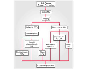
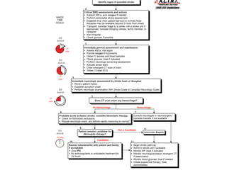
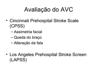
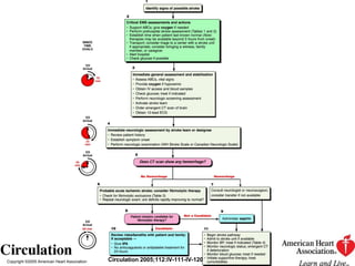
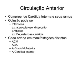
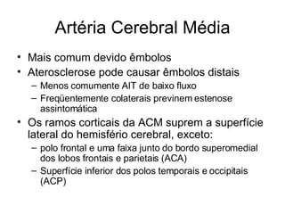
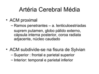
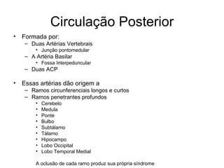
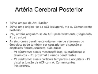
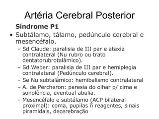
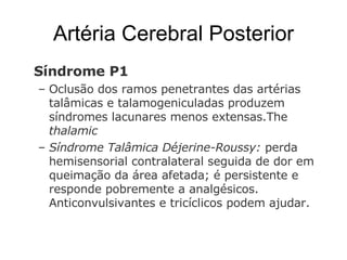
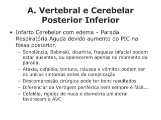
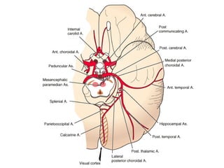
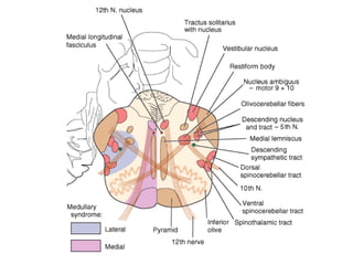
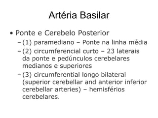
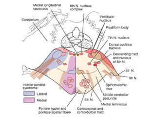
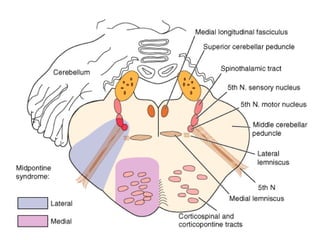
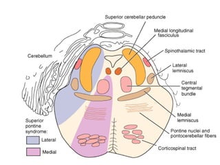
Semiologia do AVC isquêmico Tiago O. Malucelli  R2 CM
 
 
Avaliação do AVC  Cincinnati Prehospital Stroke Scale (CPSS) Assimetria facial Queda do braço Alteração da fala Los Angeles Prehospital Stroke Screen (LAPSS)
Copyright ©2005 American Heart Association Circulation 2005;112:IV-111-IV-120
Síndromes IMPORTÂNCIA: Uma história cuidadosa pode freqüentemente localizar a região da disfunção cerebral Dependendo da região afetada, determinada distribuição arterial será a provável responsável; e as possíveis causas poderão ser estreitadas, com especial importância no AIT com EF normal Se o paciente desenvolveu alteração de linguagem e hemianopsia homônima direita, uma procura por êmbolos da artéria cerebral média esquerda deverá ser realizada. Um achado de estenose da artéria carótida interna direita, por exemplo, sugere ser assintomática, e a procura por outras causas deverá prosseguir.
Síndromes São divididas em: (1) Circulação Anterior (2) Circulação Posterior  (3) Pequenos Vasos (Lacunares)
Circulação Anterior Compreende Carótida Interna e seus ramos Oclusão pode ser Intrínseca ex: aterosclerose, dissecção Embólica ex: FA, estenose carótida Cada artéria em manifestações distintas ACM ACA A Coroidal Anterior A Carótida Interna
 
Artéria Cerebral Média Mais comum devido êmbolos Aterosclerose pode causar êmbolos distais Menos comumente AIT de baixo fluxo Freqüentemente colaterais previnem estenose assintomática Os ramos corticais da ACM suprem a superfície lateral do hemisfério cerebral, exceto: polo frontal e uma faixa junto do bordo superomedial dos lobos frontais e parietais (ACA) Superfície inferior dos polos temporais e occipitais (ACP)
 
Artéria Cerebral Média ACM proximal Ramos penetrantes – a. lenticuloestriadas suprem putamen, globo pálido externo, cápsula interna posterior, coroa radiata adjacente, núcleo caudado ACM subdivide-se na fisura de Sylvian Superior : frontal e parietal superior Interior: temporal e parietal inferior
 
 
Artéria Cerebral Média Se oclusão na origem da ACM (inclusive penetrantes), c/ colaterais limitados: Hemiplegia contralateral, hemianestesia, hemianopsia homônima, um a dois dias de desvio ocular ipsilateral. Disartria é comum devido fraqueza facial.  Se hemisfério dominante, afasia global.  Se hemisfério não-dominante, anosognosia, apraxia construcional e negligência. Síndromes completas ocorrem quando êmbolos ocluem o tronco. Síndromes paciais ocorrem devido colaterais, alterações anatômicas e êmbolos que ocluem parcialmente a AMC, seus ramos, ou fragmentos que movem-se distalmente
Artéria Cerebral Média Síndromes parciais devido oclusão embólica de um único ramo: Síndrome braquial: fraqueza da mão ou braço e mão Fraqueza facial com afasia não-fluente (Broca), com ou sem fraqueza do braço (Síndrome fronto-opercular) Divisão superior proximal: distúrbio sensorial, fraqueza motora e afasia não-fluente - sugere infarto extenso dos córtex frontal e parietal Se afasia fluente (Wernicke) ocorre sem fraqueza, provável divisão inferior da ACM – córtex temporal dominante Agnosia espacial e heminegligência, sem fraqueza, sugere divisão inferior do hemisfério não-dominante.
Artéria Cerebral Média Oclusão de vasos lenticuloestriados produzem AVC de pequenos vasos (Lacunares)  Cápsula Interna. AVC motor puro AVC sensorio-motor contralateral Joelho da Cápsula Interna Fraqueza facial, seguido do MS e MI, de acordo com que a isquemia progride posteriormente Alternativamente, a mão contralateral pode tornar-se atáxica – Síndrome da Disartria Lacuna. Infartos lacunares atingindo o GP e Putâmen têm poucos achados clínicos; eventualmente parkinsonismo e hemibalismo
Artéria Cerebral Anterior Dividida em dois segmentos: A1 ou Tronco: antes do circuito de Willis, que conecta ACI a A.Comunicante Anterior Dá origem a diveersos ramos penetrantes (CI anterior, Amigdala, Hipotálamo anterior e parte da cabeça do Nu Caudado) A2:  distal às A.Comunicantes Anterior Oclusão da ACA proximal geralmente é bem tolerada devido colaterais – A.Comunicante Ant, ACM e ACP. Oclusão de um ramo A2: sintomas contralaterais como fraqueza de MMSS e MMII, sinais sensoriais de MMII... Se ambos segmentos A2 de uma única ACA (Atresia A1) afetando ambos hemisférios, causando abulia profunda e sinais piramidais com paraparesia e incontinência urinária.


Artéria Coroidal Anterior Ramo da ACI, suprindo CI posterior e substância branca sobrejacente, onde passam fibras geniculocalcarinas. Síndrome da A.Coroidal Ant. Competa Hemiplegia contralateral, hemianestesia e hemianopsia homônima Devido território ser suprido ACM, comunicantes post e coroidais post, ocorrem mínimos défcits, com recuperação substancial AVC nesses locais geralmente são causados por Trombose  in situ Local vulnerável a oclusão iatrogênica durante  clipping  de aneurismas da ACI
 
 
Artéria Carótida Interna Quadro clínico varia se a isquemia é causada por trombo, êmbolo ou baixo fluxo. O córtex suprido pela ACM é o mais afetado Se C.Willis patente, oclusão pode não ser notada Se trombo propaga ou emboliza p/ ACM, clínica de ACM Algumas vezes infarto maciço – córtex e subst. Branca Se ACM e ACA: abulia, estupor, hemiplegia, hemianestesia, afasia ou anosognosia. Se ACP for ramo da ACI (ACP fetal), sintomas de AVC posterior
Artéria Carótida Interna Também supre o n. óptico e retina via a. oftálmica. Em 25% dos sintomáticos, Amaurosis fugax Duração de poucos minutos Raramente isquemia ou infarto das artérias retinianas ocorrem no momento do AIT ou AVC. Sopro sistólico é frequentemente associado a lesões graves de ACI, que podem sumir de acordo com que a lesão avance e o fluxo distal diminui – oclusão iminente
18 December 2007 | Volume 147 Issue 12 | Pages 854-859
CLINICAL GUIDELINES Screening for Carotid Artery Stenosis: U.S. Preventive Services Task Force Recommendation Statement 18 December 2007 | Volume 147 Issue 12 | Pages 854-859
Circulação Posterior Formada por: Duas Artérias Vertebrais Junção pontomedular A Artéria Basilar Fossa Interpeduncular Duas ACP Essas artérias dão origem a Ramos circunferenciais longos e curtos Ramos penetrantes profundos Cerebelo Medula Ponte Bulbo  Subtálamo Tálamo Hipocampo Lobo Occipital Lobo Temporal Medial A oclusão de cada ramo produz sua própria síndrome
Artéria Cerebral Posterior 75%: ambas da Art. Basilar 20%: uma origina-se da ACI ipsilateral, via A. Comunicante Posterior 5%, ambas originam-se da ACI ipsilateralmente (Segmento P1 atresico) As síndromes geralmente originam-se de ateromas ou êmbolos; pode também ser causado por dissecção e displasias fibromusculares. São duas: P1 síndrome : sinais mesencefálicos, subtalâmicos e talamicos – P1 proximal e ramos penetrantes P2 síndrome : sinais corticais temporais e occipitais - P2 distal à junção da ACP com A. Comunicantes Posteriores.
Artéria Cerebral Posterior Síndrome P1  Subtálamo, tálamo, pedúnculo cerebral e mesencéfalo.  Sd Claude: paralisia de III par e ataxia contralateral (Nu rubro ou trato dentatorubrotalâmico). Sd Weber: paralisia de III par e hemiplegia contralateral (Pedúnculo cerebral). Se Nu subtalâmico: hemibalismo contralateral A. de Percheron: paresia do olhar p/ cima e sonolência, eventual abulia.  Mesencéfalo e subtálamo (ACP bilateral proximal): coma, pupilas ñ reagentes, sinais piramidais, decerebração
Artéria Cerebral Posterior Síndrome P1  Oclusão dos ramos penetrantes   das artérias talâmicas e talamogeniculadas produzem síndromes lacunares menos extensas.The  thalamic  Síndrome Talâmica Déjerine-Roussy:  perda hemisensorial contralateral seguida de dor em queimação da área afetada; é persistente e responde pobremente a analgésicos. Anticonvulsivantes e tricíclicos podem ajudar.
Artéria Cerebral Posterior Síndrome P2 Lobos Occipital e Temporal Medial Hemianopsia homônima Contralateral com preservação da mácula.  Ocasionalmente, apenas o quadrante superior está envolvido. Se as áreas de associação estão preservadas, o paciente pode estar ciente das alterações. L. Temporal medial e Hipocampo: distúrbio agudo da memória, ppl se hemisfério dominante. Melhora devido representação bilateral.  Dependendo da extensão (Corpo Caloso): alexia sem agrafia. Agnosia visual para faces, objetos, símbolos e cores; anomia com erros parafásicos (afasia amnésica) pode também ocorrer. Peduncular hallucinosis  (alucinações visuais com cenas e objetos brilhantes).
Artéria Cerebral Posterior Síndrome P2 ACP bilateral distal ( Anton's syndrome ): cegueira cortical (reação pupilar a luz preservada): inconsciência e até negação da seqüela. Pequenas porções da visão podem persistir; e raramente apenas a visão periférica é perdida - "gun-barrel" vision.  Balint's syndrome (áreas de associação visual bilat) , área entre os territórios da ACP e ACM: dificuldade visual panorâmica do ambiente, geralmente causada por baixo fluxo. Oclusão Embólica do topo das A.Basilares: tanto sintomas visuais periféricos com centrais.  O hallmark é o início súbito dos sintomas bilaterais – ptose, assimetria pupilar, falta da reação a luz, sonolência.
 
A. Vertebral e Cerebelar Posterior Inferior ª Vertebral divide-se em quatro segmentos: V1 origem até entrada no 5° ou 6° forame vertebral V2 forame vertebral de C6 to C2.  V3 até entrada do forame magno V4 até junção com ª contralateral Ramos ao tronco e cerebelo Posterior Inferior Cerebellar Artery (PICA) Proximal: Medula Lateralmente Distal: superfície inferior do Cerebelo
A. Vertebral e Cerebelar Posterior Inferior Predileção para os segmentos V1 e V4 Circulação colateral geralmente é suficiente para impedir os sintomas Sintomas: síncope, vertigem, hemiplegia alternante Se segmentos V2 e V3, pensar em dissecção, displasia fibromuscular e osteófitos. Oclusão embólica ou trombótica de V4: isquemia medular lateral – vertigem, anestesia da hemiface ipsilateral e MM contralateral, diplopia, rouquidão, disartria, disfagia e Sd Horner ipsilateral (Sd Wallenberg ou Medular Lateral). Oclusão da AV na maioria, nos outros PICA. Hemiparesia Não é um achado da oclusão da A.Vertebral
A. Vertebral e Cerebelar Posterior Inferior Infarto Cerebelar com edema – Parada Respiratória Aguda devido aumento do PIC na fossa posterior. Sonolência, Babinski, disartria, fraqueza bifacial podem estar ausentes, ou aparecerem apenas no momento da parada. Ataxia, cefaléia, tontura, náusea e vômitos podem ser os únicos sintomas antes da complicação Descompressão cirúrgica pode ter bons resultados Diferenciar da Vertigem periférica nem sempre é fácil... Cefaléia, rigidez de nuca e dismetria unilateral favorecem o AVC
 
 
Artéria Basilar Ponte e Cerebelo Posterior  (1) paramediano – Ponte na linha média (2) circumferencial curto – 23 laterais da ponte e pedúnculos cerebelares medianos e superiores (3) circumferential longo bilateral (superior cerebellar and anterior inferior cerebellar arteries) – hemisférios cerebelares.


Effect of urgent treatment of transient ischemic attack and minor stroke on early recurrent stroke (EXPRESS study): A prospective population-based sequential comparison. Lancet 370:1432, 2007 BACKGROUND: The risk of recurrent stroke is up to 10% in the week after a transient ischaemic attack (TIA) or minor stroke. Modelling studies suggest that urgent use of existing preventive treatments could reduce the risk by 80-90%, but in the absence of evidence many health-care systems make little provision. Our aim was to determine the effect of more rapid treatment after TIA and minor stroke in patients who are not admitted direct to hospital.  METHODS: We did a prospective before (phase 1: April 1, 2002, to Sept 30, 2004) versus after (phase 2: Oct 1, 2004, to March 31, 2007) study of the effect on process of care and outcome of more urgent assessment and immediate treatment in clinic, rather than subsequent initiation in primary care, in all patients with TIA or minor stroke not admitted direct to hospital. The study was nested within a rigorous population-based incidence study of all TIA and stroke (Oxford Vascular Study; OXVASC), such that case ascertainment, investigation, and follow-up were complete and identical in both periods. The primary outcome was the risk of stroke within 90 days of first seeking medical attention, with independent blinded (to study period) audit of all events.  FINDINGS: Of the 1278 patients in OXVASC who presented with TIA or stroke (634 in phase 1 and 644 in phase 2), 607 were referred or presented direct to hospital, 620 were referred for outpatient assessment, and 51 were not referred to secondary care. 95% (n=591) of all outpatient referrals were to the study clinic. Baseline characteristics and delays in seeking medical attention were similar in both periods, but median delay to assessment in the study clinic fell from 3 (IQR 2-5) days in phase 1 to less than 1 (0-3) day in phase 2 (p<0.0001), and median delay to first prescription of treatment fell from 20 (8-53) days to 1 (0-3) day (p<0.0001). The 90-day risk of recurrent stroke in the patients referred to the study clinic was 10.3% (32/310 patients) in phase 1 and 2.1% (6/281 patients) in phase 2 (adjusted hazard ratio 0.20, 95% CI 0.08-0.49; p=0.0001); there was no significant change in risk in patients treated elsewhere. The reduction in risk was independent of age and sex, and early treatment did not increase the risk of intracerebral haemorrhage or other bleeding.  INTERPRETATION: Early initiation of existing treatments after TIA or minor stroke was associated with an 80% reduction in the risk of early recurrent stroke. Further follow-up is required to determine long-term outcome, but these results have immediate implications for service provision and public education about TIA and minor stroke.
Incidence and prognosis of transient neurological attacks.   JAMA 298:2877, 2007 CONTEXT: Transient neurological attacks (TNAs) are attacks with temporary (<24 hours) neurological symptoms. These symptoms can be focal, nonfocal, or a mixture of both. The prognostic significance of TNAs with focal symptoms (better known as transient ischemic attacks [TIAs]) is well understood. Conversely, hardly anything is known about the prognostic significance of TNAs with nonfocal or mixed symptoms.  OBJECTIVE: To study the incidence and prognosis of focal TNAs (or TIAs), nonfocal TNAs, and mixed TNAs.  DESIGN, SETTING, AND PARTICIPANTS: The study population comprised 6062 community-dwelling Rotterdam Study participants who were aged 55 years or older and free from stroke, myocardial infarction, and dementia at baseline (1990-1993). They were followed up for events until January 1, 2005. We analyzed the associations between incident TNAs and subsequent adverse events with age- and sex-adjusted Cox regression models. MAIN  OUTCOME MEASURES: Stroke, ischemic heart disease, or dementia.  RESULTS: During 60 535 person-years, 548 participants developed TNA (282 focal, 228 nonfocal, and 38 mixed). The incidence rate per 1000 person-years was 4.7 (95% confidence interval [CI], 4.1-5.2) for focal TNA, 3.8 (95% CI, 3.3-4.3) for nonfocal TNA, and 0.6 (95% CI, 0.4-0.9) for mixed TNA. Participants with focal TNA were at higher risk of subsequent stroke than participants without TNA (n = 46 vs 540; hazard ratio [HR], 2.14; 95% confidence interval [CI]; 1.57-2.91) but had an equal risk of ischemic heart disease and dementia. Nonfocal TNA patients were at higher risk of stroke (27 vs 540; HR, 1.56; 95% CI, 1.08-2.28) and dementia (30 vs 552; HR, 1.59; 95% CI, 1.11-2.26) than participants without TNA. Mixed TNA patients were at higher risk of stroke (6 vs 540; HR, 2.48; 95% CI, 1.11-5.56), ischemic heart disease (8 vs 779; HR, 2.26; 95% CI, 1.07-4.78), vascular death (8 vs 594; HR, 2.54; 95% CI, 1.31-4.91), and dementia (7 vs 552; HR, 3.46; 95% CI, 1.72-6.98) than participants without TNA.  CONCLUSION: Patients who experience nonfocal TNAs, and especially those with mixed TNAs, have a higher risk of major vascular diseases and dementia than persons without TNA
 
Impact of abnormal diffusion-weighted imaging results on short-term outcome following transient ischemic attack.   Archives of Neurology 64:1105, 2007  OBJECTIVE: To characterize short-term prognoses among patients with transient ischemic attack (TIA) and normal diffusion-weighted imaging (DWI) results, TIA patients with abnormal DWI results (transient symptoms associated with infarction [TSI]), and patients with completed ischemic stroke (IS).  DESIGN: Retrospective study.  PATIENTS: We reviewed patient medical records between January 2003 and December 2004 with International Classification of Diseases, Ninth Revision codes for TIA at admission, resolution of neurological symptoms within 24 hours, magnetic resonance imaging within 48 hours, and a discharge diagnosis of TIA or IS. A random sample of 50 IS patients was selected from all IS admissions and discharges by International Classification of Diseases, Ninth Revision codes. Demographic, clinical, radiographic, and in-hospital outcome data were recorded. Three diagnostic categories were created: TIA with normal DWI results, TSI, and IS. Multivariate logistic regression was used to estimate the association between diagnostic category and rate of in-hospital stroke or recurrent TIA among the 3 groups. RESULTS: We identified 146 classic TIA (25% with TSI) and 50 IS cases. There were 4 recurrent TIAs and 6 strokes among patients with TSI (27.0%); 3 recurrent TIAs and no strokes among patients with normal DWI results (2.8%); and 1 recurrent stroke and no TIAs among IS patients (2.0%). Transient symptoms associated with infarction was independently associated with in-hospital recurrent TIA or stroke (adjusted odds ratio, 11.2; P < .01).  CONCLUSIONS: Transient symptoms associated with infarction is associated with a greater rate of early recurrent TIA and stroke than both IS and TIA with normal DWI results. These data suggest that TSI may be a separate clinical entity with unique prognostic implications.

Semiologia Do AVE IsquêMico 20.02.2008

  • 1.
    Semiologia do AVCisquêmico Tiago O. Malucelli R2 CM
  • 2.
  • 3.
  • 4.
    Avaliação do AVC Cincinnati Prehospital Stroke Scale (CPSS) Assimetria facial Queda do braço Alteração da fala Los Angeles Prehospital Stroke Screen (LAPSS)
  • 5.
    Copyright ©2005 AmericanHeart Association Circulation 2005;112:IV-111-IV-120
  • 6.
    Copyright ©2005 AmericanHeart Association Circulation 2005;112:IV-111-IV-120
  • 7.
    Copyright ©2005 AmericanHeart Association Circulation 2005;112:IV-111-IV-120
  • 8.
    Síndromes IMPORTÂNCIA: Umahistória cuidadosa pode freqüentemente localizar a região da disfunção cerebral Dependendo da região afetada, determinada distribuição arterial será a provável responsável; e as possíveis causas poderão ser estreitadas, com especial importância no AIT com EF normal Se o paciente desenvolveu alteração de linguagem e hemianopsia homônima direita, uma procura por êmbolos da artéria cerebral média esquerda deverá ser realizada. Um achado de estenose da artéria carótida interna direita, por exemplo, sugere ser assintomática, e a procura por outras causas deverá prosseguir.
  • 9.
    Síndromes São divididasem: (1) Circulação Anterior (2) Circulação Posterior (3) Pequenos Vasos (Lacunares)
  • 10.
    Circulação Anterior CompreendeCarótida Interna e seus ramos Oclusão pode ser Intrínseca ex: aterosclerose, dissecção Embólica ex: FA, estenose carótida Cada artéria em manifestações distintas ACM ACA A Coroidal Anterior A Carótida Interna
  • 11.
  • 12.
    Artéria Cerebral MédiaMais comum devido êmbolos Aterosclerose pode causar êmbolos distais Menos comumente AIT de baixo fluxo Freqüentemente colaterais previnem estenose assintomática Os ramos corticais da ACM suprem a superfície lateral do hemisfério cerebral, exceto: polo frontal e uma faixa junto do bordo superomedial dos lobos frontais e parietais (ACA) Superfície inferior dos polos temporais e occipitais (ACP)
  • 13.
  • 14.
    Artéria Cerebral MédiaACM proximal Ramos penetrantes – a. lenticuloestriadas suprem putamen, globo pálido externo, cápsula interna posterior, coroa radiata adjacente, núcleo caudado ACM subdivide-se na fisura de Sylvian Superior : frontal e parietal superior Interior: temporal e parietal inferior
  • 15.
  • 16.
  • 17.
    Artéria Cerebral MédiaSe oclusão na origem da ACM (inclusive penetrantes), c/ colaterais limitados: Hemiplegia contralateral, hemianestesia, hemianopsia homônima, um a dois dias de desvio ocular ipsilateral. Disartria é comum devido fraqueza facial. Se hemisfério dominante, afasia global. Se hemisfério não-dominante, anosognosia, apraxia construcional e negligência. Síndromes completas ocorrem quando êmbolos ocluem o tronco. Síndromes paciais ocorrem devido colaterais, alterações anatômicas e êmbolos que ocluem parcialmente a AMC, seus ramos, ou fragmentos que movem-se distalmente
  • 18.
    Artéria Cerebral MédiaSíndromes parciais devido oclusão embólica de um único ramo: Síndrome braquial: fraqueza da mão ou braço e mão Fraqueza facial com afasia não-fluente (Broca), com ou sem fraqueza do braço (Síndrome fronto-opercular) Divisão superior proximal: distúrbio sensorial, fraqueza motora e afasia não-fluente - sugere infarto extenso dos córtex frontal e parietal Se afasia fluente (Wernicke) ocorre sem fraqueza, provável divisão inferior da ACM – córtex temporal dominante Agnosia espacial e heminegligência, sem fraqueza, sugere divisão inferior do hemisfério não-dominante.
  • 19.
    Artéria Cerebral MédiaOclusão de vasos lenticuloestriados produzem AVC de pequenos vasos (Lacunares) Cápsula Interna. AVC motor puro AVC sensorio-motor contralateral Joelho da Cápsula Interna Fraqueza facial, seguido do MS e MI, de acordo com que a isquemia progride posteriormente Alternativamente, a mão contralateral pode tornar-se atáxica – Síndrome da Disartria Lacuna. Infartos lacunares atingindo o GP e Putâmen têm poucos achados clínicos; eventualmente parkinsonismo e hemibalismo
  • 20.
    Artéria Cerebral AnteriorDividida em dois segmentos: A1 ou Tronco: antes do circuito de Willis, que conecta ACI a A.Comunicante Anterior Dá origem a diveersos ramos penetrantes (CI anterior, Amigdala, Hipotálamo anterior e parte da cabeça do Nu Caudado) A2: distal às A.Comunicantes Anterior Oclusão da ACA proximal geralmente é bem tolerada devido colaterais – A.Comunicante Ant, ACM e ACP. Oclusão de um ramo A2: sintomas contralaterais como fraqueza de MMSS e MMII, sinais sensoriais de MMII... Se ambos segmentos A2 de uma única ACA (Atresia A1) afetando ambos hemisférios, causando abulia profunda e sinais piramidais com paraparesia e incontinência urinária.
  • 21.
  • 22.
  • 23.
  • 24.
    Artéria Coroidal AnteriorRamo da ACI, suprindo CI posterior e substância branca sobrejacente, onde passam fibras geniculocalcarinas. Síndrome da A.Coroidal Ant. Competa Hemiplegia contralateral, hemianestesia e hemianopsia homônima Devido território ser suprido ACM, comunicantes post e coroidais post, ocorrem mínimos défcits, com recuperação substancial AVC nesses locais geralmente são causados por Trombose in situ Local vulnerável a oclusão iatrogênica durante clipping de aneurismas da ACI
  • 25.
  • 26.
  • 27.
    Artéria Carótida InternaQuadro clínico varia se a isquemia é causada por trombo, êmbolo ou baixo fluxo. O córtex suprido pela ACM é o mais afetado Se C.Willis patente, oclusão pode não ser notada Se trombo propaga ou emboliza p/ ACM, clínica de ACM Algumas vezes infarto maciço – córtex e subst. Branca Se ACM e ACA: abulia, estupor, hemiplegia, hemianestesia, afasia ou anosognosia. Se ACP for ramo da ACI (ACP fetal), sintomas de AVC posterior
  • 28.
    Artéria Carótida InternaTambém supre o n. óptico e retina via a. oftálmica. Em 25% dos sintomáticos, Amaurosis fugax Duração de poucos minutos Raramente isquemia ou infarto das artérias retinianas ocorrem no momento do AIT ou AVC. Sopro sistólico é frequentemente associado a lesões graves de ACI, que podem sumir de acordo com que a lesão avance e o fluxo distal diminui – oclusão iminente
  • 29.
    18 December 2007| Volume 147 Issue 12 | Pages 854-859
  • 30.
    CLINICAL GUIDELINES Screeningfor Carotid Artery Stenosis: U.S. Preventive Services Task Force Recommendation Statement 18 December 2007 | Volume 147 Issue 12 | Pages 854-859
  • 31.
    Circulação Posterior Formadapor: Duas Artérias Vertebrais Junção pontomedular A Artéria Basilar Fossa Interpeduncular Duas ACP Essas artérias dão origem a Ramos circunferenciais longos e curtos Ramos penetrantes profundos Cerebelo Medula Ponte Bulbo Subtálamo Tálamo Hipocampo Lobo Occipital Lobo Temporal Medial A oclusão de cada ramo produz sua própria síndrome
  • 32.
    Artéria Cerebral Posterior75%: ambas da Art. Basilar 20%: uma origina-se da ACI ipsilateral, via A. Comunicante Posterior 5%, ambas originam-se da ACI ipsilateralmente (Segmento P1 atresico) As síndromes geralmente originam-se de ateromas ou êmbolos; pode também ser causado por dissecção e displasias fibromusculares. São duas: P1 síndrome : sinais mesencefálicos, subtalâmicos e talamicos – P1 proximal e ramos penetrantes P2 síndrome : sinais corticais temporais e occipitais - P2 distal à junção da ACP com A. Comunicantes Posteriores.
  • 33.
    Artéria Cerebral PosteriorSíndrome P1 Subtálamo, tálamo, pedúnculo cerebral e mesencéfalo. Sd Claude: paralisia de III par e ataxia contralateral (Nu rubro ou trato dentatorubrotalâmico). Sd Weber: paralisia de III par e hemiplegia contralateral (Pedúnculo cerebral). Se Nu subtalâmico: hemibalismo contralateral A. de Percheron: paresia do olhar p/ cima e sonolência, eventual abulia. Mesencéfalo e subtálamo (ACP bilateral proximal): coma, pupilas ñ reagentes, sinais piramidais, decerebração
  • 34.
    Artéria Cerebral PosteriorSíndrome P1 Oclusão dos ramos penetrantes das artérias talâmicas e talamogeniculadas produzem síndromes lacunares menos extensas.The thalamic Síndrome Talâmica Déjerine-Roussy: perda hemisensorial contralateral seguida de dor em queimação da área afetada; é persistente e responde pobremente a analgésicos. Anticonvulsivantes e tricíclicos podem ajudar.
  • 35.
    Artéria Cerebral PosteriorSíndrome P2 Lobos Occipital e Temporal Medial Hemianopsia homônima Contralateral com preservação da mácula. Ocasionalmente, apenas o quadrante superior está envolvido. Se as áreas de associação estão preservadas, o paciente pode estar ciente das alterações. L. Temporal medial e Hipocampo: distúrbio agudo da memória, ppl se hemisfério dominante. Melhora devido representação bilateral. Dependendo da extensão (Corpo Caloso): alexia sem agrafia. Agnosia visual para faces, objetos, símbolos e cores; anomia com erros parafásicos (afasia amnésica) pode também ocorrer. Peduncular hallucinosis (alucinações visuais com cenas e objetos brilhantes).
  • 36.
    Artéria Cerebral PosteriorSíndrome P2 ACP bilateral distal ( Anton's syndrome ): cegueira cortical (reação pupilar a luz preservada): inconsciência e até negação da seqüela. Pequenas porções da visão podem persistir; e raramente apenas a visão periférica é perdida - &quot;gun-barrel&quot; vision. Balint's syndrome (áreas de associação visual bilat) , área entre os territórios da ACP e ACM: dificuldade visual panorâmica do ambiente, geralmente causada por baixo fluxo. Oclusão Embólica do topo das A.Basilares: tanto sintomas visuais periféricos com centrais. O hallmark é o início súbito dos sintomas bilaterais – ptose, assimetria pupilar, falta da reação a luz, sonolência.
  • 37.
  • 38.
    A. Vertebral eCerebelar Posterior Inferior ª Vertebral divide-se em quatro segmentos: V1 origem até entrada no 5° ou 6° forame vertebral V2 forame vertebral de C6 to C2. V3 até entrada do forame magno V4 até junção com ª contralateral Ramos ao tronco e cerebelo Posterior Inferior Cerebellar Artery (PICA) Proximal: Medula Lateralmente Distal: superfície inferior do Cerebelo
  • 39.
    A. Vertebral eCerebelar Posterior Inferior Predileção para os segmentos V1 e V4 Circulação colateral geralmente é suficiente para impedir os sintomas Sintomas: síncope, vertigem, hemiplegia alternante Se segmentos V2 e V3, pensar em dissecção, displasia fibromuscular e osteófitos. Oclusão embólica ou trombótica de V4: isquemia medular lateral – vertigem, anestesia da hemiface ipsilateral e MM contralateral, diplopia, rouquidão, disartria, disfagia e Sd Horner ipsilateral (Sd Wallenberg ou Medular Lateral). Oclusão da AV na maioria, nos outros PICA. Hemiparesia Não é um achado da oclusão da A.Vertebral
  • 40.
    A. Vertebral eCerebelar Posterior Inferior Infarto Cerebelar com edema – Parada Respiratória Aguda devido aumento do PIC na fossa posterior. Sonolência, Babinski, disartria, fraqueza bifacial podem estar ausentes, ou aparecerem apenas no momento da parada. Ataxia, cefaléia, tontura, náusea e vômitos podem ser os únicos sintomas antes da complicação Descompressão cirúrgica pode ter bons resultados Diferenciar da Vertigem periférica nem sempre é fácil... Cefaléia, rigidez de nuca e dismetria unilateral favorecem o AVC
  • 41.
  • 42.
  • 43.
    Artéria Basilar Pontee Cerebelo Posterior (1) paramediano – Ponte na linha média (2) circumferencial curto – 23 laterais da ponte e pedúnculos cerebelares medianos e superiores (3) circumferential longo bilateral (superior cerebellar and anterior inferior cerebellar arteries) – hemisférios cerebelares.
  • 44.
  • 45.
  • 46.
  • 47.
  • 48.
    Effect of urgenttreatment of transient ischemic attack and minor stroke on early recurrent stroke (EXPRESS study): A prospective population-based sequential comparison. Lancet 370:1432, 2007 BACKGROUND: The risk of recurrent stroke is up to 10% in the week after a transient ischaemic attack (TIA) or minor stroke. Modelling studies suggest that urgent use of existing preventive treatments could reduce the risk by 80-90%, but in the absence of evidence many health-care systems make little provision. Our aim was to determine the effect of more rapid treatment after TIA and minor stroke in patients who are not admitted direct to hospital. METHODS: We did a prospective before (phase 1: April 1, 2002, to Sept 30, 2004) versus after (phase 2: Oct 1, 2004, to March 31, 2007) study of the effect on process of care and outcome of more urgent assessment and immediate treatment in clinic, rather than subsequent initiation in primary care, in all patients with TIA or minor stroke not admitted direct to hospital. The study was nested within a rigorous population-based incidence study of all TIA and stroke (Oxford Vascular Study; OXVASC), such that case ascertainment, investigation, and follow-up were complete and identical in both periods. The primary outcome was the risk of stroke within 90 days of first seeking medical attention, with independent blinded (to study period) audit of all events. FINDINGS: Of the 1278 patients in OXVASC who presented with TIA or stroke (634 in phase 1 and 644 in phase 2), 607 were referred or presented direct to hospital, 620 were referred for outpatient assessment, and 51 were not referred to secondary care. 95% (n=591) of all outpatient referrals were to the study clinic. Baseline characteristics and delays in seeking medical attention were similar in both periods, but median delay to assessment in the study clinic fell from 3 (IQR 2-5) days in phase 1 to less than 1 (0-3) day in phase 2 (p<0.0001), and median delay to first prescription of treatment fell from 20 (8-53) days to 1 (0-3) day (p<0.0001). The 90-day risk of recurrent stroke in the patients referred to the study clinic was 10.3% (32/310 patients) in phase 1 and 2.1% (6/281 patients) in phase 2 (adjusted hazard ratio 0.20, 95% CI 0.08-0.49; p=0.0001); there was no significant change in risk in patients treated elsewhere. The reduction in risk was independent of age and sex, and early treatment did not increase the risk of intracerebral haemorrhage or other bleeding. INTERPRETATION: Early initiation of existing treatments after TIA or minor stroke was associated with an 80% reduction in the risk of early recurrent stroke. Further follow-up is required to determine long-term outcome, but these results have immediate implications for service provision and public education about TIA and minor stroke.
  • 49.
    Incidence and prognosisof transient neurological attacks. JAMA 298:2877, 2007 CONTEXT: Transient neurological attacks (TNAs) are attacks with temporary (<24 hours) neurological symptoms. These symptoms can be focal, nonfocal, or a mixture of both. The prognostic significance of TNAs with focal symptoms (better known as transient ischemic attacks [TIAs]) is well understood. Conversely, hardly anything is known about the prognostic significance of TNAs with nonfocal or mixed symptoms. OBJECTIVE: To study the incidence and prognosis of focal TNAs (or TIAs), nonfocal TNAs, and mixed TNAs. DESIGN, SETTING, AND PARTICIPANTS: The study population comprised 6062 community-dwelling Rotterdam Study participants who were aged 55 years or older and free from stroke, myocardial infarction, and dementia at baseline (1990-1993). They were followed up for events until January 1, 2005. We analyzed the associations between incident TNAs and subsequent adverse events with age- and sex-adjusted Cox regression models. MAIN OUTCOME MEASURES: Stroke, ischemic heart disease, or dementia. RESULTS: During 60 535 person-years, 548 participants developed TNA (282 focal, 228 nonfocal, and 38 mixed). The incidence rate per 1000 person-years was 4.7 (95% confidence interval [CI], 4.1-5.2) for focal TNA, 3.8 (95% CI, 3.3-4.3) for nonfocal TNA, and 0.6 (95% CI, 0.4-0.9) for mixed TNA. Participants with focal TNA were at higher risk of subsequent stroke than participants without TNA (n = 46 vs 540; hazard ratio [HR], 2.14; 95% confidence interval [CI]; 1.57-2.91) but had an equal risk of ischemic heart disease and dementia. Nonfocal TNA patients were at higher risk of stroke (27 vs 540; HR, 1.56; 95% CI, 1.08-2.28) and dementia (30 vs 552; HR, 1.59; 95% CI, 1.11-2.26) than participants without TNA. Mixed TNA patients were at higher risk of stroke (6 vs 540; HR, 2.48; 95% CI, 1.11-5.56), ischemic heart disease (8 vs 779; HR, 2.26; 95% CI, 1.07-4.78), vascular death (8 vs 594; HR, 2.54; 95% CI, 1.31-4.91), and dementia (7 vs 552; HR, 3.46; 95% CI, 1.72-6.98) than participants without TNA. CONCLUSION: Patients who experience nonfocal TNAs, and especially those with mixed TNAs, have a higher risk of major vascular diseases and dementia than persons without TNA
  • 50.
  • 51.
    Impact of abnormaldiffusion-weighted imaging results on short-term outcome following transient ischemic attack. Archives of Neurology 64:1105, 2007  OBJECTIVE: To characterize short-term prognoses among patients with transient ischemic attack (TIA) and normal diffusion-weighted imaging (DWI) results, TIA patients with abnormal DWI results (transient symptoms associated with infarction [TSI]), and patients with completed ischemic stroke (IS). DESIGN: Retrospective study. PATIENTS: We reviewed patient medical records between January 2003 and December 2004 with International Classification of Diseases, Ninth Revision codes for TIA at admission, resolution of neurological symptoms within 24 hours, magnetic resonance imaging within 48 hours, and a discharge diagnosis of TIA or IS. A random sample of 50 IS patients was selected from all IS admissions and discharges by International Classification of Diseases, Ninth Revision codes. Demographic, clinical, radiographic, and in-hospital outcome data were recorded. Three diagnostic categories were created: TIA with normal DWI results, TSI, and IS. Multivariate logistic regression was used to estimate the association between diagnostic category and rate of in-hospital stroke or recurrent TIA among the 3 groups. RESULTS: We identified 146 classic TIA (25% with TSI) and 50 IS cases. There were 4 recurrent TIAs and 6 strokes among patients with TSI (27.0%); 3 recurrent TIAs and no strokes among patients with normal DWI results (2.8%); and 1 recurrent stroke and no TIAs among IS patients (2.0%). Transient symptoms associated with infarction was independently associated with in-hospital recurrent TIA or stroke (adjusted odds ratio, 11.2; P < .01). CONCLUSIONS: Transient symptoms associated with infarction is associated with a greater rate of early recurrent TIA and stroke than both IS and TIA with normal DWI results. These data suggest that TSI may be a separate clinical entity with unique prognostic implications.