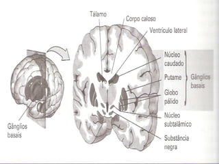
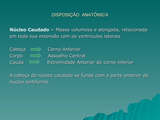
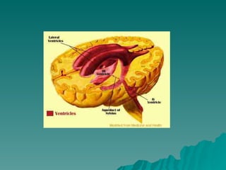
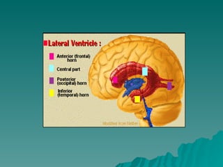
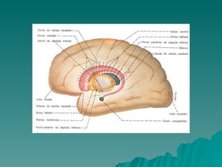
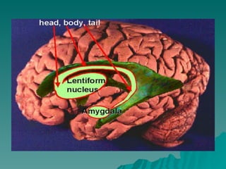
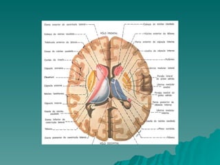
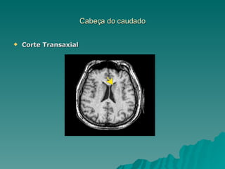
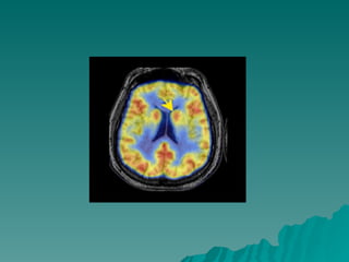
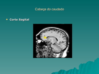
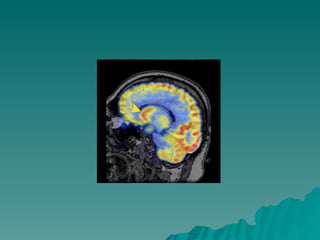
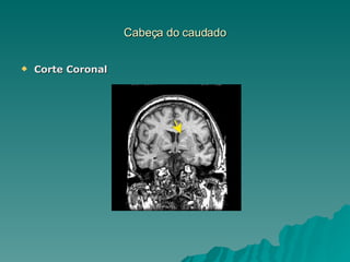
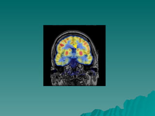
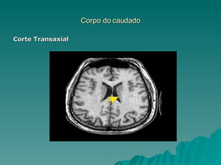
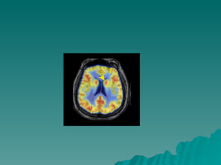
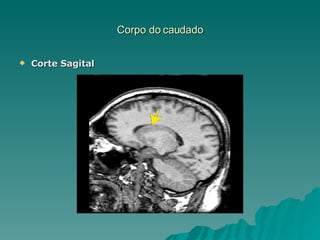
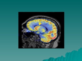
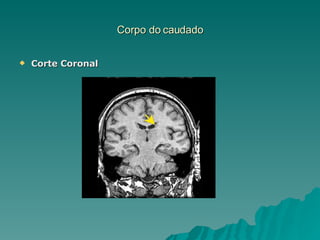
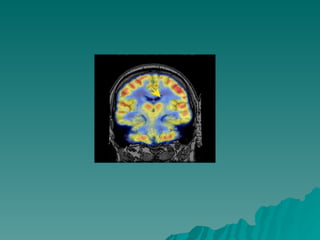
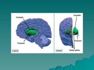
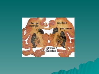
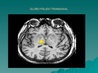
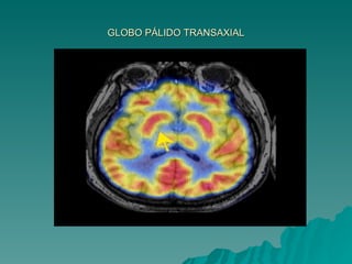
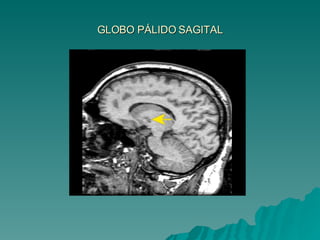
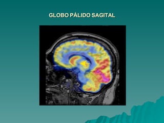
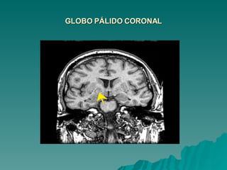
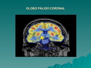
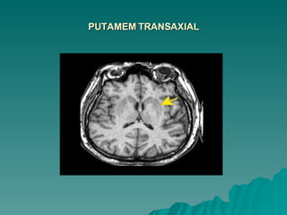
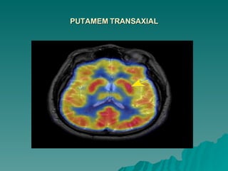
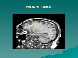
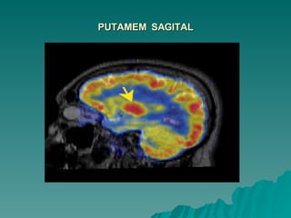
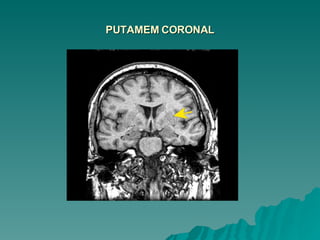
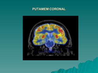
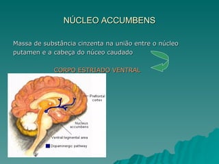
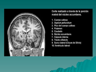
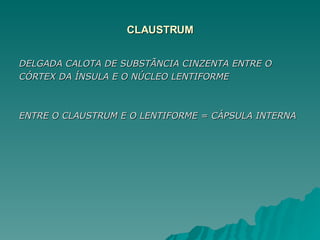
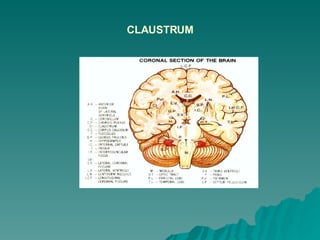
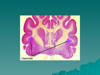
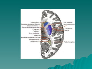
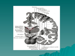
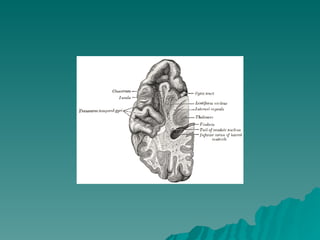
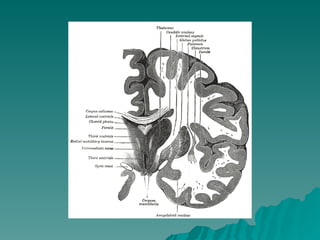
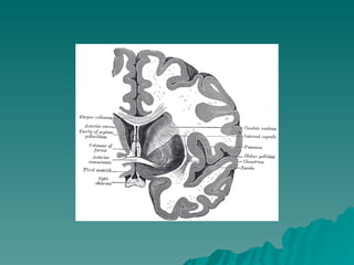
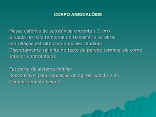
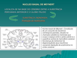
NÚCLEOS DA BASE Localizam-se profundamente no prosencéfalo, atrás da extensão rostral dos ventrículos laterais e coordenam os  movimentos do corpo. Ligam-se ao sistema límbico através das amigdalas (regula a percepção e a produção de emoções Segundo Andreasen N. C. ...Os gânglios basais (“nós nervosos inferiores”) também são  importantes para a expressão e regulação da emoção e da  cognição.
Ex: Coréia de Huntington – atrofia do núcleo caudado – primeiros  sintomas são semelhantes aos da esquizofrenia e aos do  transtorno de humor = pensamentos delirantes, depressão ou  comportamento impulsivo inadequado Doença de Parkinson (perda de neurônios DA na substância  negra que envia projeções para o núcleo caudado) –  embotamento da capacidade de produzir emoções, diminuição da capacidade de lembrança e da volição - semelhantes a esquizofrenia
 
ESTRUTURA E FUNÇÃO DOS GÂNGLIOS DA BASE CLAUSTRUM CORPO AMIGDALÓIDE NÚCLEO CAUDADO PUTAMEM  ESTRIADO  LENTIFORME  GLOBO PÁLIDO ESTUDOS MAIS RECENTES Núcleo basal de Meynert Núcleo Accubens (integrante do corpo estriado ventral) ALGUNS AUTORES Substância Negra  Subtálamo
DISPOSIÇÃO  ANATÕMICA Núcleo Caudado  = Massa volumosa e alongada, relacionada  em toda sua extensão com os ventrículos laterais Cabeça  Corno Anterior Corpo  Assoalho Central Cauda  Extremidade Anterior do corno inferior A cabeça do núcleo caudado se funde com a parte anterior do núcleo lentiforme


Cabeça do caudado Corte Transaxial
 
Cabeça do caudado Corte Sagital
 
Cabeça do caudado Corte Coronal
 
Corpo do caudado Corte Transaxial
 
Corpo do caudado Corte Sagital
 
Corpo do caudado Corte Coronal
 
LENTIFORME = FORMA DE UMA CASTANHA DO PARÁ Não aparece na superfície ventricular – Profundo Medialmente se relaciona com a cápsula interna  Separa do caudado e do tálamo Lateralmente  córtex da ínsula, separado pela  substância branca e pelo claustrum DIVIDIDO – Putamem = lâmina medial Globo pálido = lâmina mais lateral e branca  Coloração mais clara = fibras mielínicas que a atravessam PARTE EXTERNA GLOBO PÁLIDO  lâmina medular  medial PARTE INTERNA
 
 
GLOBO PÁLIDO TRANSAXIAL
GLOBO PÁLIDO SAGITAL
GLOBO PÁLIDO CORONAL
PUTAMEM TRANSAXIAL
PUTAMEM TRANSAXIAL
PUTAMEM  SAGITAL
PUTAMEM  SAGITAL
PUTAMEM CORONAL
PUTAMEM CORONAL
Caudado + Putamen = neoestritum  Globo Palium = Paleoestriatum  FILOGENÉTICO  Caudado +  Putâmem  muitas fibras   Globo pálido  Impulso aferente  NEOESTRIATUM  PALEOESTRIATUM Fibras Eferentes (maior nº.) do corpo estriado  Corpo Estriado Ventral = extensões ventrais do  striatum  e  Paluidum  – conexões com áreas corticais do sistema límbico = Participam  na regulação do comportamento emocional
Pálido Ventral = fica logo abaixo da comissura anterior
Striatum Ventral  = núcleo accubens – entre a cabeça do  caudado e o putâmen
NÚCLEO ACCUMBENS Massa de substância cinzenta na união entre o núcleo putamen e a cabeça do núceo caudado  CORPO ESTRIADO VENTRAL
 
CLAUSTRUM DELGADA CALOTA DE SUBSTÃNCIA CINZENTA ENTRE O CÓRTEX DA ÍNSULA E O NÚCLEO LENTIFORME ENTRE O CLAUSTRUM E O LENTIFORME = CÁPSULA INTERNA
Within the context of the neurobiological theories of consciousness  mentioned in the introduction, the highly networked nature of the claustrum raises the question of whether it acts as a sort of ‘ Cartesian theatre’. This is a metaphor, introduced and ridiculed by the philosopher Dennett (1991), for the  Fictitious centre where the mind and brain meet ,  where ‘it all comes together’ and consciousness occurs. We think that a more appropriate analogy for the claustrum  is that of a conductor coordinating a group of players in the orchestra, the various cortical regions. Without the conductor, the players can still play but they fall increasingly out of synchrony with each other. The result is a cacophony of sounds.  This metaphor would suggest that  different attributes of objects, both within (e.g. colour and motion) and across modalities (e.g. visual form and sound location), are rapidly combined and bound in the claustrum.
Without this structure— and that of its twin in the other Hemisphere — the subject may still be Able to respond to simple, isolated or to highly familiar stimuli, but not to complex or unfamiliar ones.  Objects or events in the real world have many simultaneous  attributes : colour, shape, distance, velocity, smell, sound, feel and so on. In the absence of both claustra, these attributes may not be experienced in an integrated manner and the subject may fail to  altogether perceive these objects or events or only be consciously aware of some isolated  attribute.
Thus, if the claustrum is critical to binding information within and across sensory and motor modalities, certain anatomical constraints would have to be met.  In particular, the information from say, a visual cortical region would need to be combined with information from somatosensory, auditory or motor cortices. This demands some sort of intermixing of the associated signals within the claustrum.  Several, non-exclusive, anatomical and biophysical substrates for such widespread intra-claustral interactions are possible. Francis C. Crick1,† and Christof Koch2,*
 
CLAUSTRUM


CORPO AMIGDALÓIDE Massa esférica de substância cinzenta ( 2 cm) Situada no pólo temporal do Hemisfério cerebral Em relação estreita com o núcleo caudado Discretamente saliente no tecto da parede terminal do corno  Inferior ventrolateral. Faz parte do sistema límbico Responsável pela regulação da agressividade e do  Comportamento sexual
Foto: Medical College of Georgia


NUCLEO BASAL DE MEYNERT LOCALIZA-SE NA BASE DO CÉREBRO ENTRE A SUBSTÃNCIA PERFURADA ANTERIOR E O GLOBO PÁLIDO  SUBSTÂNCIA INONIMADA Produção de Acetilcolina
CONEXÕES
Córtex pré-frontal e pré-motor estão envolvidos na concepção dos atos motores. Giro pré-central = neurônios que se projetam ao longo de  toda a medula espinhal Todas as áreas corticais (exceto córtex auditivo e visual  Primário)  caudado, putamen e gânglios basais Caudado  córtex de associação multimodal Lida mais com informações cognitivas envolvida na realização  do movimento. Putamem  áreas somatossensoriais primárias, auditivas e  visuais secundárias. Processa informação sobre o contexto sensorial no qual o movimento  ocorre
Nigro-estriatal-nigral = DA e exercem função moduladora sobre o circuito básico – conexões com neurônios espinhosos do neoestriado = lesão deste circuito =  Doença de Parkinson Participa de movimentos já iniciados e no planejamento do  ato motor  projeção para áreas motoras secundárias NEURÔNIOS DO ESTRIATUM SÃO ATIVADOS NO MOMENTO DO MOVIMENTO E ATÉ MESMO ANTES DESTE O Circuito Pálido–subtálamo–palidal influencia o circuito  básico, agindo sobre a motricidade básica = lesão deste  circuito = hemibalismo
Córtex envia input glutamatérgicos para o neoestriado Os Neurônios do neoestriato utilizam GABA e recebem input de DA da Substância Negra compacta Os Neurônios GABA do neoestriato projetam-se para o globo pálido interno e substância negra reticulada (neurônios GABA) Projetam-se para o colículo superior  e tálamo VA/VL As projeções da SNr para o colículo superior  e pedunculopontino influenciam os movimentos oculares e do tronco
Rota Direta Plano gerado pelo córtex frontal – glutamato estimulando os  neurônios GABA do neoestriado que se projetam para inibir  GPi/SNr de modo que não liberem mais GABA para o tálamo (VA/VL) – iniciado um novo comportamento motor
Neocórtex envia informações para um 2º  conjunto de neurônios GABA no Neoestriado Neurônios GABA se projetam para o Globo Pálido Externo que se projeta para núcleos subtalâmicos -(glutamatérgicos) Núcleos Subtalâmicos projetam fibras para o Globo Pálido  Interno e Substância Negra Reticular
Rota Indireta que serve como freio Quando neurônios estriatais inibem Globo Pálido Externo, este fica impedido de inibir os Núcleos Subtalâmicos que passa a  liberar Glutamato  no Globo Pálido Interno e Substância Negra Reticular, estimulando-a
O input glutamatérgico do neocortex não é suficiente para  interromper a influência inibidora GPi/SNr A DA da SNc é fundamental para estimular os neu8rônios  estriatais que se projetam pela via direta (via D1),  Como também inibe os neurônios GABA estriatais da via Indireta (via D2)  DA reforça a ação do glutamato na rota direta i inibe o efeito do glutamato na rota indireta
Caudate, Putamen, and Globus Pallidus Volume in Schizophrenia: A Quantitative MRI Study   Hiroto Hokama, M.D.1, Martha E. Shenton, Ph.D.1, Paul G. Nestor, Ph.D.1, Ron Kikinis, M.D.2, James J. Levitt, M.D.1, David Metcalf, M.A.2, Cynthia G. Wible, Ph.D.1, Brian F. O'Donnell, Ph.D.1, Ferenc A. Jolesz, M.D.2 and Robert W. McCarley, M.D.3 ABSTRACT Basal ganglia structures have been reported to be abnormal in schizophrenia, However, while component structures of the basal ganglia are functionally  differentiated, there have been no evaluations of their separate MRI volumes using small voxel (1.5 mm3) SPGR techniques and multi-plane assessments. We examined MRI scans from 15 male, right-handed neuroleptic-medicated schizophrenics and 15 age, handedness, and gender-matched normal controls. Compared to controls, schizophrenics showed enlarged volumes: 14.2% for total basal ganglia, 27.4% for globus pallidus, 15.9% for putamen and 9.5% for caudate. Increased volumes, especially caudate, were ssociated with poorer neuropsychological test performance on finger tapping and Hebb's Recurring Digits. These findings indicate abnormalities throughout all basal ganglia structures in at least a subgroup of schizophrenics
14.2% para o total dos ganglios da base; 27.4% para o globo pallido; 15.9% para o putamen – consistente com defeito na  neurogênese – fase de pruning; 9.5% para o caudate; O caudado pode estar reduzido em pacientes que  desenvolveram discinesia tardia comparado aos que não Desenvolveram; O Aumento do volume do  caudado, em especial, foi associado Com teste neuropsicológico pobre
NEUROCIÊNCIA: APLICAÇÕES NA CLÍNICA (atualização e revisão bibliográfica) 1 Dr.Luiz Miller de Paiva 2 Hiperatividade dos gânglios da base (neoestriatum e  accumbens), com aumento de dopamina e diminuição de  receptores D2 principalmente no accubens. Amigdala encontra-se diminuída
Structural and Functional Abnormalities of the Amygdala in Schizophrenia  STEPHEN M. LAWRIE, HEATHER C. WHALLEY, DOMINIC E. JOB AND EVE C. JOHNSTONE  Edinburgh University Department of Psychiatry, Kennedy Tower, Royal Edinburgh Hospital,  Edinburgh,Scotland, UK  Address for correspondence: Dr. Stephen M. Lawrie, Edinburgh University Department of Psychiatry, Kennedy Tower, Royal Edinburgh Hospital, Edinburgh, Scotland, UK. Voice: 0131 537 6671;  Schizophrenia is characterized by delusions and hallucinations, which tend  to respond to treatment with dopamine receptor blockers, and a loss of motivation and affect, which do not. Structural magnetic resonance imaging (sMRI) has convincingly demonstrated reduced volumes of the amygdala  -hippocampal complex (AHC)  and other limbic and paralimbic structures, on both manual tracing and automated analyses. The Edinburgh High-Risk Study (EHRS) of initially healthy adolescents with at least two affected relatives has found that, and functional imaging studies of patients with schizophrenia.
AHC volumes appear to be genetically mediated in families with a  dominant pattern of transmission, whereas prefrontal lobe and basal  ganglia volumes are related to genetic liability to schizophrenia in the generality of high-risk subjects. Temporallobe volumes may fall as  psychotic symptoms develop, in the context of drug abuse and  stress. Neuropsychological testing has also demonstrated pre-morbid impairments and symptom-related deterioration. More detailed  analyses of the temporal lobe changes on sMRI and fronto-tempora dysconnectivity on fMRI are in progress. These findings are  discussed with reference to other indications of pre-morbid  developmental disturbance in our high-risk subjects, animal models  of schizophrenia, and reliable findings from neuropathological,  neuropsychological, and functional imaging studies of patients with  schizophrenia.

CóPia De Sn Central MóDulo 8

  • 1.
    NÚCLEOS DA BASELocalizam-se profundamente no prosencéfalo, atrás da extensão rostral dos ventrículos laterais e coordenam os movimentos do corpo. Ligam-se ao sistema límbico através das amigdalas (regula a percepção e a produção de emoções Segundo Andreasen N. C. ...Os gânglios basais (“nós nervosos inferiores”) também são importantes para a expressão e regulação da emoção e da cognição.
  • 2.
    Ex: Coréia deHuntington – atrofia do núcleo caudado – primeiros sintomas são semelhantes aos da esquizofrenia e aos do transtorno de humor = pensamentos delirantes, depressão ou comportamento impulsivo inadequado Doença de Parkinson (perda de neurônios DA na substância negra que envia projeções para o núcleo caudado) – embotamento da capacidade de produzir emoções, diminuição da capacidade de lembrança e da volição - semelhantes a esquizofrenia
  • 3.
  • 4.
    ESTRUTURA E FUNÇÃODOS GÂNGLIOS DA BASE CLAUSTRUM CORPO AMIGDALÓIDE NÚCLEO CAUDADO PUTAMEM ESTRIADO LENTIFORME GLOBO PÁLIDO ESTUDOS MAIS RECENTES Núcleo basal de Meynert Núcleo Accubens (integrante do corpo estriado ventral) ALGUNS AUTORES Substância Negra Subtálamo
  • 5.
    DISPOSIÇÃO ANATÕMICANúcleo Caudado = Massa volumosa e alongada, relacionada em toda sua extensão com os ventrículos laterais Cabeça Corno Anterior Corpo Assoalho Central Cauda Extremidade Anterior do corno inferior A cabeça do núcleo caudado se funde com a parte anterior do núcleo lentiforme
  • 6.
  • 7.
  • 8.
  • 9.
  • 10.
  • 11.
  • 12.
    Cabeça do caudadoCorte Transaxial
  • 13.
  • 14.
    Cabeça do caudadoCorte Sagital
  • 15.
  • 16.
    Cabeça do caudadoCorte Coronal
  • 17.
  • 18.
    Corpo do caudadoCorte Transaxial
  • 19.
  • 20.
    Corpo do caudadoCorte Sagital
  • 21.
  • 22.
    Corpo do caudadoCorte Coronal
  • 23.
  • 24.
    LENTIFORME = FORMADE UMA CASTANHA DO PARÁ Não aparece na superfície ventricular – Profundo Medialmente se relaciona com a cápsula interna Separa do caudado e do tálamo Lateralmente córtex da ínsula, separado pela substância branca e pelo claustrum DIVIDIDO – Putamem = lâmina medial Globo pálido = lâmina mais lateral e branca Coloração mais clara = fibras mielínicas que a atravessam PARTE EXTERNA GLOBO PÁLIDO lâmina medular medial PARTE INTERNA
  • 25.
  • 26.
  • 27.
  • 28.
  • 29.
  • 30.
  • 31.
  • 32.
  • 33.
  • 34.
  • 35.
  • 36.
  • 37.
  • 38.
  • 39.
    Caudado + Putamen= neoestritum Globo Palium = Paleoestriatum FILOGENÉTICO Caudado + Putâmem muitas fibras Globo pálido Impulso aferente NEOESTRIATUM PALEOESTRIATUM Fibras Eferentes (maior nº.) do corpo estriado Corpo Estriado Ventral = extensões ventrais do striatum e Paluidum – conexões com áreas corticais do sistema límbico = Participam na regulação do comportamento emocional
  • 40.
    Pálido Ventral =fica logo abaixo da comissura anterior
  • 41.
    Striatum Ventral = núcleo accubens – entre a cabeça do caudado e o putâmen
  • 42.
    NÚCLEO ACCUMBENS Massade substância cinzenta na união entre o núcleo putamen e a cabeça do núceo caudado CORPO ESTRIADO VENTRAL
  • 43.
  • 44.
    CLAUSTRUM DELGADA CALOTADE SUBSTÃNCIA CINZENTA ENTRE O CÓRTEX DA ÍNSULA E O NÚCLEO LENTIFORME ENTRE O CLAUSTRUM E O LENTIFORME = CÁPSULA INTERNA
  • 45.
    Within the contextof the neurobiological theories of consciousness mentioned in the introduction, the highly networked nature of the claustrum raises the question of whether it acts as a sort of ‘ Cartesian theatre’. This is a metaphor, introduced and ridiculed by the philosopher Dennett (1991), for the Fictitious centre where the mind and brain meet , where ‘it all comes together’ and consciousness occurs. We think that a more appropriate analogy for the claustrum is that of a conductor coordinating a group of players in the orchestra, the various cortical regions. Without the conductor, the players can still play but they fall increasingly out of synchrony with each other. The result is a cacophony of sounds. This metaphor would suggest that different attributes of objects, both within (e.g. colour and motion) and across modalities (e.g. visual form and sound location), are rapidly combined and bound in the claustrum.
  • 46.
    Without this structure—and that of its twin in the other Hemisphere — the subject may still be Able to respond to simple, isolated or to highly familiar stimuli, but not to complex or unfamiliar ones. Objects or events in the real world have many simultaneous attributes : colour, shape, distance, velocity, smell, sound, feel and so on. In the absence of both claustra, these attributes may not be experienced in an integrated manner and the subject may fail to altogether perceive these objects or events or only be consciously aware of some isolated attribute.
  • 47.
    Thus, if theclaustrum is critical to binding information within and across sensory and motor modalities, certain anatomical constraints would have to be met. In particular, the information from say, a visual cortical region would need to be combined with information from somatosensory, auditory or motor cortices. This demands some sort of intermixing of the associated signals within the claustrum. Several, non-exclusive, anatomical and biophysical substrates for such widespread intra-claustral interactions are possible. Francis C. Crick1,† and Christof Koch2,*
  • 48.
  • 49.
  • 50.
  • 51.
  • 52.
  • 53.
  • 54.
  • 55.
  • 56.
    CORPO AMIGDALÓIDE Massaesférica de substância cinzenta ( 2 cm) Situada no pólo temporal do Hemisfério cerebral Em relação estreita com o núcleo caudado Discretamente saliente no tecto da parede terminal do corno Inferior ventrolateral. Faz parte do sistema límbico Responsável pela regulação da agressividade e do Comportamento sexual
  • 57.
  • 58.
  • 59.
  • 60.
  • 61.
    NUCLEO BASAL DEMEYNERT LOCALIZA-SE NA BASE DO CÉREBRO ENTRE A SUBSTÃNCIA PERFURADA ANTERIOR E O GLOBO PÁLIDO SUBSTÂNCIA INONIMADA Produção de Acetilcolina
  • 62.
  • 63.
    Córtex pré-frontal epré-motor estão envolvidos na concepção dos atos motores. Giro pré-central = neurônios que se projetam ao longo de toda a medula espinhal Todas as áreas corticais (exceto córtex auditivo e visual Primário) caudado, putamen e gânglios basais Caudado córtex de associação multimodal Lida mais com informações cognitivas envolvida na realização do movimento. Putamem áreas somatossensoriais primárias, auditivas e visuais secundárias. Processa informação sobre o contexto sensorial no qual o movimento ocorre
  • 64.
    Nigro-estriatal-nigral = DAe exercem função moduladora sobre o circuito básico – conexões com neurônios espinhosos do neoestriado = lesão deste circuito = Doença de Parkinson Participa de movimentos já iniciados e no planejamento do ato motor projeção para áreas motoras secundárias NEURÔNIOS DO ESTRIATUM SÃO ATIVADOS NO MOMENTO DO MOVIMENTO E ATÉ MESMO ANTES DESTE O Circuito Pálido–subtálamo–palidal influencia o circuito básico, agindo sobre a motricidade básica = lesão deste circuito = hemibalismo
  • 65.
    Córtex envia inputglutamatérgicos para o neoestriado Os Neurônios do neoestriato utilizam GABA e recebem input de DA da Substância Negra compacta Os Neurônios GABA do neoestriato projetam-se para o globo pálido interno e substância negra reticulada (neurônios GABA) Projetam-se para o colículo superior e tálamo VA/VL As projeções da SNr para o colículo superior e pedunculopontino influenciam os movimentos oculares e do tronco
  • 66.
    Rota Direta Planogerado pelo córtex frontal – glutamato estimulando os neurônios GABA do neoestriado que se projetam para inibir GPi/SNr de modo que não liberem mais GABA para o tálamo (VA/VL) – iniciado um novo comportamento motor
  • 67.
    Neocórtex envia informaçõespara um 2º conjunto de neurônios GABA no Neoestriado Neurônios GABA se projetam para o Globo Pálido Externo que se projeta para núcleos subtalâmicos -(glutamatérgicos) Núcleos Subtalâmicos projetam fibras para o Globo Pálido Interno e Substância Negra Reticular
  • 68.
    Rota Indireta queserve como freio Quando neurônios estriatais inibem Globo Pálido Externo, este fica impedido de inibir os Núcleos Subtalâmicos que passa a liberar Glutamato no Globo Pálido Interno e Substância Negra Reticular, estimulando-a
  • 69.
    O input glutamatérgicodo neocortex não é suficiente para interromper a influência inibidora GPi/SNr A DA da SNc é fundamental para estimular os neu8rônios estriatais que se projetam pela via direta (via D1), Como também inibe os neurônios GABA estriatais da via Indireta (via D2) DA reforça a ação do glutamato na rota direta i inibe o efeito do glutamato na rota indireta
  • 70.
    Caudate, Putamen, andGlobus Pallidus Volume in Schizophrenia: A Quantitative MRI Study Hiroto Hokama, M.D.1, Martha E. Shenton, Ph.D.1, Paul G. Nestor, Ph.D.1, Ron Kikinis, M.D.2, James J. Levitt, M.D.1, David Metcalf, M.A.2, Cynthia G. Wible, Ph.D.1, Brian F. O'Donnell, Ph.D.1, Ferenc A. Jolesz, M.D.2 and Robert W. McCarley, M.D.3 ABSTRACT Basal ganglia structures have been reported to be abnormal in schizophrenia, However, while component structures of the basal ganglia are functionally differentiated, there have been no evaluations of their separate MRI volumes using small voxel (1.5 mm3) SPGR techniques and multi-plane assessments. We examined MRI scans from 15 male, right-handed neuroleptic-medicated schizophrenics and 15 age, handedness, and gender-matched normal controls. Compared to controls, schizophrenics showed enlarged volumes: 14.2% for total basal ganglia, 27.4% for globus pallidus, 15.9% for putamen and 9.5% for caudate. Increased volumes, especially caudate, were ssociated with poorer neuropsychological test performance on finger tapping and Hebb's Recurring Digits. These findings indicate abnormalities throughout all basal ganglia structures in at least a subgroup of schizophrenics
  • 71.
    14.2% para ototal dos ganglios da base; 27.4% para o globo pallido; 15.9% para o putamen – consistente com defeito na neurogênese – fase de pruning; 9.5% para o caudate; O caudado pode estar reduzido em pacientes que desenvolveram discinesia tardia comparado aos que não Desenvolveram; O Aumento do volume do caudado, em especial, foi associado Com teste neuropsicológico pobre
  • 72.
    NEUROCIÊNCIA: APLICAÇÕES NACLÍNICA (atualização e revisão bibliográfica) 1 Dr.Luiz Miller de Paiva 2 Hiperatividade dos gânglios da base (neoestriatum e accumbens), com aumento de dopamina e diminuição de receptores D2 principalmente no accubens. Amigdala encontra-se diminuída
  • 73.
    Structural and FunctionalAbnormalities of the Amygdala in Schizophrenia STEPHEN M. LAWRIE, HEATHER C. WHALLEY, DOMINIC E. JOB AND EVE C. JOHNSTONE Edinburgh University Department of Psychiatry, Kennedy Tower, Royal Edinburgh Hospital, Edinburgh,Scotland, UK Address for correspondence: Dr. Stephen M. Lawrie, Edinburgh University Department of Psychiatry, Kennedy Tower, Royal Edinburgh Hospital, Edinburgh, Scotland, UK. Voice: 0131 537 6671; Schizophrenia is characterized by delusions and hallucinations, which tend to respond to treatment with dopamine receptor blockers, and a loss of motivation and affect, which do not. Structural magnetic resonance imaging (sMRI) has convincingly demonstrated reduced volumes of the amygdala -hippocampal complex (AHC) and other limbic and paralimbic structures, on both manual tracing and automated analyses. The Edinburgh High-Risk Study (EHRS) of initially healthy adolescents with at least two affected relatives has found that, and functional imaging studies of patients with schizophrenia.
  • 74.
    AHC volumes appearto be genetically mediated in families with a dominant pattern of transmission, whereas prefrontal lobe and basal ganglia volumes are related to genetic liability to schizophrenia in the generality of high-risk subjects. Temporallobe volumes may fall as psychotic symptoms develop, in the context of drug abuse and stress. Neuropsychological testing has also demonstrated pre-morbid impairments and symptom-related deterioration. More detailed analyses of the temporal lobe changes on sMRI and fronto-tempora dysconnectivity on fMRI are in progress. These findings are discussed with reference to other indications of pre-morbid developmental disturbance in our high-risk subjects, animal models of schizophrenia, and reliable findings from neuropathological, neuropsychological, and functional imaging studies of patients with schizophrenia.