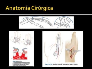
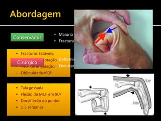
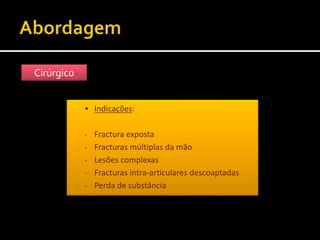
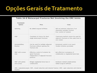
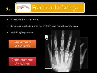
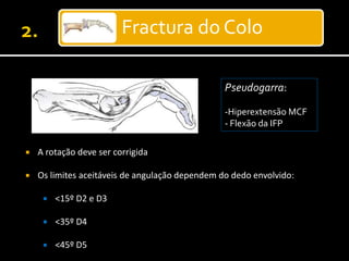
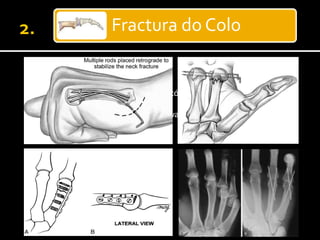
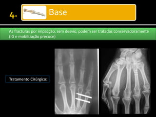
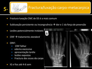
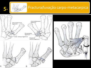
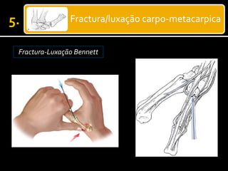
 A maioria ocorre na população activa e trabalhadora;
 Adolescentes e Jovens adultos
 Fracturas dos dedos da mão:
 Falanges  46%
 Metacarpos  36%
 Fracturas do colo do 5º metacarpo ± 10%
 Incidência ≈ 2,5%
Fractura da Cabeça
Fractura do Colo
Fractura da Diáfise
Base
Extra-articulares
Intra-articulares
Fractura/luxação carpo-
metacarpica
 Traumatismo
 Directo
 Indirecto
 Idade
 Profissão
 Dominância (Esq/Drt)
 Hobbies
Axial
Angulação
Torsão
Alta Energia
 Fracturas de compressão articular ou metafisária
 Suspeitar de fracturas proximais (rádio, carpo)
 Luxação ou Fractura diafisária
 Fractura em espiral/obliqua
 Lesões complexas (pele, vasos, nervos)
 Dor, edema, crepitação, deformidade…
 Feridas (incisas, contusas, esfacelo…)
 Avaliação neuro-vascular
 Radiografia (AP e Perfil)
 TC
 RMN
 Ecografia
Brewerton Mehara
Obliqua inversa Skyline
 Fracturas Estáveis:
- <50% descoaptação
- <40º de angulação
- Obliquidade<60º
Conservador
 Maioria das Fracturas do colo e diáfise
 Fracturas estáveis e coaptadas da cabeça e base
Cirúrgico
 Deformidade rotacional
 Encurtamento
 Tala gessada
 Flexão da MCF em 90º
 Dorsiflexão do punho
  3 semanas
Cirúrgico
 Indicações:
- Fractura exposta
- Fracturas múltiplas da mão
- Lesões complexas
- Fracturas intra-articulares descoaptadas
- Perda de substância
 Definir os limites de deformidade para cada tipo de fractura é
sede de controvérsia
 Deformidade vs Sintomas
 Angulação é + tolerada nas #s do colo
  flexão com angulação > 30º no colo do 5º metacarpo
Fractura da Cabeça
 A maioria é intra-articular
 Se descoaptação importante  ORIF para redução anatómica
 Mobilização precoce
Parcialmente
Articulares
Completamente
Articulares
Fractura do Colo
Pseudogarra:
-Hiperextensão MCF
- Flexão da IFP
 A rotação deve ser corrigida
 Os limites aceitáveis de angulação dependem do dedo envolvido:
 <15º D2 e D3
 <35º D4
 <45º D5
Fractura do Colo
 Cirurgia:
 Redução e/ou angulação insatisfatórias
 Rotação
 Cominução volar (indicação relativa)
Fractura do Colo
Fractura do Boxer
Fractura da Diáfise
 Menor angulação tolerada
 <10º D2 e D3
 <20º D4
 <30º D5
 Transversal
 Oblíqua
 Cominutiva
 Encurtamento < 5mm
  Rotação
Fractura da Diáfise
Fractura da Diáfise
Fractura da Diáfise
Fractura da Diáfise
Osteossíntese em ponte
Base
As fracturas por impacção, sem desvio, podem ser tratadas conservadoramente
(IG e mobilização precoce)
Tratamento Cirúrgico:
Fractura/luxação carpo-metacarpica
 Fractura-luxação CMC de D5 é a mais comum
 Subluxação persistente ou incongruência  dor e  da força de preensão
 Lesões potencialmente instáveis
 CRIF  tratamento standard
 ORIF:
 CRIF falhar
 edema excessivo
 apresentação tardia
 lesões expostas
 Fractura dos ossos do carpo
 IG e fios até 4-6 sem
Fractura/luxação carpo-metacarpica
Fractura-Luxação Bennett
Fractura/luxação carpo-metacarpica
Fractura-Luxação Bennett CRIFORIF
Fractura/luxação carpo-metacarpica
Fractura-Luxação Rolando Lesões cominutivas!
Fractura/luxação carpo-metacarpica
Fractura-Luxação Rolando Lesões cominutivas!
1. Redução incruenta e imobilização gessada
2. Redução incruenta e fixação com fios de Kirschner ao 2º
metacarpo
3. Redução cruenta e osteossintese com placa (risco de
desvascularização dos fragmentos)
4. Fixador externo
Fractura/luxação carpo-metacarpica
Fractura-Luxação Rolando Lesões cominutivas!
 Infecção
 Rigidez
 Hipersensibilidade
 Deformidade e Pseudartrose
 Instabilidade
 Artrose pós-traumática
 Complicações de hardware
 Ruptura de tendão
 As fracturas e luxações da mão constituem um grupo diverso
e complicado de lesões
 Objectivo: recuperação funcional
 Quanto mais agressivo o tratamento cirúrgico, mais
“agressiva” deve ser a reabilitação
 Alertar os doentes para as possíveis complicações
 Técnicas diversas disponíveis ao Cirurgião
1. Rockwood & Green's Fractures in Adults, 6th Edition 2007
2. Orthopaedics and Trauma Fractures of the metacarpals and
phalanges, 25:1, 43-56, 2010
3. Green's Operative Hand Surgery, 5th edition
4. www.maitrise-orthop.com – Metacarpal Fractures
5. www.emedicine.com – Metacarpal Fractures

Fractura dos metacarpos slideshare

Notas do Editor

  • #3 As fracturas da mão constituem um custo acrescido na sociedade actual em termos de cuidados médicos e diminuição da produtividade no local de trabalho. Foi realizado um estudo prospectivo na Noruega que mostrou a prevalencia de fracturas das falanges em 46% e do metacarpo em 36%, não muito diferente dos estados unidos.
  • #4 Dividir as fracturas consoante localização anatómica e referir que as fracturas do 1º metacarpo são faladas à parte
  • #5 Extra-articulares e Intra-articulares Dividir as fracturas consoante localização anatómica e referir que as fracturas do 1º metacarpo são faladas à parte
  • #8 A rotação é melhor avaliada através do alinhamento do segmento distal mais próximo (melhor observado quando flexão articular a 90º), sendo 10º o limite do tolerável. A full history should be obtained, including such details as the mechanism of injury and whether the injury was a result of high or low energy. The hand and fingers should be fully examined for open wounds and rotational deformities. A full neurovascular examination should be done, checking for capillary refill and two-point sensation. Rotational deformity can only be determined by physical examination. The fingertips should be examined with the fingers flexed, paying close attention to the alignment of the nailbeds.
  • #9 Specialized views may help to define injury patterns in the metacarpal including (A) the Brewerton view for the metacarpal bases, (B) the Mehara view for the index CMC relationships, (C) the reverse oblique view for angulation in the index metacarpal neck, and (D) the skyline view for vertical impaction fractures of the metacarpal head Vista de Brewerton e de Mehara: podem mostrar fracturas ocultas da base do metacarpo Projecção obliqua inversa: estimar a angulação a nivel do colo do 2º metacarpo Vista skyline: impacção vertical nas fracturas da cabeça dos metacarpos. TC: planeamento cirurgico em fracturas intra-articulares ou caracterização de fractura não caracterizada por Rx RMN: detectar fracturas ocultas, particularmente no carpo.
  • #10 The metacarpals are the key skeletal elements participating in the formation of the three arches of the hand (Fig. 24-85). There are two transverse arches that exist at the CMC and MP joints. The metacarpals themselves are longitudinally arched with a fairly broad convex dorsal surface. Intramedullary geometry is highly variable but with a consistently 20% thicker volar cortex Surgical access to the metacarpals is easily achieved through incisions placed over the inter-metacarpal valleys and curved distally to avoid entering the digital web commissures. The metacarpals are held tightly bound to each other by strong interosseous ligaments at their bases and by the deep transverse inter-metacarpal ligaments distally. These connections help to maintain the transverse arches of the hand, but flattening can occur with multiple metacarpal fractures or crushing injuries. Shortening of individual metacarpal fractures is limited by these same ligaments (more effectively for the central metacarpals than for the border metacarpals) (244).
  • #11 A: Schematic of the Jahss maneuver for closed reduction of a metacarpal neck fracture. B: Schematic of cast immobilization after closed reduction of a metacarpal neck fracture. Note the extended posture of the interphalangeal (IP) joints. The arrows indicate the line of force, and the asterisk marks the loading point on the metacarpal head that facilitates reduction of the fracture. MP, metacarpophalangeal
  • #12 Most metacarpal injuries are managed by closed reduction and immobilization or sometimes controlled mobilization utilizing a dorsal block splint. Indications for operative treatment include the following: Failure to achieve or maintain acceptable reduction using closed techniques Open fractures Multiple hand fractures Complex injuries Displaced intra-articular fractures Fractures with severe soft-tissue loss requiring a stable skeleton
  • #16 Except for some collateral ligament avulsion fractures, metacarpal head fractures are intra-articular and, if displaced, should be treated by open reduction and internal fixation (ORIF), with the goal being anatomic reduction and early mobilization The plate is contoured and placed on the lateral aspect of the metacarpal bone (Figs. 21 & 22) to avoid potential impingement upon the extensor muscle. Partial release of the interosseous is necessary. Reduction is maintained with a clamp. The plate is secured with two cortical screws either side of the fracture. For partial articular metacarpal head fractures screw only fixation is the treatment of choice. If sufficient interlock of bone spicules occurs, a single 1.3 to 1.5 mm countersunk screw can control rotation of the fragment. If interlock is not effective, two screws are preferred even if this means downsizing the screw diameter to accommodate both of them in the fragment without causing comminution For complete articular head fractures the condylar blade plate is usually required, but it is the most technically demanding plate to apply (Fig. 24-98). This plate is applied laterally with the blade dorsal and the condylar screw hole volar. The drill hole for the blade is round but the blade is rectangular in cross-sectional area. This makes manual insertion of the blade into its hole difficult, and a fine touch is required to not further comminute the metacarpal head fragments. More importantly, the exact angle of the drill path for the blade must be determined in advance relative to the cortical surface of the more proximal shaft against which the plate will be applied. The condylar screw hole of the plate usually needs to be bent to avoid prominence on its volar edge.
  • #17 A pseudogarra é indicação para redução da deformidade. Funcionalmente é inaceitável.
  • #18 1- Técnica do bouquet (controlo rotacional) 2 – transfixação de fios de Kirschner até ao metacarpo adjacente 3- A redução cruenta e fixação interna é o metodo de eleição para fracturas com angulação e rotação acentuadas
  • #19  Mencionar que a fractura do colo do 5º metacarpo a nivel do colo se chama fractura do boxer
  • #20 Metacarpal shaft fractures tend to angulate apex dorsal with the head displaced palmarly due to the deforming pull of the interossei muscles. Only small amounts of angulation (≤ 10°) are acceptable in the 2nd and 3rd metacarpals. The 4th and 5th finger metacarpals are much more mobile, and angulation of 20° and 30°, respectively, can be accepted. The more proximal the fracture, the more pronounced the deformity and the less angulation that can be accepted. Any malrotation is an indication for surgical intervention. Shortening from 3-4 mm is well tolerated. In addition to fractures that cannot be reduced and stabilized closed, fractures such as open fractures, multiple fractures, and malreduced subacute fractures are best addressed surgically. As fracturas obliquas têm probabilidade elevada de deformidade rotacional e encurtamento
  • #21 A resistencia biomecanica dos fios de kirschner é inferior ao dos parafusos interfragmentários ou das placas.
  • #22 Os parafusos interfragmentários conseguem resistir a forças de carga, mas devem ser aplicados 2 ou mais parafusos Tratamento de eleição para fracturas em espiral; o comprimento da fractura é pelo menos 3x o diametro do metacarpo.
  • #23 As fracturas obliquas curtas podem ser tratadas com parafuso interfragmentário em associação com placa de neutralização dorsal.
  • #25 Impaction fractures of the metacarpal bases that are not significantly displaced can be treated with splinting, followed by early mobilization.
  • #26 CMC dislocations and fracture-dislocations, especially when multiple, are unstable injuries. In the past, these fractures were managed by closed reduction and external immobilization, which frequently lead to grip weakness and residual pain. The current literature supports closed reduction, if joint congruity can be obtained, but with the addition of internal fixation. Displaced fracture-dislocations of the 4th and 5th metacarpals, which are accompanied by fracture of the dorsal hamate, require ORIF. Index finger CMC joint dislocations more commonly require open reduction because of extensor carpi radialis brevis interposition.
  • #27 A fractura-luxação de Bennet é parcialmente articular: fragmento volar cubital que permanece na sua posição atraves do ligamento volar oblíquo; o fragmento metacarpico distal apresenta um desvio proximal dorsal e supinação atraves do longo abdutor do polegar. Incidencia de Robert – permite uma melhor visualização da lesão. Fractura-luxação de Rolando é uma lesão completamente articular e a cominução é geralmente a regra.
  • #28 Manobra de redução: tracção longitudinal, extensão, abdução e pronação com pressão na base do metacarpo. FIGURE 24-107 The radial artery, branches of both the superficial radial and lateral antebrachial cutaneous nerves, and the thumb extensor tendons all pose soft tissue risks for damage during closed pinning strategies around the thumb metacarpal base.
  • #29 Redução fechada, fixação com fios e imobilização gessada durante 4 a 6 semanas.