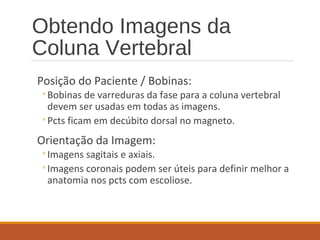
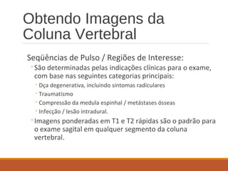
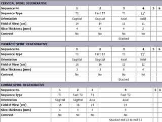
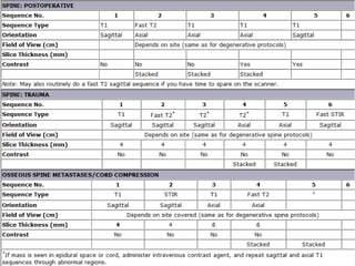
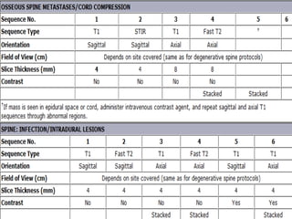
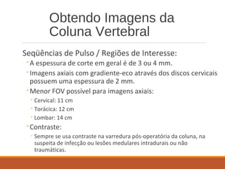
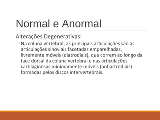
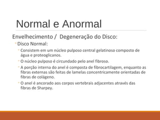
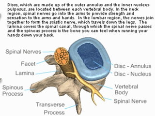
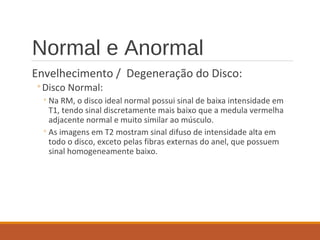
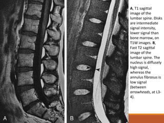
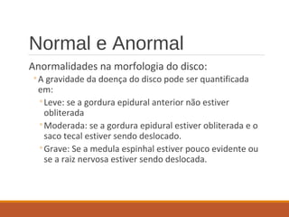
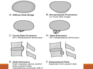
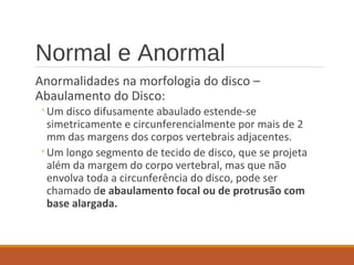
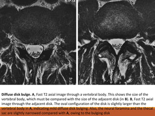
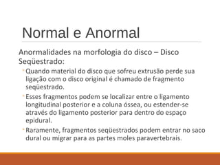
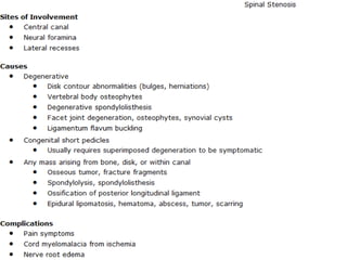
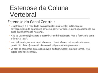
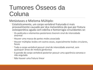
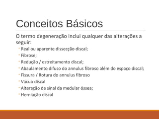
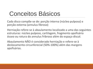
Este documento fornece informações sobre a avaliação por ressonância magnética da coluna vertebral. Ele descreve os protocolos de imagem, alterações degenerativas normais e anormais dos discos intervertebrais e das articulações facetárias, e anormalidades morfológicas dos discos como abaulamentos, protrusões e extrusões. Também discute hematomas epidurais e mielopatia compressiva relacionada aos discos.
2. Obtendo Imagens da
Coluna Vertebral
Posição do Paciente / Bobinas:
◦Bobinas de varreduras da fase para a coluna vertebral
devem ser usadas em todas as imagens.
◦Pcts ficam em decúbito dorsal no magneto.
Orientação da Imagem:
◦Imagens sagitais e axiais.
◦Imagens coronais podem ser úteis para definir melhor a
anatomia nos pcts com escoliose.
3. Obtendo Imagens da
Coluna Vertebral
Seqüências de Pulso / Regiões de Interesse:
◦São determinadas pelas indicações clínicas para o exame,
com base nas seguintes categorias principais:
◦Dça degenerativa, incluindo sintomas radiculares
◦Traumatismo
◦Compressão da medula espinhal / metástases ósseas
◦Infecção / lesão intradural.
◦Imagens ponderadas em T1 e T2 rápidas são o padrão para
o exame sagital em qualquer segmento da coluna
vertebral.
4.
5.
6.
7. Obtendo Imagens da
Coluna Vertebral
Seqüências de Pulso / Regiões de Interesse:
◦A espessura de corte em geral é de 3 ou 4 mm.
◦Imagens axiais com gradiente-eco através dos discos cervicais
possuem uma espessura de 2 mm.
◦Menor FOV possível para imagens axiais:
◦Cervical: 11 cm
◦Torácica: 12 cm
◦Lombar: 14 cm
◦Contraste:
◦Sempre se usa contraste na varredura pós-operatória da coluna, na
suspeita de infecção ou lesões medulares intradurais ou não
traumáticas.
8. Normal e Anormal
Alterações Degenerativas:
◦Na coluna vertebral, as principais articulações são as
articulações sinoviais facetadas emparelhadas,
livremente móveis (diatrodiais), que correm ao longo da
face dorsal da coluna vertebral e nas articulações
cartilaginosas minimamente móveis (anfiartrodiais)
formadas pelos discos intervertebrais.
9. Normal e Anormal
Envelhecimento / Degeneração do Disco:
◦Disco Normal:
◦Consistem em um núcleo pulposo central gelatinoso composto de
água e proteoglicanos.
◦O núcleo pulposo é circundado pelo anel fibroso.
◦A porção interna do anel é composta de fibrocartilagem, enquanto as
fibras externas são feitas de lamelas concentricamente orientadas de
fibras de colágeno.
◦O anel é ancorado aos corpos vertebrais adjacentes através das
fibras de Sharpey.
10.
11. Normal e Anormal
Envelhecimento / Degeneração do Disco:
◦Disco Normal:
◦Na RM, o disco ideal normal possui sinal de baixa intensidade em
T1, tendo sinal discretamente mais baixo que a medula vermelha
adjacente normal e muito similar ao músculo.
◦As imagens em T2 mostram sinal difuso de intensidade alta em
todo o disco, exceto pelas fibras externas do anel, que possuem
sinal homogeneamente baixo.
12. A, T1 sagittal
image of the
lumbar spine. Disks
are intermediate
signal intensity,
lower signal than
bone marrow, on
T1W images. B,
Fast T2 sagittal
image of the
lumbar spine. The
nucleus is diffusely
high signal,
whereas the
annulus fibrosus is
low signal
(between
arrowheads, at L3-
4).
13. Fast T2 axial image,
L3-4 disk. The nucleus
pulposus (NP) is high
signal, whereas the
annulus fibrosus (AF)
around the periphery
of the disk is low
signal.
14. Normal e Anormal
Envelhecimento / Degeneração do Disco:
◦Núcleo Anormal:
◦Com o envelhecimento e degeneração, os discos intervertebrais perdem a
hidratação e proteoglicanos e ganham colágeno conforme se tornam mais
fibrosos.
◦Uma fissura fibrosa intranuclear orientada horizontalmente se desenvolve no
núcleo.
◦A RM demonstra uma fissura intranuclear como uma linha horizontal, com
sinal de baixa intensidade, que divide o disco em metades superior e inferior
nas imagens sagitais em T2.
◦Eventualmente, há sinal difuso de intensidade diminuída nas imagens
ponderada em T2.
◦O disco progressivamente perde altura com graus crescentes de
degeneração.
15. Disk degeneration
and aging. A, Fast T2
sagittal image of the
lumbar spine.
Horizontal low signal
fibrous intranuclear
clefts at each level
divide the disks into
upper and lower
halves as an early
manifestation of
degeneration. B,
Fast T2 sagittal
image of the lumbar
spine (different
patient than in A).
Diffuse low signal
intensity throughout
the disks is a more
advanced change of
degeneration and
aging
16. Normal e Anormal
Envelhecimento / Degeneração do Disco:
◦Anel Anormal:
◦Envelhecimento e alterações bioquímicas nos discos são associados ao
desenvolvimento de múltiplas fraturas focais anulares.
◦Rupturas radiais (ou fissuras) envolvem todas ou parte da espessura do
anel desde o núcleo até as fibras anulares externas.
◦Rupturas radiais correm perpendicularmente ao eixo longitudinal do
anel e ocorrem mais comumente na metade posterior do disco,
usualmente em L4-L5 e L5-S1.
17. Normal e Anormal
Envelhecimento / Degeneração do Disco:
◦Anel Anormal:
◦ A RM de rupturas anulares mostra áreas focais de sinal de alta intensidade nas
imagens em T2 ou em imagens realçadas por contraste em T1.
◦ Rupturas radiais podem ser vistas nas imagens sagitais em T2 no interior do anel
posterior como linhas globulares ou horizontais de sinal de alta intensidade.
◦ Nas imagens axiais, rupturas radiais podem ser vistas como áreas focais de sinal de
alta intensidade, paralelas à margem externa do disco com pequena distância.
18. Annular tears. A, Fast T2 sagittal image of the lumbar spine. There is a focal line of increased signal in the
posterior midline of the L5-S1 annulus (arrow), representing a radial tear/fissure or high intensity zone. The
disk is protruding posteriorly slightly. B, Fast T2 sagittal image of the lumbar spine (different patient than in
A). A focal high intensity zone (arrow) from a radial tear of the L4-5 annulus is seen in the region of the left
neural foramen.
19. Fast T2 axial image of
L4-5 (same patient as
in B). Short, linear
segments of high
signal (arrows) are
present in the
posterolateral L4-5
disk from annular
tears in the foraminal
regions. The disks are
protruding at the
sites of the tears,
resulting in mild
bilateral foraminal
narrowing.
20. Normal e Anormal
Anormalidades na morfologia do disco:
◦Podem ser descritas em:
◦Abaulamento difuso do disco
◦Protrusão de base alargada
◦Protrusão focal do disco
◦Extrusão do disco
◦Seqüestro do disco
◦Anormalidades focais do disco ocorrem quando material
proveniente do núcleo estende-se parcial ou
completamente através das rupturas radiais no anel.
21. Normal e Anormal
Anormalidades na morfologia do disco:
◦A gravidade da doença do disco pode ser quantificada
em:
◦Leve: se a gordura epidural anterior não estiver
obliterada
◦Moderada: se a gordura epidural estiver obliterada e o
saco tecal estiver sendo deslocado.
◦Grave: Se a medula espinhal estiver pouco evidente ou
se a raiz nervosa estiver sendo deslocada.
22.
23. Normal e Anormal
Anormalidades na morfologia do disco –
Abaulamento do Disco:
◦Um disco difusamente abaulado estende-se
simetricamente e circunferencialmente por mais de 2
mm das margens dos corpos vertebrais adjacentes.
◦Um longo segmento de tecido de disco, que se projeta
além da margem do corpo vertebral, mas que não
envolva toda a circunferência do disco, pode ser
chamado de abaulamento focal ou de protrusão com
base alargada.
24. Diffuse disk bulge. A, Fast T2 axial image through a vertebral body. This shows the size of the
vertebral body, which must be compared with the size of the adjacent disk (in B). B, Fast T2 axial
image through the adjacent disk. The oval configuration of the disk is slightly larger than the
vertebral body in A, indicating mild diffuse disk bulging. Also, the neural foramina and the thecal
sac are slightly narrowed compared with A, owing to the bulging disk
25. Focal disk
bulge/broad-based
protrusion. Tl axial
image of L4-5.
There is extension
of disk beyond the
margin of the
vertebral body
(between
arrowheads) that is
relatively long,
referred to as
either a broad-
based protrusion or
a focal disk bulge
26. Normal e Anormal
Anormalidades na morfologia do disco – Protrusão do
Disco:
◦É uma extensão focal e assimétrica de tecido do disco além
da margem do corpo vertebral, usualmente para o interior do
canal vertebral ou forames neurais, o que frequentemente
não causa sintomas.
◦A base (dimensão mediolateralao longo da margem posterior
do disco) está mais larga que qualquer outra dimensão.
◦A RM mostra a maioria das protrusões do disco e seus disco
originais com sinal de baixa intensidade tanto nas imagens
em T1 quanto em T2
27. Disk protrusion. T1
axial image of L4-5. A
short segment of disk
protrudes into the left
neural foramen
(arrowhead), narrowing
it. The base of the disk
abnormality is greater
than its anteroposterior
dimension, typical of a
focal disk protrusion
28. Normal e Anormal
Anormalidades na morfologia do disco – Extrusão do
Disco:
◦É uma versão mais pronunciada de uma protrusão e
freqüentemente responsável por sintomas.
◦A anormalidade do disco usualmente é maior em sua
dimensão ântero-posterior que na sua base (dimensão
mediolateral).
◦O disco que sofre extrusão pode migrar para cima ou para
baixa por trás dos corpos vertebrais adjacentes, mas mantém
continuidade com seu disco original, diferentemente da
protrusão que não se estende em direção cranial ou caudal.
29. Disk extrusion. A, T1
axial image of L5-S1. A
large piece of disk
extends into the spinal
canal in the right
paracentral region. Its
base is shorter than its
anteroposterior
dimension (between
arrowheads), making
this a disk extrusion.
The descending S1
nerve is not identified
because of
displacement by the
disk.
30. T1 sagittal image of L5-S1. The disk extends superior and inferior to the level of the parent disk
(arrow), which is an additional criterion for calling this a disk extrusion. C, T1 sagittal image with
contrast enhancement of L5-S1. There is a peripheral rim of high signal surrounding the disk
extrusion from enhancement of inflammatory reactive tissue.
31. Normal e Anormal
Anormalidades na morfologia do disco – Disco
Seqüestrado:
◦Quando material do disco que sofreu extrusão perde sua
ligação com o disco original é chamado de fragmento
seqüestrado.
◦Esses fragmentos podem se localizar entre o ligamento
longitudinal posterior e a coluna óssea, ou estender-se
através do ligamento posterior para dentro do espaço
epidural.
◦Raramente, fragmentos seqüestrados podem entrar no saco
dural ou migrar para as partes moles paravertebrais.
32. Sequestered disk. A, T1 sagittal image of the lumbar spine. There is a fragment of disk (arrow) posterior to the L4 vertebral
body. There is a discrete line separating it from the L4-5 disk. Because this fragment has no attachment to a disk, it is a
sequestered disk fragment. It may have originated from the L3-4 disk, which is narrowed and degenerated. B, Fast T2
sagittal image of the lumbar spine. The sequestered fragment (arrow) is much higher signal than any of the lumbar disks
because of inflammatory reaction in and around the fragment. C, T1 sagittal image of the lumbar spine (different patient
than in A and B). There is a large fragment of disk (arrow) in the posterior epidural space at the L4-5 level that is
compressing the thecal sac. This was a sequestered fragment at surgery. MRI done 2 weeks earlier showed a disk extrusion
at L4-5, but no abnormality posteriorly. MRI was repeated because of acute onset of severe back pain and radicular
symptoms. Incidentally noted is Baastrup's disease involving the spinous processes (see text).
33. Normal e Anormal
Localização das Anormalidades Focais do Disco:
◦Mais de 90% das anormalidades focais de discos
lombares afetam o canal vertebral (regiões central e
paracentral), ao passo que aproximadamente 4%
ocorrem no forame neural, e 4% nas regiões
extraforaminais.
34. Location of disk
abnormalities. Diagram
depicting how to describe
the location of disk
abnormalities. Focal disk
contour abnormalities may
be central (C); left or right
paracentral (PC); left or right
foraminal (F); left or right
extraforaminal (EF), which
also may be called far lateral
disk abnormalities; or
anterior (A). Of focal disk
contour abnormalities, 90%
affect the central and
paracentral regions.
35. Normal e Anormal
Hematoma Epidural / Mielopatia Compressiva Relacionada ao Disco:
◦ Áreas com sinal de alta intensidade nas imagens em T2 podem ser vistas no
interior da medula espinhal no ponto da estenose da coluna secundária ao
abaulamento ou extrusão do disco.
◦ Isto pode ser proveniente da mielomalacia resultante da isquemia causada à
medula, podendo não desaparecer após cirurgia descompressiva.
36. Disk-related myelopathy. Fast T2
sagittal image of the cervical spine.
The C3-4 disk is protruding into the
spinal canal, and there is compression
of the cord with high signal within it
(arrowhead). The cord abnormality is
the result of cord ischemia with
myelomalacia. The disks are
protruding to a lesser extent at lower
levels, causing multilevel canal
stenosis
37. Normal e Anormal
Hematoma Epidural / Mielopatia Compressiva Relacionada
ao Disco:
◦Pequenos hematomas epidurais espontâneos podem
ocorrer ocasionalmente em associação com herniações
de disco, provenientes de rupturas de vasos epidurais
frágeis.
◦Isto pode ser impossível de distinguir seqüestro ou
extrusão de disco localizado posteriormente ao corpo
vertebral.
38. Spontaneous epidural hematoma. A, T1 sagittal image of the lumbar spine. There is an intermediate signal
mass with a convex posterior margin posterior to L3 (arrow) that causes narrowing of the thecal sac. B, Fast
T2 sagittal image of the lumbar spine. The mass becomes high signal (arrow). This is compatible with a
spontaneous epidural hematoma. It also could be an extruded disk, but the symptoms resolved rapidly, and
there is no narrowing of the disks to indicate that a large amount of disk material has been extruded
39. Normal e Anormal
Vácuo Discal e nos Corpos Vertebrais:
◦A degeneração do disco pode levar a dessecação do mesmo
com formação de fissuras ou fendas no material nuclear,
que pode encher-se de nitrogênio.
◦Quando este achado está presente, é essencial excluir a
possibilidade de infecção ou tumor superposto envolvendo
o disco.
◦A RM mostra o vácuo discal como espaço vazio orientado
horizontalmente com sinal linear em todas as seqüências de
pulso.
40. Vacuum disk. A, T1 sagittal
image of the lumbar spine.
There is horizontal, very low
signal in the L2-3 disk
(arrowheads) compatible
with nitrogen in a vacuum
disk from degeneration. The
adjacent vertebral bodies
have large areas of low signal
marrow adjacent to the
degenerated disk. B, Fast T2
sagittal image of the lumbar
spine. This sequence was
obtained later than the T1
sequence. High signal fluid is
now filling most of the cleft
in the disk. Low signal
nitrogen is still present
anteriorly (arrowhead) in the
nondependent portion of the
disk. The marrow changes
remain low signal, indicating
diskogenic sclerosis (type 3
marrow signal changes). This
appearance could be
confused with disk infection
if the signal is not analyzed
carefully.
41. Normal e Anormal
Discos Calcificados:
◦Pode ocorrer calcificação dos discos devido a alterações
degenerativas e envelhecimento, mobilidade limitada da
coluna, etc.
◦A RM pode mostrar pequenas quantidades de cálcio nos discos
que não são evidentes na TC, as quais possuem sinal de alta
intensidade nas imagens em T1.
◦A aparência nas imagens em T2 é variável.
◦À medida que mais cálcio é depositado nos discos, estes
mostram sinal de baixa intensidade nas imagens em T1 e T2.
42. Calcified disks. T1 sagittal image
of the lumbar spine. The lower
three lumbar disks have large
areas of heterogeneous high signal
(arrows). This occurs when calcium
is present in certain quantities.
43. Alterações Ósseas
Degenerativas
Corpos vertebrais:
◦Os corpos vertebrais respondem às alterações degenerativas
nos discos adjacentes de duas formas principais:
◦Formação de osteófitos
◦Alterações de medula paralela às epífises.
◦Na coluna cervical, as anormalidade do disco são tão
comumente acompanhadas por osteófitos que nos referimos à
combinação de osteófitos e disco como material
discoosteofítico.
◦Desse modo, abaulamento disco-osteofítico difuso ou
protrusão disco-osteofítica focal são comuns na coluna cervical.
44. Alterações Ósseas
Degenerativas
Corpos vertebrais:
◦A medula nos corpos vertebrais adjacentes aos discos
degenerados pode ser alterada em resposta à dça do
disco.
◦Bandas paralelas de sinal anormal nas epífises dos corpos
vertebrais têm sido divididas em 3 tipos por Modic.
◦Estas alterações da medula podem ser focais ou difusas
ao longo destas epífises, mas tendem a ser lineares, e
sempre paralelas a estas.
45. Alterações Ósseas
Degenerativas
Corpos vertebrais:
◦Alterações do tipo 1:
◦São as mais precocemente encontradas.
◦Consistem em tecido inflamatório e de granulação na medula com
sinal de baixa intensidade em T1 que passam a ter alta intensidade
em T2.
◦Esta aparência pode levar a dúvida de espondilodiscite, mas a
infecção no disco demonstra sinal de alta intensidade dentro do
disco nas imagens em T2, ao passo que seria incomum sinal de alta
intensidade em T2 em um disco degenerado e não infeccioado.
47. Osseous degenerative
changes (type 1 and 2
changes). A, T1 sagittal
image of the lumbar
spine. Linear high signal
fat parallels the inferior
end plate of L4 and
superior end plate of L5
from type 2 marrow
signal changes
associated with adjacent
degenerative disk
disease. B, Fast T2
sagittal image of the
lumbar spine. High signal
fat in the L4 and L5 end
plates is still evident
because fat is not
suppressed on fast T2
sequences. There also is
linear high signal
paralleling the inferior
end plate of L5 and
superior end plate of S1
that was not evident on
the T1 sequence,
compatible with type 1
marrow signal changes
from the degenerative
disk disease
49. Vacuum disk. A, T1 sagittal
image of the lumbar spine.
There is horizontal, very low
signal in the L2-3 disk
(arrowheads) compatible
with nitrogen in a vacuum
disk from degeneration. The
adjacent vertebral bodies
have large areas of low signal
marrow adjacent to the
degenerated disk. B, Fast T2
sagittal image of the lumbar
spine. This sequence was
obtained later than the T1
sequence. High signal fluid is
now filling most of the cleft
in the disk. Low signal
nitrogen is still present
anteriorly (arrowhead) in the
nondependent portion of the
disk. The marrow changes
remain low signal, indicating
diskogenic sclerosis (type 3
marrow signal changes). This
appearance could be
confused with disk infection
if the signal is not analyzed
carefully.
50. Alterações Ósseas
Degenerativas
Facetas Articulares:
◦São formadas pelo processo articular inferior da vértebra
superior, que se articula com o processo articular
superior da vértebra inferior.
◦Alterações degenerativas das facetas articulares
manifestam-se por fibrilação da cartilagem com
estreitamento do espaço articular, esclerose subcondral,
cistos subcondrais, e formação de osteófito que resulta
em supercrescimento ou hipertrofia das porções ósseas
das articulações.
51. Alterações Ósseas
Degenerativas
Facetas Articulares:
◦A RM da dça degenerativa das facetas articulares é típica das
alterações degenerativas em qualquer articulação:
◦ esclerose subcondral possui sinal de baixa intensidade em todas as
seqüências de pulso;
◦ Cistos possuem baixa intensidade em T1 e alta intensidade em T2
◦Os osteófitos e as alterações ósseas hipertróficas criam uma
aparência circular e alargadas dos processos articulares das
facetas nas imagens axiais que pode afetas a aparência do canal
vertebral adjacente, recessos laterais ou forames neurais.
◦Alterações de intensidade de sinal (alterações de Modic) nos
pedículos adjacentes à faceta articula em degeneração podem ser
vistas.
52. Degenerative facet joint disease. A, T1 axial image of the
lumbar spine. The right facet joint shows mild hypertrophic
changes of the bones from osteophyte formation, subchondral
sclerosis (arrowheads), and ligamentum flavum thickening
(arrow), all of which are different in appearance than on the
normal left side. B, T1 sagittal image of the lumbar spine.
Severe degenerative disease of the L5-S1 facet is seen (open
arrow) with hypertrophic changes, a large inferior recess, and
inward buckling of the ligamentum flavum into the neural
foramen. Marrow signal intensity changes are evident in the
pedicles, which are associated with adjacent degenerative facet
joint disease. High signal fat (type 2 changes) is seen in the
pedicles of L4, L5, and S1 (arrows). The L3 pedicle has normal
signal that matches the signal in the adjacent vertebral body.
53. Alterações Ósseas
Degenerativas
Facetas Articulares:
◦Cistos sinoviais são massas circulares de tamanhos variados e
ocasionalmente de sinal de intensidade variável.
◦Eles geralmente possuem baixo sinal em T1, exceto nos casos de
hemorragia no seu interior, quando podem ter sinal relativamente alto em
T1.
◦Nas imagens em T2, geralmente demonstram sinal de alta intensidade, o
sinal de instensidade mista relacionado a presença de calcificações (em até
30%) e fenômeno do vácuo.
◦Imagens com contraste mostram realce periférico com aparência similar à
do disco seqüestrado, exceto pelo fato que a maioria dos discos
seqüestrados não se localiza posteriormente ao canal vertebral e não
possui sinal difusamente alto nas imagens em T2.
54. Synovial cyst from degenerative facet joint disease. A, T1 sagittal image of the lumbar spine.
There is an intermediate signal mass (arrow) in the posterior epidural space compressing the thecal
sac at the L4-5 level. B, Fast T2 sagittal image of the lumbar spine. The mass becomes mainly high
signal with a low signal rim. C, T1 contrast-enhanced axial image of the lumbar spine. There is
peripheral rim enhancement of the left-sided mass (arrow). It is immediately adjacent to the
degenerated left facet joint, but a communication cannot be seen. The thecal sac and nerve are
displaced
55. Alterações Ósseas
Degenerativas
Processos Espinhosos Posteriores:
◦Alterações degenerativas dos processos espinhosos e das
partes moles interespinhais posteriores (doença de Baastrup)
podem ocorrer como resultado de hiperlordose da coluna
cervical ou lombar ou por associação de dça degenerativa do
disco e dça da faceta articular.
◦A aposição mínima dos processos espinhosos adjacentes causa
frouxidão do ligamento supra-espinhal subjacente e e dado
aos ligamentos interespinhais interpostos.
◦O ligamento interespinhal se torna fibrilado e roto, produzindo
espaços no ligamento que podem levar à formação de bolsas
ou, eventualmente, verdadeiras articulações sinoviais entre os
processos espinhosos.
56. Alterações Ósseas
Degenerativas
Processos Espinhosos Posteriores:
◦A principal aparência na RM é a coleção de líquido na
bolsa com sinal de alta intensidade entre os processos
espinhosos nas imagens em T2.
◦Também se percebem ausência de espaço entre os
processos espinhosos adjacentes, retificação das
superfícies superior e inferior (aparência facetada),
eburnação (esclerose) com sinal de intensidade baixa em
todas as seqüências de pulso.
57. Baastrup's disease
(kissing spine). A,
T1 sagittal image of
the lumbar spine.
The spinous
processes of L3 and
L4 are closer
together than at the
other levels. There
is sclerosis in the
bones (arrowheads)
where they abut one
another, and the
bones are angled
and faceted from
chronic wear against
each other. B, Fast
T2 sagittal image of
the lumbar spine.
There is high signal
between the L3 and
L4 spinous
processes from
breakdown of the
interspinous
ligament and
formation of a bursa
(arrow).
58. Estenose da Coluna
Vertebral
Estenose da Coluna Vertebral:
Estenose da coluna vertebral é o estreitamento do canal
vertebral central, forame neural, recesso lateral ou
qualquer combinação destas regiões anatômicas, por partes
moles ou estruturas ósseas que comprimem os elementos
neurais.
59.
60. Estenose da Coluna
Vertebral
Estenose do Canal Central:
◦Usualmente é o resultado dos osteófitos das facetas articulares e
envergamento do ligamento amarelo posteriormente, com abaulamento do
disco anteriormente no canal.
◦Não se usa medições para determinar se há estenose, mas a forma do canal
e do saco tecal.
◦Normalmente, o canal central e o saco tecal são estruturas circulares ou
quase circulares (uma estrutura oval roliça) nas imagens axiais
◦Se elas se tornarem aplainadas ovais ou triangulares em sua forma, isso
indica estenose central.
61. Central canal stenosis: cervical (acquired). A, T2*
axial image of the cervical spine. There is
diffuse disko-osteophytic bulging into the central canal, causing the thecal sac to lose its rounded
appearance. This is mild in extent because there is still cerebrospinal fluid present between the
osteophyte and the cord. The neural foramina are normal and unaffected by the degenerative
process. B, T2*
axial image of the cervical spine (same patient as in A but different level). The
central canal is markedly narrowed with essentially no cerebrospinal fluid seen, and the cord is
flattened by the diffuse disko-osteophytic bulge. Both neural foramina are narrowed from
osteophytes, worse on the right than on the left side
62. Fast T2 sagittal image of the
cervical spine (different patient
than in A and B). There is focal
high signal in the cord (open
arrows) at the level of the
bulging disk and osteophytes
from myelomalacia.
63. Central canal stenosis: lumbar (acquired). A, Fast T2
sagittal image of the lumbar spine. Disks and ligamentum
flavum protrude into the spinal canal, causing multilevel
canal stenosis with narrowing of the thecal sac at the disk
levels. B, Fast T2 axial image of the lumbar spine. The
central canal is markedly narrowed and has a triangular
shape, rather than the normal plump oval. The central
stenosis is from a diffusely bulging disk in concert with
bilateral facet degenerative joint disease.
64. Estenose da Coluna
Vertebral
Estenose do Recesso Lateral:
◦Usualmente causada por alterações degenerativas hipertróficas das
facetas articulares, ou menos comumente por fibrose pós-operatória
de fragmento de disco.
◦Lembre-se que os recessos laterais estão localizados nas regiões
mediais dos pedículos.
◦Se houver deformidade no formato do recesso e o nervo
descendente estiver deslocado ou comprimido, há estenose do
recesso lateral.
65. Lateral recess stenosis. A, Fast T2 axial image of the lumbar spine. Both lateral
recesses are narrowed as the result of osteophytes from facet degenerative joint
disease. The left side is more severe than the right (arrow), and the nerve that runs in
the lateral recess is compressed between osteophyte and the vertebral body. B, T1 axial
image of the lumbar spine (different patient than in A). There is a large, extruded disk
fragment (arrowhead) narrowing the left lateral recess (arrow) and compressing the
nerve in it.
66. Estenose da Coluna
Vertebral
Estenose do Forame Neural:
◦ Ocorre devido a:
◦ osteófitos degenerativos das facetas articulares ou das articulares uncovertebrais na coluna
cervical;
◦ Envergamento do ligamento amarelo
◦ Um protrusão, extrusão ou seqüestro foraminal do disco;
◦ Abaulamento difuso do disco;
◦ Fibrose pós-operatória.
◦ O estreitamento dos forames pode ser avaliado tantos nas imagens axiais
quanto sagitais.
67. Estenose da Coluna
Vertebral
Estenose do Forame Neural:
◦Nas imagens sagitais, o forame neural normal possui uma
aparência vertical oval.
◦Se material do disco se estender para dentro do forame,
a estrutura oval estreita-se inferiormente, criando um
formato de buraco de fechadura.
68. Foraminal stenosis. Normal
neural foramina on sagittal
images should have a vertical
oval appearance (open arrows).
Stenosis from disk abnormalities
creates narrowing of the lower
portion of the foramen so that it
has a keyhole appearance (solid
arrows). The dorsal root ganglion
is evident in the superior portion
of the lumbar foramina.
69. Foraminal stenosis: lumbar. A, Fast T2 axial image of the
lumbar spine. There is a large L3-4 intraforaminal disk
extrusion that essentially obliterates the left neural foramen.
B, T1 sagittal image of the lumbar spine. The disk extrusion
seen on axial images was not evident on any of the sagittal
images. It is essential to use axial and sagittal images to
evaluate the neural foramina and extraforaminal regions
because they are sometimes complementary to one
another.
70. Alterações Inflamatórias
Espondilodiscite:
◦A infecção da coluna vertebral geralmente ocorre por
disseminação hematogênica de S. aureus proveniente de um local
à distância.
◦Em adultos, a M.O. na região da epífise de um corpo vertebral
usualmente é afetada primeiramente (osteomielite ou
espondilite), e a infecção rapidamente se dissemina para o disco
adjacente (discite) e para o corpo vertebral mais próximo.
◦Nas crianças, os discos apresentam vascularização proeminente,
de modo que a infecção inicial pode ocorrer no disco e então se
disseminar secundariamente para o osso adjacente.
71. Alterações Inflamatórias
Espondilodiscite:
◦Pcts usualmente não se apresentam para o exame até que a
infecção tenha se disseminado através do disco e envolvido no
mínimo dois corpos vertebrais adjacentes.
◦Os achados na RM consistem em uma tríade:
◦Sinal de baixa intensidade em T1 na medula do corpo vertebral;
◦Realce pelo contraste da medula nas imagens em T1 e possivelmente do
disco se um abscesso não tiver se formado
◦Sinal de alta intensidade do disco nas imagens ponderadas em T2.
72. Spondylodiskitis. A, T1 sagittal image of thoracic spine. There is abnormal low signal in two adjacent vertebral
bodies. The end plates show destruction. Soft tissue masses extend anterior and posterior to the vertebral
bodies. The posterior mass is displacing the cord. B, T1 contrast-enhanced sagittal image of the thoracic spine.
The marrow shows contrast enhancement, as does the mass in the anterior epidural space and the mass anterior
to the spine. The diffuse enhancement of the soft tissue masses indicates that these are phlegmonous masses
rather than abscesses. The disk shows rim enhancement only, which means there is an abscess in the disk. C,
T2 sagittal image of the thoracic spine. The vertebral bodies show heterogeneous high signal. The soft tissue
phlegmon is difficult to see compared with the contrast-enhanced images. The pus in the disk is diffusely high
signal
73. Alterações Inflamatórias
Espondilodiscite:
◦Anormalidades associadas que podem ser detectadas incluem:
◦Altura diminuída do disco;
◦Destruição da epífise do osso do corpo vertebral de sinal de baixa
intensidade
◦Abscesso ou fleimção subligamentar, epidural ou paravertebral.
◦Se um abscesso se formou, a massa de partes moles possui
sinal de baixa intensidade em T1, alta intensidade em T2, e
borda periférica realçada nas imagens contrastadas em T1.
74.
75. Alterações Inflamatórias
Abscesso Epidural:
◦Extensão direta da infecção por espondilodiscite (80%
dos casos) por causar um abscesso epidural.
◦Outras vezes, há disseminação hematogênica para o
espaço epidural a partir de uma infecção local distante,
ou pode ocorrer implantação direta de bactérias através
de instrumentalização.
◦Dois estágios podem ser evidenciados:
◦ Fleimão (inflamação difusa das partes moles)
◦ Abscesso (coleção de líquido focal).
76. Epidural abscess.
A, Fast T2 sagittal
image of the lumbar
spine. Fever and
neurologic
symptoms occurred
in this patient after
instrumentation for
placement of an
epidural catheter.
There is a high
signal mass (arrow)
displacing the dura
and cauda equina
anteriorly. B, T1
sagittal image with
fat suppression and
contrast
enhancement of the
lumbar spine.
There is peripheral
rim enhancement
(arrow) of the
mass, which
indicates it is cystic
(abscess).
77. Alterações Inflamatórias
Achados Atípicos:
◦Baixo sinal em T2:
◦Pode ser explicado pela presença de esclerose óssea no interior
dos corpos vertebrais; quando a esclerose trabecular substitui a
medula;
◦Sinal isointenso no interior dos corpos vertebrais, com falta de
esclerose dos platôs vertebrais em T1 e T2, sendo o achado de
realce do disco e a medula óssea adjacente sugestivo de
espondilodiscite.
78. Atypical signal characteristics of pyogenic spondylitis involving only one vertebral body
in a 36-year-old man. (a, b) Sagittal T1-weighted (550.2/12) (a) and T2-weighted
(4084.9/120) (b) MR images show nearly isointense signal in bone marrow in the L3
vertebral body (arrow) relative to that of adjacent vertebral bodies. (c) Sagittal
contrast-enhanced fat-suppressed T1-weighted MR image (727.9/12) shows
homogeneous enhancement of the entire L3 body (arrow). No significant abnormalities
are seen in the adjacent intervertebral disks or vertebral bodies.
79. Alterações Inflamatórias
Achados Atípicos:
◦Envolvimento de apenas um corpo vertebral ou um corpo
vertebral e um disco ou dois corpos vertebrais sem
comprometimento do disco interposto;
◦Infecção de apenas um disco intervertebral pode ser
interpretado como infecção incipiente.
◦Quando dois corpos vertebrais estão envolvidos, porém não o
disco, pode ser difícil de diferenciar espondilodiscite de
comprometimento neoplásico;
◦A presença de lesão óssea destrutiva com espaço discal bem
preservado e platôs vertebrais bem definidos e com sinal
preservado, sugere neoplasia.
80. Tuberculous spondylitis involving two vertebral bodies but not the disk in a 20-year-
old man. Sagittal T1-weighted (700/13.8) (a), T2-weighted (2850/129.6) (b), and
contrast-enhanced fat-suppressed T1-weighted (600/12.1) (c) MR images show
signal abnormalities and contrast enhancement (arrows) of both the L5 and S1
vertebral bodies. The L5-S1 disk shows no significant abnormalities. The most
important differential diagnosis for this pattern of infectious spondylitis is a neoplasm.
81. Alterações Inflamatórias
Aracnoidite:
◦Processo inflamatório que pode ocorrer após a cirurgia,
causado por agentes injetados dentro do espaço
aracnóide (ex.: anestésicos), por infecção ou por
hemorragia intratecal.
◦Ocorre uma resposta inflamatória e formam-se
aderências; massas inflamatórias fibrosas podem ser
ocasionalmente evidentes.
82. Alterações Inflamatórias
Aracnoidite:
◦Na RM, os achados são mais bem visualizados em T2:
◦ As raízes nervosas podem estar unidas ao invés de igualmente distribuídas
através do saco tecal.
◦ Nervos podem ficar aderidos à dura-máter de modo que pareça que o saco
tecal esteja vazio, sem raízes nervosas presentes.
◦ Nas imagens sagitais, os nervos da cauda eqüina podem ter uma aparência
irregular, angulada ou ondulada, em lugar da curva descendente suave
normal.
83. Arachnoiditis:
normal and
abnormal nerves,
sagittal plane. A,
Fast T2 sagittal
image of the
lumbar spine. This
is an example of
the normal
appearance of the
cauda equina
nerves descending
in the dural sac
with a gentle
curve. Compare
with arachnoiditis
in B. B, Fast T2
sagittal image of
the lumbar spine
(different patient
than in A). The
nerves of the
cauda equina have
a wavy, angled,
and irregular
appearance typical
of arachnoiditis in
the sagittal plane.
84. Arachnoiditis: axial plane. A, Fast T2 axial image of the lumbar spine. The nerve roots are
clumped and show an uneven distribution in the thecal sac from arachnoiditis. B, Fast T2 axial
image of the lumbar spine (different patient than in A). The nerves adhere to the dura, creating the
empty thecal sac appearance of arachnoiditis
85. Alterações Traumáticas
Espondilólise e Espondilolistese:
◦A espondilólise é um defeito ósseo (fratura) da parte
interarticular da coluna vertebral.
◦Grandes quantidades de material ósseo, cartilaginoso e
fibroso podem se acumular ao redor do defeito,
resultando em estenose da columa, e pseudoartrose
pode ocorrer.
◦O corpo vertebral acima pode ser deslocado para a frente
sobre o corpo vertebral que está embaixo
(espondilolistese) em extensão variável e pode levar à
estenose da coluna.
86. Alterações Traumáticas
Espondilólise e Espondilolistese:
◦Achados de espondilólise podem ser difíceis de detectar
na RM.
◦Nas imagens sagitais de RM, a interrupção na parte pode
ser vista como focal de sinal de baixa intensidade.
◦Uma parte intacta ocasionalmente possui esclerose com
sinal de baixa intensidade em todas as seqüências de
pulso, o que parece ser um defeito espondilolítico na RM
quando nada está presente.
87. Spondylolysis:
sagittal plane. T1
sagittal image of the
lumbar spine. The
normal pars
interarticularis at L5 is
shown for reference
(solid white arrow).
The pars at L4 is in
two separate
fragments (1, 2), and
a large spondylolytic
defect in the pars is
shown (open arrow).
The gap created by
the L4 pars defect is
filled by inferior and
superior articular
processes that are
resting on each other
(arrowheads), so that
the foot of the “Scottie
dog” above is resting
on the ear of the one
below.
88. Alterações Traumáticas
Espondilólise e Espondilolistese:
◦É útil identificar diretamente um defeito da parte nas
imagens axiais, onde um corte através do corpo vertebral
médio (na região inferior dos pedículos) mostra ruptura
da parte no local onde normalmente há um anel ósseo
intacto feito pelo pedículos e lâminas.
89. Spondylolysis:
axial plane. T1
axial image of the
lumbar spine.
Oblique, irregular
defects (open
arrows) are
evident bilaterally
in the posterior
ring of L4 from
spondylolysis. A
cut through the
mid–vertebral body
at the inferior
aspect of the
pedicles should
have a solid ring of
bone, without any
defects from facet
joints,
spondylolysis, or
other entities. The
anteroposterior
diameter of the
canal is elongated
from displacement
of bone as the
result of the lysis.
90. Alterações Traumáticas
Espondilólise e Espondilolistese:
◦Devido à dificuldade em fazer o diagnóstico de espondilólise
pela RM, vários sinais secundários tem sido descritos:
◦O forame neural no nível afetado torna-se algo horizontal em sua
orientação e pode ter uma aparência lobulada, oblíqua e em forma de
oito.
◦Os pedículos e processos articulares adjacentes ao defeito da parte
podem apresentar alterações reacionais da M.O. (alterações de Modic)
devido ao estresse anormal.
◦Encunhamento, com altura diminuída do corpo vertebral no nível da
espondilólise.
◦Alargamento do diâmetro ântero-posterior do canal no nível afetado,
mesmo que não haja espondilolistese anterior.
91. Spondylolysis: secondary signs. A, T1 sagittal image of the lumbar spine. A spondylolytic defect is seen
between the arrowheads at L5. Secondary evidence includes the lobulated, oblique figure-of-eight L5-S1 neural
foramen (solid arrows) and the high signal fat from reactive type 2 marrow changes in the pedicle and superior
articular process of L5 (open arrow). B, T1 sagittal image of the lumbar spine. There is wedging of the posterior
L5 vertebral body. The spinal canal at L5 (5; arrows) is more than 25% wider compared with the anteroposterior
diameter of the canal at L1 (1; arrows
92. Alterações Traumáticas
Herniações de Disco Intra-Ósseas:
◦Múltiplos nódulos torácicos de Schmorl podem ocorrer
em adolescentes muito ativos devido ao estresse axial e
resultar em:
◦Irregularidade de várias epífises vertebrais;
◦Perda da altura do disco
◦Estreitamento da altura dos corpos vertebrais anteriores afetados
pelas fraturas, com cifose resultante (dça de Scheurmann).
93. Scheuermann's disease. T1 sagittal
image of the lumbar spine. The end
plates are irregular at multiple levels in
the lower thoracic and throughout the
lumbar spine from intraosseous disk
herniations. There also is loss of the
normal lumbar lordosis and loss of
disk and vertebral body height.
94. Alterações Traumáticas
Herniações de Disco Intra-Ósseas:
◦Uma resposta inflamatória, do tipo corpo estranho, a
uma herniação de disco intra-óssea pode ocorrer, com
vascularização ao redor do material do disco e edema da
M.O circundante, podendo causar grave dor.
95. Vascularized (painful) Schmorl's nodes. A, T1 sagittal image of the lumbar spine. The inferior
end plate of L5 is irregular from intraosseous disk herniation (arrows). B, T1 contrast-enhanced
sagittal image of the lumbar spine. An enhancing rim is present in the marrow surrounding the
Schmorl's node (arrowheads), and there is peripheral enhancement around the herniated
intraosseous disk itself. This patient had disk surgery in the remote past, which accounts for the
enhancing posterior periphery of the L5-S1 disk.
96. Tumores Ósseos da
Coluna
Tumores Ósseos Benignos:
◦Os tumores ósseos mais comuns da coluna são os
hemangiomas, osteoma osteóide, osteoblastoma, tumor
de céls gigantes e cisto ósseo aneurismático.
◦Osteomas osteóides, osteoblastomas e cistos ósseos
aneurismáticos são mais prováveis de ocorrer nos
elementos posteriores da coluna.
97.
98. Tumores Ósseos da
Coluna
Hemangiomas Intra-Ósseos:
◦São comuns e freqüentemente múltiplos.
◦Os corpos vertebrais são mais comumente afetados que os
elementos posteriores.
◦Nas imagens em T1, eles são lesões circulares com sinal de
alta intensidade, causado pelo grande componente
gorduroso dos típicos hemangiomas.
◦Nas imagens em T2, eles também possuem sinal de alta
intensidade (mais alta que a da gordura) devido ao fluxo
lento de sangue nas lesões.
◦Trabéculas verticais, espessas, de sinal baixo, podem ser
vistas no interior das lesões.
99. Benign bone tumors:
hemangioma. T1 sagittal image of
the lumbar spine. There is a large,
round, high signal fat lesion in the L2
vertebral body with thick, low signal
vertical struts in its center, typical of
a hemangioma
100. Tumores Ósseos da
Coluna
Tumores Ósseos Malignos:
◦As lesões malignas mais comuns que afetam a coluna
usualmente ocorrem após os 30 anos de idade e incluem:
◦ Metástases
◦ Mieloma múltiplo
◦ Linfoma
◦ Cordoma
◦ Sarcomas (Ewing, osteossarcoma e condrossarcoma, em ordem
decrescente de freqüência).
101. Tumores Ósseos da
Coluna
Metástases e Mieloma Múltiplo:
◦Metástases líticas geralmente possuem sinal de baixa
intensidade nas imagens em T1 e se tornam brilhantes
nas imagens em T2.
◦A maioria das metástases escleróticas possuem sinal de
baixa intensidade tanto em T1 quanto em T2.
◦Fraturas osteoporóticas agudas por compressão na
coluna podem ter aspectos similares à dça metastática.
102. Marrow replacement
disorders: diffuse
metastases. A, T1 sagittal
image of the cervical and
upper thoracic spine. This
is a patient with prostate
cancer and diffuse skeletal
metastases. The image
gives an appearance
identical to many marrow
proliferative disorders. The
metastases are sclerotic,
which gives the very low
signal on the MRI. B, Fast
T2 sagittal image of the
spine. The diffuse
metastases remain low
signal on the T2
sequence, which is typical
of most sclerotic
metastases
103. Tumores Ósseos da
Coluna
Metástases e Mieloma Múltiplo:
◦Estatisticamente, um corpo vertebral fraturado é mais
provavelmente causado por dça metastática do que por fratura
osteoporótica aguda com edema e hemorragia circundantes se:
◦ Os pedículos e elementos posteriores tiverem sinal de intensidade
anormal
◦ Houver uma massa de partes moles associada
◦ Houver múltiplas lesões em outros ossos, especialmente lesões circulares,
focais
◦ Todo o corpo vertebral possuir sinal de intensidade anormal, sem
quaisquer áreas de medula gordurosa
◦ A parede do corpo vertebral posterior possuir uma aparência convexa e
não angulada
◦ Não houver uma fratura linear
105. Conceitos Básicos
O termo degeneração inclui qualquer das alterações a
seguir:
◦Real ou aparente dissecção discal;
◦Fibrose;
◦Redução / estreitamento discal;
◦Abaulamento difuso do annulus fibroso além do espaço discal;
◦Fissura / Rotura do annulus fibroso
◦Vácuo discal
◦Alteração de sinal da medular óssea;
◦Herniação discal
106. Conceitos Básicos
Cada disco compõe-se de: porção interna (núcleo pulposo) e
porção externa (annulus fibroso)
Herniação refere-se à abaulamento localizado a uma das seguintes
estruturas: núcleo pulposo, cartilagem, fragmento apofisário
ósseo ou rotura do annulus fribroso além do espaço discal.
Abaulamento NÃO é considerado herniação e refere-se à
deslocamento circunferecial (50%-100%) além das margens
apofisárias.