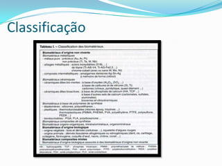
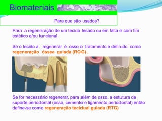
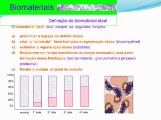
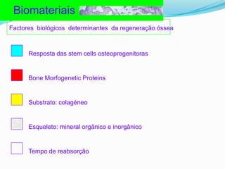
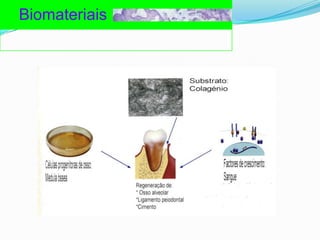
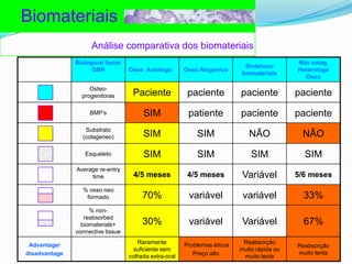
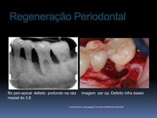
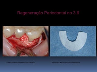
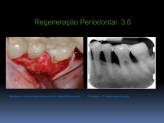
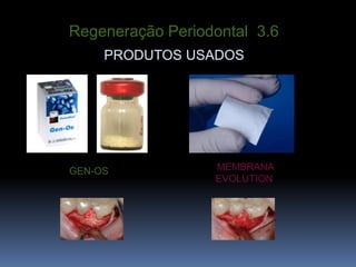
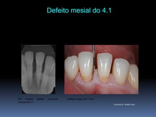
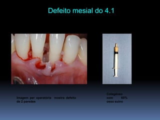
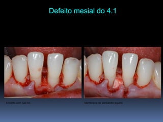
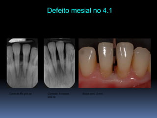
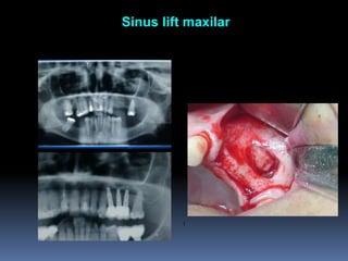
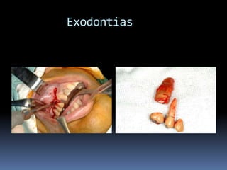
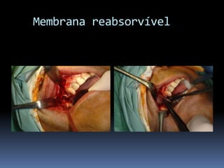
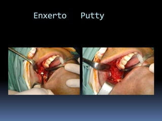
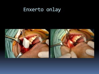
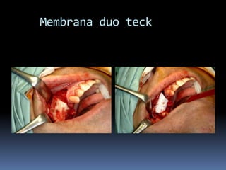
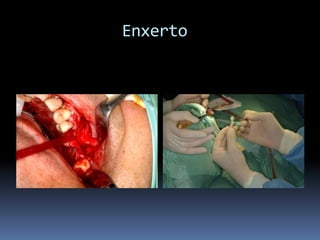
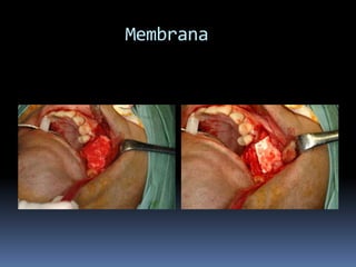
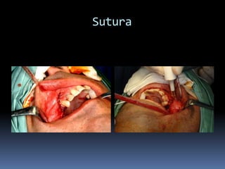
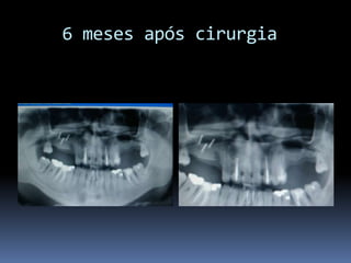
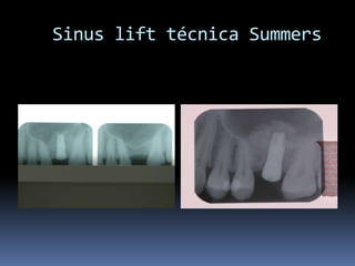
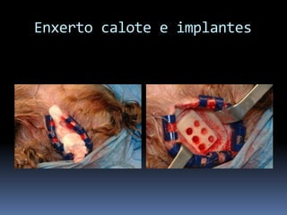
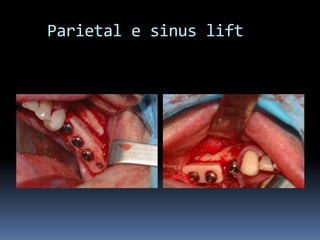
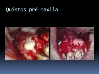
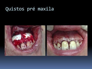
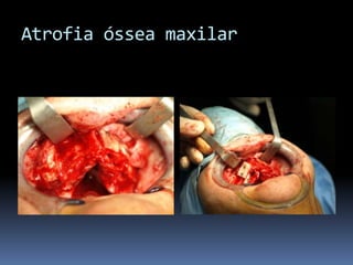
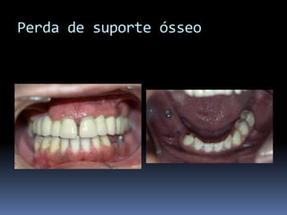
BoneregenerationanditsimpactinAntiagingÓscar Prim da Costa      Serviço de Cirurgia Maxilofacial  - Hospital de São JoséClínica  Maxfac – Cirurgia Estética da Face
Chester (1991):São materiais destinados a estar em contacto com os tecidos  vivos e/ou fluidos biológicos para avaliar, tratar, e modificar  as formas ou substituir todo um tecido, orgão  ou  função  do corpo.
Classificação
BiomateriaisPara que são usados?Para  a regeneração de um tecidolesadoouemfalta e com fimestético e/oufuncionalSe o tecido a   regenerar  é  osso o  tratamento é definidocomoregeneraçãoósseaguiada (ROG) .Se for necessárioregenerar, paraalém de osso, a estutura de suporte periodontal (osso, cemento e ligamento periodontal) então define-se comoregeneraçãotecidualguiada (RTG)
BiomateriaisDefinição do biomaterial idealO biomaterial idealdevecumprir  as  seguintesfunções: preencher o espaço do defeitoósseocriar  o “ambiente”  favorávelpara a regeneraçãoóssea(biocompatível)estimular a regeneraçãoóssea(substrato)Reabsorverem tempo semelhanteao tempo necessáriopara a neo formaçãoósseafisiológica(tipo de material , granulometria e processoproductivo)Manter o volume  original do enxerto100%80%60%40%20%0%1° mês2° mês3° mês4° mêsenxerto
BiomateriaisFactoresbiológicosdeterminantesdaregeneraçãoósseaResposta das stem cells osteoprogenitorasBone Morfogenetic ProteinsSubstrato: colagéneoEsqueleto: mineral orgânico e inorgânicoTempo de reabsorção
Biomateriais
BiomateriaisAnálisecomparativa dos biomateriais
Colagéneo: o substrato idealAfinidade natural pelas  BMPsProtege BMPs de proteólisenãoespecificaPrevine a difusãoprematura das BMPsÉ completamentereabsorvidoÉ substituidopor novo tecidoósseoformadoExcelentesubstratopara o recrutamento,  ancorageme diferenciação de célulasprogenitoras de osso
Bibliografia sobre colagéneo	Bruce E, Steven P, Louis M. Use of microcristalline collagen for hemostasis after oral surgery in a hemophylic. J Oral Surg 1979 37(2): 126-128	Capuano A, Bargelli F, Fadda GM, Parrini S. Collagene eterologo in chirurgia odontostomatologica. Il dentista moderno. Dicembre 2002	Centrella M, McCarthy TL, Canalis E. Trasforming growth factor beta is a bifunctional regular of replication and collagen synthesis in osteoblast-enriched cell cultures from fetal rat bone. J Biol Chem, 1987; 262: 2869-2874	Charulatha V, Rajaram A. Influence of different crosslinking treatments on the physical properties of collagen membranes. Biomater, 2003; 24: 759-767	Hsu FY, Chueh SC, Wang YJ. Microspheres of hydroxyapatite/reconstituted collagen as supports for osteoblast cell growth. Biomater, 1999; 20: 1931-1936	Kadler KE, Holmes DF, Trotter JA, Chapman JA. Collagen fibril formation. Biochem J, 1996; 316: 1-11	Liu LS, Thomson AY, Heidaran MA, et al. An osteoconductive collagen/hyaluronate matrix for bone regeneration. Biomater, 1999; 12: 1097-1108	Lucas PA, Syftestad GT, Goldberg VM, Caplan AI. Ectopic induction of cartilage and bone by water soluble proteins from bovine bone using a collagenous delivery vehicle. J Biomed Mater Res, 1989; 23(A1): 23-29 	Meyer U et al. Microstructural investigations of strain-related collagen mineralization. British J Oral  Maxillofacial Surgery, 2001; 39: 381-389	Patino MG, Neiders ME, Andreana S, Noble B, Cohen RE. Collagen as an implantable material in medicine and dentistry. J Oral Implantology, 2002; 28(5): 220-225	Stein MD, Salkin LM, Freedman AL, Glushko V. Collagen sponge as a topical hemostatic agent in mucogingival surgery. J Periodontol 1985; 56: 35-38  	Salasznyk RM, Williams WA, Boskey A, Batorsky A,, Plopper GE. Adhesion to vitronectin and collagen I promotes osteogenic differentiation of human mesenchymal stem cells. Journal of Biomedicine and Biotechnology, 2004; 1: 24-34	Wallace DG, Rhee W, et al. Injectable cross-linked collagen with imporved flow properties. J Biomed Mater Res, 1989; 23: 931-945	Werkmeister JA, Tebb TA, White JF, Ramshaw JAM. Collagenous tissue formation in association with medical implants. Current Opinion in Solid State and Materials Science, 2001; 5: 185-191
Int J Oral Maxillofac Implants. 2004 Mar-Apr;19(2):199-207  Histomorphometric analysis of natural bone mineral for maxillary sinus augmentation.John HD, Wenz B.PURPOSE: Lack of bone height in the posterior maxilla often necessitates augmentation prior to or simultaneously with dental implant placement. The purpose of this clinical study was to evaluate the use of the natural bone mineral Bio-Oss alone or in combination with autogenous bone in sinus floor elevations performed as 1- or 2-step procedures. MATERIALS AND METHODS: Thirty-eight patients required sinus augmentation. Natural bone mineral alone was used in sinus floor augmentation in 21 patients. In 13 patients, a mixture of the bone substitute and autogenous bone was used, and in 4 patients autogenous bone alone was used. In all of the patients, samples were taken for biopsy 3 to 8 months postoperatively, and bone regeneration was evaluated histologically and histomorphometrically. RESULTS: In all patients, the amount of new bone significantly increased over the observation time, while marrow areas decreased. There was no statistically significant difference in the amount of new bone formation between the Bio-Oss group (new bone 29.52% +/- 7.43%) and the Bio-Oss/autogenous bone group (new bone 32.23% +/- 6.86%). In the 4 patients treated with autogenous bone alone, a greater amount of newly formed bone was found; however, in these cases the area volume filled was smaller than in the other 2 groups. DISCUSSION: The data showed that new bone formation takes place up to 8 months after sinus floor elevation and that there is no difference in the amount of bone formation between procedures done with the bone substitute alone or with the mixture of the substitute and autogenous bone. CONCLUSION: These data suggest that predictable bone formation can be achieved with the use of Bio-Oss.
Casos clínicos
Regeneração Periodontal Rx peri-apical: defeitoprofundonaraizmesial do 3.6 Imagem  per op: Defeito infra ósseoCortesia Drs. Giuseppe Corrente e Roberto Abundo
Regeneração Periodontal no 3.6Preenchimento do defeito com Gen-Os.Membrana X-Fine Evolution membrane
Regeneração Periodontal  3.6Membranacolocadaparaprotecção do material de enxertoControlerx  6 mesesapóscirurgia
Regeneração Periodontal  3.6PRODUTOS USADOSMEMBRANAEVOLUTIONGEN-OS
Defeito mesial do 4.1 RX mostradefeitoprofundomesialdo 4.1.Defeitoósseo de 7 mmCortesia Dr. WalterRao
Defeito mesial do 4.1 Colagéneo com 60% ossosuinoImagem per operatóriamostradefeito de 2 paredes
Defeito mesial do 4.1 Membrana de pericárdioequinoEnxerto com Gel 40.
Defeito mesial no 4.1 Controle Rx pós op Bolsa com  2 mm.Controle  8 mesespós op
 Sinus lift maxilarI
Sinus lift maxilarDefeitoósseomaxilarRemoçãopontefixaCortesia Dr. Roberto Rossi
Sinus lift maxilar técnica de summersSinus lift Técnica de SummersColocação de implantes
 Sinus liftmaxilar8 mesesapóscargaRX pós op
Atrofia óssea maxilar e ramo horizontal mandibula
Sinuslift
Implantes após enxerto
Colheita enxerto ramo horizontal
Enxerto ramo horizesqmand
      1 ano após carga
      1 ano após carga
  Perda de suporte ósseo
   Perda de suporte ósseo
Exodontias
   Membrana reabsorvível
     Enxerto   Putty
      Enxerto onlay
    Membrana duo teck
           Enxerto
          Membrana
            Sutura
    6 meses após cirurgia
Sinuslift técnica Summers
  Enxerto calote e implantes
   Parietal e sinuslift
Osteotomia e expansão com     implantes
Expansão com osteotomia e implantes
  Enxerto ósseo com PRGF
Traumatismo alveolo dentárioEnxerto com putty e fixação com parafuso osteossintese
Traumatismo alveolo dentário
Quistos da pré maxila
Quistos pré maxila
Quistos pré maxila
Quistos pré maxila
Atrofia óssea maxilar
Perda de suporte ósseo
Atrofia mandibula
Atrofia mandibula
IMPLANTES ZIGOMÁTICOS     INDICAÇÕESMaxilar Edéntulo com Grande Reabsorção óssea		Pneumatização do Seio Maxilar	Status Pós-Maxilectomia	Fenda Lábio-Palatina
IMPLANTES ZIGOMÁTICOSFRACASSO FACIALHipoplasia maxilarFalso prognatismo
IMPLANTES ZIGOMÁTICOS
Obrigado

Bone Regeneration Impact In Anti Aging