Captura imagens macrofungos
•
0 gostou•30 visualizações

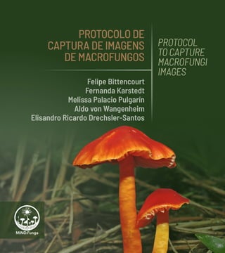
PROTOCOLO DE CAPTURA DE IMAGENS DE MACROFUNGOS (Felipe Bittencourt, Fernanda Karstedt, Melissa Palacio Pulgarín, Aldo von Wangenheim e Elisandro Ricardo Drechsler-Santos)

Recomendados
Mais conteúdo relacionado
Semelhante a Captura imagens macrofungos
Semelhante a Captura imagens macrofungos (20)
Mais de Jerbialdo
Mais de Jerbialdo (20)
Captura imagens macrofungos
- 1. PROTOCOLO DE CAPTURA DE IMAGENS DE MACROFUNGOS PROTOCOL TO CAPTURE MACROFUNGI IMAGES Felipe Bittencourt Fernanda Karstedt Melissa Palacio Pulgarín Aldo von Wangenheim Elisandro Ricardo Drechsler-Santos
- 2. PROTOCOLO DE CAPTURA DE IMAGENS DE MACROFUNGOS PROTOCOL TO CAPTURE MACROFUNGI IMAGES
- 3. PROTOCOLO DE CAPTURA DE IMAGENS DE MACROFUNGOS PROTOCOL TO CAPTURE MACROFUNGI IMAGES Felipe Bittencourt Fernanda Karstedt Melissa Palacio Pulgarín Aldo von Wangenheim Elisandro Ricardo Drechsler-Santos — 2 0 2 2 —
- 4. A P R E S E N T A Ç Ã O Antes do advento de microscópios e biologia molecular, a identificação de espécies era realizada por meio de características macroscópicas. Atualmente, faz parte de uma identificação precisa uma combinação de características macro e micromorfológicas, químicas e moleculares. Existem espécies similares e microscopicamente, metabolicamente ou geneticamente distintas. Assim como existem organismos com morfolo- gia variada, mas que molecularmente compreendem uma única espécie. Estas dificuldades nos processos de identificação são reconhecidas por toda a comunidade científica. Mesmo frente a essas dificuldades é de conhecimento comum que existem muitas espécies neotropicais que podem ser reconhecidas pela morfologia capturada em uma fotografia, sendo essas em nível de espécie ou enquadrada em algum outro grupo taxonômico. Deste modo, este guia tem como objetivo propor uma metodologia padrão para a captura de imagens que possibilitem o reconhecimento de espécies de macrofungos por meio de fotografias em um aplicativo, app MIND.Funga. Este aplicativo está ligado à uma base de dados que são utilizados para treinar uma rede neural artificial (inteligência artificial) com o objetivo de reconhecimento de fungos através de imagens. Este livro também é parte dos seguintes projetos de pesquisa: this book is part of the follow research projects: » CNPq (457451/2012-9); » PQ311158/2018-8; » CNPq/Capes/FAPs/BC - Fundo Newton/PELD nº 15/2016, » FAPESC/2018TR0928 » FAPESC/CNPq PRONEM 04/2019. » CNPq/MCTI/CONFAP-FAPS - PROTAX (CNPq 441821/2020-0, FAPESC2021TR390) A P O I O / S U P P O R T INCT-HerbárioVirtual da Flora e dos Fungos R E A L I Z A Ç Ã O / C O O R D I N A T I O N Dados Internacionais de Catalogação na Publicação (CIP) (Câmara Brasileira do Livro, SP, Brasil) Protocolo de captura de imagens de macrofungos = Protocol to capture macrofungi images [livro eletrônico] / Felipe Bittencourt ... [et al.]. -- 1. ed. -- Florianópolis, SC : Officio, 2022. PDF Outros autores : Fernanda Karstedt, Melissa Palacio Pulgarín, Aldo von Wangenheim, Elisandro Ricardo Drechsler-Santos. Bibliografia. ISBN 978-65-87710-12-9 1. Biologia 2. Fotografia 3. Fungos 4. Imagens fotográficas I. Bittencourt, Felipe. II. Karstedt, Fernanda. III. Pulgarín, Melissa Palacio. IV. Wangenheim, Aldo von. V. Drechsler- Santos, Elisandro Ricardo. VI. Título : Protocol to capture macrofungi images. 21-92690 CDD-579.5 Índices para catálogo sistemático: 1. Fungos : Microbiologia 579.5 Aline Graziele Benitez - Bibliotecária - CRB-1/3129
- 5. Assim, os passos a seguir sugerem boas práticas de fotografia de ma- crofungos para que as imagens contenham um número mínimo de infor- mações importantes, possibilitando uma maior eficiência do algoritmo do aplicativo no reconhecimento de espécies e/ou grupos taxonômicos mais inclusivos. P R E S E N T A T I O N Before the arrival of microscopes and molecular biology, species identi fication was carried out using macroscopic characteristics. Currently, a proper identification is a combination of macro-and micro-morphological, chemical and molecular characteristics. There are similar and microscop ically, metabolically or genetically distinct species; as well as there are organisms with diverse morphology, but that molecularly comprise a single species. These difficulties in the identification processes are recognized by the entire scientific community. Even with these difficulties, is common knowledge that there are many neotropical species that can be recognized by the morphology captured in a photograph, at species level or framed in some other taxonomic group. Thus, this guide aims to propose a standard methodology for capturing images that make it possible to recognize macrofungi species through pho tographs in an application, app MIND.Funga. This application is connected to a dataset used to train an artificial neural network (artificial intelligence) with the purpose of recognizing fungi through images. Thus, the following steps suggest good practices of macrofungi photogra phy to obtain images containing a minimum number of important informa tion; this will allow a greater efficiency of the application algorithm in the recognition of species and/or more inclusive taxonomic groups.
- 6. O S M A C R O F U N G O S Os fungos assim como os animais e as plantas, são seres vivos que fazem parte do planeta e estão presentes em todos os ambientes. De modo geral a fauna e a flora de um determinado local são bem conhecidas, mas e a Funga? Na maior parte do tempo os fungos passam despercebidos, ob- viamente porque muitos são microscópios, mas mesmo os macrofungos em boa parte do seu ciclo de vida ficam escondidos nos substratos que consomem. Os macrofungos, são assim nomeados, porque são capazes de produzir estruturas reprodutivas (produtora de esporos), os esporomas e/ou estromas, que são visíveis a olho nu. Estes esporomas possuem as mais diversas formas, tamanhos e cores, sendo popularmente conheci- das, por exemplo, como cogumelos, bolas-da-terra, dedos-de-defunto, estrelas-da-terra, orelha-de-geleia, orelhas-de-pau. Como já dito, muitas vezes uma boa fotografia pode ser suficiente para o reconhecimento da espécie ou grupo taxonômico desses macrofungos. Apresentamos a seguir o que foi avaliado como importante no ato de re- alizar fotografias para ter boas imagens e suficientemente informativas para o reconhecimento de espécies via aplicativo MIND.Funga. Fungos possuem hábitos diversos. Hygrocybe sp. é um cogumelo que cresce no solo das florestas. Fungi have different habits. Hygrocybe sp. is a mushroom that grows in the forests soils.
- 7. [11] PROTOCOL TO CAPTURE MACROFUNGI IMAGES M I N D. F U N G A [10] PROTOCOLO DE CAPTURA DE IMAGENS DE MACROFUNGOS M I N D. F U N G A Alguns fungos se associam a outros organismos formando simbioses, como é o caso dos fungos liquenizados, que formam associações com algas. Some fungi associate with other organisms forming symbioses, as is the case of lichenized fungi, which form associations with algae.
- 8. M A C R O F U N G I Fungi, like animals and plants, are living beings that are part of the planet and are present in all types of environments. In general, fauna and flora of a given place are well known, but what about Funga? Fungi are not easily seen as some of them are microscopic and others, such as macrofungi, spend much of their life cycle hidden in the substrates they consume. Macrofungi are so called because they can produce reproductive structures (spore-bearing), sporomata and/or stromata, which are visible to the naked eye. These sporomata have many variable shapes, sizes, and colours; com monly known, for instance, as mushrooms, puffballs, earthstars, jellyfungi, and brackets. As already said, a good photography can often be enough to recognize species or taxonomic group of these macrofungi. Below we present what was considered important in the act of taking photographs to have good images and sufficiently informative for the species recognition through the MIND.Funga application. Sumário/Contents Preparo do cenário / Scenario planning.............................................................................................15 1. Limpe a área / Clean the area.................................................................................................................16 2. Enquadramento / Framing.........................................................................................................................18 3. Cuidado com a luz / Take care of the light..........................................................................20 4. Escala / Scale............................................................................................................................................................. 22 5. Composição / Composition...................................................................................................................... 24 O fungo (esporoma)/ The fungi (sporoma).....................................................................................27 1. Visão superior/ Top view ............................................................................................................................ 32 2. Visão inferior / Underside view.......................................................................................................... 34 3. Visão de perfil/ Side view (profile)................................................................................................. 36 4. Aspecto geral e substrato/ General appearance and substrate.......... 38 Informações adicionais/ Additional information.................................................................... 41 A inteligência artificial pode ajudar você a conhecer os fungos Artificial Intelligence can help you to recognize Fungi.................................................... 47 Colaboração na construção do aplicativo/ Collaboration to build the app........................................................................................................................ 63
- 9. Preparo docenário Scenario planning Comoemtodasas fotografias,opreparo docenárioéimportante. Vejaabaixoalgumasdicas quandonosreferimos aosmacrofungos. Aswithallphotographs, scenarioplanningis important.Seebelowfor sometipswhenreferring tomacrofungi. O himenóforo dos fungos, geralmente localizado na parte de baixo, carrega as hifas produtoras de esporos. Além disso, possuem diversas características importantes para a identificação das espécies. The fungal hymenophore, usually located at the bottom, carries the spore-producing hyphae. In addition, they have several important characteristics for species identification. VOLTAR AO SUMÁRIO
- 10. [17] PROTOCOL TO CAPTURE MACROFUNGI IMAGES M I N D. F U N G A [16] PROTOCOLO DE CAPTURA DE IMAGENS DE MACROFUNGOS M I N D. F U N G A 1. Limpe a área Limpe a área: remova cuida do sa menteelementosqueestiveremso- breeaoredordoesporoma(fungo) aserfotografadocomo,porexemplo, folhas,galhos,raízes.Deixetodasas estruturasdoesporomabemexpos- tas(Figuras1e2).Àsvezes,éneces- sárioremoveroesporomadosubs trato,mas evite danificá-lo,princi pal mente na base, a qual pode ter alguma informação importante. 1. Clean the area Clean the area: remove carefully elements on or around the sporo ma (fungi), such as leaves, branch es, and roots. Leave all sporoma structures well exposed (Figure 1 and 2). Sometimes it is necessary to remove the sporoma from the substrate, but avoid damaging it, especially at the base, which may have some important information. FIGURA 1. AeC.Fotografiadeesporo mas sem limpeza e sem preparo do cenário. BeD.Fotografiaapóslimpezadoambiente. FIGURE 1. A and C. Sporomata photograph without cleaning or planning scenario. B and D. Photograph after cleaning. A C B D VOLTAR AO SUMÁRIO
- 11. [19] PROTOCOL TO CAPTURE MACROFUNGI IMAGES M I N D. F U N G A [18] PROTOCOLO DE CAPTURA DE IMAGENS DE MACROFUNGOS M I N D. F U N G A 2. Enquadramento O enquadramento pode ser de um esporoma em seu ambiente, de modo que ele ocupe o centro da imagem,depreferênciaocupando pelomenos60-75%dela(Figura2). Senãoforpossívelaproximartanto do esporoma sem perder o foco, priorize o foco! 2. Framing Theframingmaybeofasporomain its environment, so that it occupies the image center, preferably occu pyingatleast60-75%ofit(Figure2). Ifitisnotpossibletogetsocloseto the sporoma without losing focus, prioritize the focus! FIGURA 2. Exemplos de fotografias em que o esporoma está enquadrado. A. Asorea rubra group. B. Cyathus sp. A-B. Fotografados de cima. C. Boletinellus exiguus, fotografia da parte de baixo. D. Ionomidotis sp., fotografado de perfil. FIGURE 2. Examples of photographs in which the sporoma is framed. A. Asorea rubra group. B. Cyathus sp. A-B. Photo- graphed from above. C. Boletinellus exiguus, underside photograph. D. Ionomidotis sp., profile photograph. VOLTAR AO SUMÁRIO A C B D
- 12. [21] PROTOCOL TO CAPTURE MACROFUNGI IMAGES M I N D. F U N G A [20] PROTOCOLO DE CAPTURA DE IMAGENS DE MACROFUNGOS M I N D. F U N G A 3. Cuidado com a luz Certifique-se de que o lugar onde o esporoma está tenha uma lumi- nosidade adequada e homogênea, sem focos de luz/sombra (Figura 3). Se necessário transporte o es- pécime para uma área com ilumi- nação mais adequada ou utilize um guarda-chuvas para homoge- neizar a luz do ambiente e tripé para uma exposição 3. Take care of the light Make sure that the place where the sporomais,hasadequateandhomo geneous brightness, without light / shadow(Figure3A).Ifnecessary,take the specimen to an area with more adequatelightingoruseanumbrella or other object to homogenize the ambientlight.Indarkerenvironments it is easier the photo to come out blurry, so it is recommended to use atripodtosetupalargerexposureof thecameraandthuscompensatefor lowlight;anotheralternativeistouse flash, but use it sparingly, there may be a significant natural coloration lossofthesporoma. FIGURA 3. A-C. Fotografia inadequada, com manchas de luz ou ausência de luz prejudicando a melhor imagem dos esporomas. D-F. Fotografia adequada, com uma iluminação homogênea. FIGURE 3. A-C. Improper photographs, with overexposed light spots or light absence impairing the photograph. D-F. Proper photographs with homogeneous lighting. A B C D E F VOLTAR AO SUMÁRIO
- 13. [23] PROTOCOL TO CAPTURE MACROFUNGI IMAGES M I N D. F U N G A [22] PROTOCOLO DE CAPTURA DE IMAGENS DE MACROFUNGOS M I N D. F U N G A 4. Escala Um esporoma pode medir milí- metros ou atingir quase um me- tro, caso seja possível, inclua em uma das fotografias algo que atue como escala, por exemplo, uma moeda ou uma régua (Figura 4). Mas mantenha a escala distante do espécime para que possa ser excluída e não prejudicar a esté- tica de sua foto. FIGURA 4. A. Exemplos de esporomas de macrofungos e uma pequena amostra de diversidade de tamanhos. B. Exemplo de fungofotografadosemescala,Favolaschiasp. C.Exemplodefotografiacomescaladecor, Geastrum sp. D. Exemplo de fotografia com escalademedida,Agaricussp. 4. Scale A sporoma can measure millime ters or reach almost a meter, if possible, include in one photograph something as a scale, for example, acoinoraruler(Figure4).But,keep the scale away from the specimen so that it can be deleted and not affect your photo aesthetics. FIGURE 4. A. Examples of macrofungi sporomata as a little sample of size diversity. B. Example of fungi photographed without scale, Favolaschia sp. C. Example of photograph with colored scale, Geastrum sp. D. Example of photograph with scale, Agaricus sp. A D C B VOLTAR AO SUMÁRIO
- 14. [25] PROTOCOL TO CAPTURE MACROFUNGI IMAGES M I N D. F U N G A [24] PROTOCOLO DE CAPTURA DE IMAGENS DE MACROFUNGOS M I N D. F U N G A 5. Composição Procure por outros exemplares de esporomas da mesma espécie até cerca de dois metros e agrupe-os, deste modo terás uma ideia me- lhor dos formatos e estado de ma- turação dos fungos encontrados (Figura 3.B e 5). Você pode incluir mais de um esporoma na mesma foto, posicionando-os em diferen- tes ângulos, porém mantendo dis- tância para que a imagem possa ser fragmentada para possíveis enquadramentos individuais. 4. Composition Look for other sporomata of the same species up to about two me ters and group them, this way you willhaveabetterideaoftheshapes and maturation status of the fungi found (Figure 3.B and 5). You can include more than one sporoma in the same photo, positioning them at different angles, but maintaining a distance so that the image can be fragmented for possible individu al framings. But be careful: avoid grouping sporomata that may rep resent different species! FIGURA 5. A-D. Exemplos de imagens compostas.A.Pluteuscrinitus.B.Trametessp. C. Xylariaceae. D. Calvatia rugosa. FIGURE 5. A-D. Examples of composite photos. A. Pluteus crinitus. B. Trametes sp. C. Xylariaceae. D. Calvatia rugosa. A C B D VOLTAR AO SUMÁRIO
- 15. [27] PROTOCOL TO CAPTURE MACROFUNGI IMAGES M I N D. F U N G A [26] PROTOCOLO DE CAPTURA DE IMAGENS DE MACROFUNGOS M I N D. F U N G A Ofungo (esporoma) Thefungi (sporoma) Oregistrodascaracterísticas doesporomaqueirãoauxiliaro reconhecimentodotáxoné tãooumaisimportantequea qualidadedoregistrofotográfico. Therecordingofthesporoma characteristicswillassistthe taxonrecognitionisasormore importantthanthequalityof thephotographicrecord. Fungos são os grandes responsáveis pela reciclagem da matéria orgânica dos ecossistemas. Este Hymenochaete sp. está decompondo um tronco morto. Fungi are largely responsible for recycling organic matter from ecosystems. This Hymenochaete sp. is rotting a dead trunk. VOLTAR AO SUMÁRIO
- 16. [29] PROTOCOL TO CAPTURE MACROFUNGI IMAGES M I N D. F U N G A [28] PROTOCOLO DE CAPTURA DE IMAGENS DE MACROFUNGOS M I N D. F U N G A O registro ideal, considerando o reconhecimento via aplicativo MIND.Funga, é de duas a quatro fotografias para o mesmo espo- roma, de modo que forneçam in- formações como o substrato em que o fungo está se desenvolven- do, formato geral e das partes que compõem o esporoma, coloração, texturas e possíveis detalhes de ornamentação. É importante ressaltar que cada grupo taxonômico de macrofun- gos possui suas particularidades, algunsrequeremmaisdetalhesna horaderealizaracoletaefotogra- fias de campo, outros mais testes químicos,masdemodogeralpen- se em um conjunto de fotografias quepermitiriamreproduziroespé- Theidealrecord,consideringrecog nition through the MIND.Funga ap plication, is two to four quality pho tographs for the same sporoma, so that they provide information such as the substrate on which the fungi is growing, the general format and all sporoma components, coloring, textures and possible ornamenta tion details. Itisimportanttonotethateachtax onomic group of macrofungi has its particularities, some require more details when collecting and taking field photographs, others require more chemical tests, but in general thinkofaphotographssetthatwould allowyoutoreproducethespecimen inathree-dimensionalway.Thisway Alguns belos detalhes dos esporomas só podem ser enxergados através das lentes. Na foto, um espécime de Aegis luteocontexta. Some beautiful sporomata details can only be seen through the lens. In the photo, a specimen of Aegis luteocontexta. VOLTAR AO SUMÁRIO
- 17. [31] PROTOCOL TO CAPTURE MACROFUNGI IMAGES M I N D. F U N G A [30] PROTOCOLO DE CAPTURA DE IMAGENS DE MACROFUNGOS M I N D. F U N G A cimedemodotridimensional.Des- te modo terás contemplado um número mínimo ideal de imagens. Propomos que sejam realizadas pelomenostrêsfotografiasdecada esporoma, uma de visão superior, umadevisãolateral(perfil)eumade visãoinferior(Figura6-9)eemuma destas imagens recomendamos queestejaevidenciadoosubstrato. Procuresempretirarafotodofun- go“depé”ounamesmadireçãodo seu crescimento (a base do fungo napartedebaixodafoto,apartede cimadelenapartedecimadafoto). you will have contemplated an ideal minimum number of images. Weproposethatatleastthreepho tographsbetakenofeachsporoma, onefromthetopview,onefromthe side view (profile) and one from un derside view (Figure 6-9) and in one of these images we recommend that the substrate is highlighted. Alwaystrytotakethepictureofthe fungus “standing”, or in the same direction of its growth (the lower part of the fungus at the bottom of the picture, and the upper part at the top). FIGURA 6.A-C. Esquema de como foto grafar.D-L. Exemplos de fotografias. A, D, G. Fotografia de visão superior, B, E, H. Fotografia de visão inferior. C, F, I. Fotografia de perfil. D-F. Polyporus sp. G-I. Geastrum sp. FIGURE 6. A-C. Layout of how to shoot. D-L. Photographs examples. A, D, G. from the top view. B, E, H. underside view. C, F, I. side view (profile). D-F. Polyporus sp. G-I. Geastrum sp. A D G B E H C F I VOLTAR AO SUMÁRIO
- 18. [33] PROTOCOL TO CAPTURE MACROFUNGI IMAGES M I N D. F U N G A [32] PROTOCOLO DE CAPTURA DE IMAGENS DE MACROFUNGOS M I N D. F U N G A 1. Visão superior Uma foto de cima. Nesta imagem serão coletadas as características como aspecto da superfície pilear para orelha-de-pau e cogumelos, assim como número de “ovinhos” nos ninhos-de-passarinho ou nú- mero de braços nas estrelas-da- -terra (Figura 2.A-B, 6.A,D,G, J e 7). Para alguns grupos de fungos, como os crostosos, está será a única foto possível (Figura 7.E). FIGURA 7. Exemplos de fotografias vista de cima. A. Arambarria cognatas. B. Amanita coacta. C. Cookeina tricholoma. D. Geastrum sp. E. Aleurodiscus miriabilis. F. Parmotrema tinctorum (Foto:E.Gumboski). 1. Top view A photo from above. In this image, thecharacteristicssuchasthepilear surface appearance for brackets and mushrooms will be collected, as well as the number of “eggs” in the bird’s nests or the number of arms in the earthstars (Figure 2.AB, 6 .A, D, G, J and 7). For some fungi groups,suchascorticioid,thiswillbe the only possible photo (Figure 7.E). FIGURE 7. Examples of top view photo- graphs. A. Arambarria destruens. B. Amanita coacta. C. Cookeina tricholoma. D. Geastrum sp. E. Aleurodiscus miriabilis. F.Parmotrematinctorum(Photo:E.Gumboski). E C A F D B VOLTAR AO SUMÁRIO
- 19. [35] PROTOCOL TO CAPTURE MACROFUNGI IMAGES M I N D. F U N G A [34] PROTOCOLO DE CAPTURA DE IMAGENS DE MACROFUNGOS M I N D. F U N G A FIGURA 8. Exemplos de fotografias da parte inferior de cogumelos e orelhas- de-pau. A. Pleurotus sp. B. Geastrum sp. C. Polyporales. D. Pluteus sp. 2. Visão inferior Umafotodapartedebaixodoespo roma, podendo representar de- talhes das estrelas-da-terra e/ ou ninhos-de-passarinho ou da região fértil (himenóforo) no caso de cogumelos e orelhas-de-pau (Figura 6.B, E, H, K e 8). FIGURE 8. Examples of underside view photographs of mushrooms and brackets. A. Pleurotus sp. B. Geastrum sp. C. Poliporales. D. Pluteus sp. 2. Underside view A photo of the sporoma underside, which may represent details of the earthstar and/or bird’s nests or the fertile part (hymenophore) in the case of mushrooms and brackets (Figure 6.B, E , H, K and 8). A C B D VOLTAR AO SUMÁRIO
- 20. [37] PROTOCOL TO CAPTURE MACROFUNGI IMAGES M I N D. F U N G A [36] PROTOCOLO DE CAPTURA DE IMAGENS DE MACROFUNGOS M I N D. F U N G A 3. Side view (profile) A profile photo, side, from top to bottom, is essential for fungi that grow and stretch away from the substrate. In addition, to showing the substrate, this photograph may register specific characteristics of taxonomic groups such as, the presenceorabsenceofscales,ring in the stipe, and volva or mycelium in the stipe base. (Figure 6.C, F, I, L and 9). FIGURE 9. A-D.Examplesofprofilephoto graph. A. Volvariella perciliata. B. Calvatia rugosa. C. Phallus indusiatus group. D. Mutinus argentinus. E. Ramaria sp. F.Cladoniasp. (Photo: Emerson Gumboski). FIGURA 9. A-D. Exemplos de imagens em perfil. A. Volvariella perciliata. B. Calvatia rugosa. C. Phallus indusiatus group. D. Mutinus argentinus. E. Ramaria sp. F. Cladonia sp. (Foto F: Emerson Gumboski). 3. Visão de perfil Uma foto em perfil, lateral, do topo até a base, é essencial para os fungos que crescem e se alongam para longe do substrato. Além de evidenciar o substrato, esta fo- tografia poderá registrar carac- terísticas específicas de grupos taxonômicos como a presença ou ausência de escamas, anel no es- tipe e volva ou micélio na base do estipe (Figura 6.C,F, I,L e 9). E C A F D B VOLTAR AO SUMÁRIO
- 21. [39] PROTOCOL TO CAPTURE MACROFUNGI IMAGES M I N D. F U N G A [38] PROTOCOLO DE CAPTURA DE IMAGENS DE MACROFUNGOS M I N D. F U N G A 4.Aspectogeralesubstrato Evidencieofungoeosubstratoem que está, por exemplo: se estiver sobreumtronco,seestáinseridona madeira (viva ou morta) ou somente entreosmusgosnasuperfíciedeste tronco; se estiver no chão, se este estánaserapilheira,emalgumgalho ouinsetoouemmeioaosolo.Note que nesta fotografia o esporoma tambémestarápresente,assimela poderá representar uma das fotos sugeridas nos tópicos anteriores, não necessariamente uma quarta foto (Figura 10). FIGURA 10. A. Cogumelo crescendo no chão, em areia. B. Cogumelo crescendo no chão, na serapilheira. C. Cogumelo cres cendo no chão, em bainha de palmeira. D. Orelha-de-pau crescendo em galho. E. Ninhos-de-passarinho crescendo em esterco de gado. F. Fungo crescendo em lagarta enterrada. 4. General appearance and substrate Show the fungi and it’s substrate; forinstance:ifitisonatrunk,ifit is inserted in the wood (live or dead) or only between mosses on the trunk surface; if it is on the ground, if it is on the litter, on a branch or insect or in the middle of the soil. Note that in this photo the sporoma will also be present, so it can repre sentoneofthesuggestedphotosin the previous topics, not necessarily a fourth photo (Figure 10). FIGURE 10. A. Mushroom growing on the ground, in sand. B. Mushroom growing on the ground, in leaf litter. C. Mushroom growing on the ground, in leaf sheath palm. D. Bracket growing on a branch. E. Bird’s nests growing on cow dung. F. Fungi growing on buried caterpillar. A D E F B C VOLTAR AO SUMÁRIO
- 22. [41] PROTOCOL TO CAPTURE MACROFUNGI IMAGES M I N D. F U N G A [40] PROTOCOLO DE CAPTURA DE IMAGENS DE MACROFUNGOS M I N D. F U N G A Informações adicionais Além das fotografias, outras informações associadas ao material fotografado também são importantes. Additional information Inadditiontophotographs, otherinformationassociated withthephotographed material are also important. Photographer’s name It is always important to recognize the image author. Date of the photograph record Over time, will allow seasonality analysis. Geographic location City, state, country – make it pos sible to know the most real species distribution. Nome do fotógrafo É sempre importante reconhecer o autor da imagem. Data do registro fotográfico Ao longo do tempo, possibilitará uma análise de sazonalidade. Localização geográfica Cidade, estado, país – possibilitará conhecer a distribuição mais real da espécie. VOLTAR AO SUMÁRIO Fungos possuem colorações variadas e às vezes muito vivas. Na foto, apotécios de Chlorociboria sp. Fungi have varied and sometimes very vivid colors. This photo shows apothecia of Chlorociboria sp.
- 23. [43] PROTOCOL TO CAPTURE MACROFUNGI IMAGES M I N D. F U N G A [42] PROTOCOLO DE CAPTURA DE IMAGENS DE MACROFUNGOS M I N D. F U N G A Collector, collection number and herbarium In which it was or will be deposited (if any) – will allow access to those interested in studying the photo graphed. Fungi Identification species, genuso or family as far as possible. Through this information, the spe cies bank will become more robust, reliable,anditwillbepossibletouse it in the future as a tool to better understandthedistribution,habitat, seasonality and phenology of spe cies, as well as their monitoring in theNeotropicalregion,considering changes in space and time. Coletor, número de coletor e herbário No qual foi ou será depositado (se houver) – possibilitará o acesso a interessados em estudar o mate- rial fotografado. Identificação Do fungo, em espécie, gênero ou família, na medida do possível. Através destas informações o banco de espécies se tornará mais robusto, confiável, sendo possível utilizá-lo no futuro como uma ferramenta conhecer melhor a distribuição, habitat, sazonali- dade e fenologia das espécies, bem como seu monitoramento na Região Neotropical, frente às mudanças no espaço e no tempo. Muitas pessoas lembram apenas dos cogumelos, mas o reino dos fungos é infinitamente mais rico em formas. Alguns são chamados de coraloides, como este Clavulina fuscolilacinea. Many people only remember mushrooms, but the kingdom Fungi is infinitely richer in shapes. Some are called coralloids, like this Clavulina fuscolilacinea.
- 24. [45] PROTOCOL TO CAPTURE MACROFUNGI IMAGES M I N D. F U N G A [44] PROTOCOLO DE CAPTURA DE IMAGENS DE MACROFUNGOS M I N D. F U N G A Trametes sp. crescendo em um tronco à luz do sol. Trametes sp. growing on a log in the sunlight.
- 25. [47] PROTOCOL TO CAPTURE MACROFUNGI IMAGES M I N D. F U N G A [46] PROTOCOLO DE CAPTURA DE IMAGENS DE MACROFUNGOS M I N D. F U N G A O registro fotográfico é essencial para muitas espécies de fungos serem identificadas e estudadas. Muitas características se perdem durante o estudo dos espécimes. The photographic record is essential for many fungal species to be identified and studied. Many features are lost during the study of specimens. Ainteligência artificialpodeajudar vocêaconhecer osfungos O MIND.Funga App emprega uma técnica de Visão Robótica para classificar os fungos. Artificial Intelligencecanhelp youtorecognize Fungi The MIND.Funga App employs a technique of Robotic Vision in order to classify fungi specimens. VOLTAR AO SUMÁRIO
- 26. [49] PROTOCOL TO CAPTURE MACROFUNGI IMAGES M I N D. F U N G A [48] PROTOCOLO DE CAPTURA DE IMAGENS DE MACROFUNGOS M I N D. F U N G A Você já viu que este protocolo de fotografia aqui, além de servir para você fotografar melhor ma- crofungos que você for encontrar na natureza, seja você um pesqui- sadorounaturalistaamador,serve também para alimentar o nosso aplicativo MIND.Funga App de reconhecimento de fungos com imagens padronizadas e com boa qualidade de registro científico. O MIND.Funga App emprega uma técnica da área da Visão Robótica chamada de Inteligência Artificial com Redes Neurais de Aprendizado Profundo para classificar imagens de espécimes de macrofungos que você enviar para ele e tam- bém para aprender novas imagens e novas espécies que forem ali- mentadas por pesquisadores que participam do nosso projeto. Com o MIND.Funga App você pode fotografar e enviar imagens de macrofungos para classificação automática pela Inteligência Artifi- cial que nós desenvolvemos e,as- sim, ficar sabendo que fungo você fotografou. Você pode também, se desejar,participardoprojetocomo pesquisador ou naturalista ama- dor e coletar fotos de novos espé- cimes e contribuir para o acervo de imagens do nosso Projeto. FIGURA 11. O mindFunga App FIGURE 11. The mindFunga App The MIND.Funga App employs a technique of the field of Robotic Vision called Artificial Intelligence with Deep Learning Neural Net works in order to classify the im ages of specimens of macrofungi that you will be sending to it. It also employs Deep Learning to learn new images and new species fed toitbyresearchersthatparticipate on our project. With the MIND.Funga App you can photograph and upload images of macrofungi for automatic recog nition by the artificial intelligence that we developed and, so, know which fungus you have photo graphed. You can also, if you want, participate in our project as a re searcher or amateur naturalist and collect photographs of new speci mens and contribute to the image collection of our Project. You must have seen that this pho tographic protocol here, besides serving as a guide for you to pho tograph better macrofungi that you will eventually find in nature, regardless if you are a researcher or an amateur naturalist, serves also to feed our fungi recognition MIND.Funga App with standard ized images of a good scientific record quality. VOLTAR AO SUMÁRIO
- 27. [51] PROTOCOL TO CAPTURE MACROFUNGI IMAGES M I N D. F U N G A [50] PROTOCOLO DE CAPTURA DE IMAGENS DE MACROFUNGOS M I N D. F U N G A O que é a Visão Robótica? Também chamada de Visão Com- putacional, é o conjunto de téc- nicas de programação e modelos matemáticos que nós utilizamos para desenvolver programas de computador que são capazes de “enxergar”, isto é, compreender e classificar o conteúdo de imagens ou vídeos. A ideia básica da Visão Computa- cional é você tomar uma imagem e gradativamente ir simplificando e extraindo características impor- tantes dessa imagem até você ter uma versão abstrata da imagem, que descreve só os objetos que você está procurando, através de alguns poucos parâmetros ma- temáticos, ou características. A visão computacional começou a ser desenvolvida na década de 1960 mas, longe de produzir robôs FIGURA 12. Visão computacional clássica: vários passos para extrair características. FIGURE 12. Classic computer vision: several steps to extract features. What is Robotic Vision? Also called Computer Vision, it is the set of programming techniques and mathematical models that we use in order to develop computer programs that are able to “see”, that is, understand and classify the content of images or videos. Thebasicideaofcomputervisionis that you take an image and gradu allysimplifyitandextractimportant features from this image until you have an abstract version of the im age that describes only the objects you’re looking for, through only a few mathematical parameters or characteristics. Computer vision began to be developed in the 1960s but, far from producing robots that see everything and will dominate the world, during many decades VOLTAR AO SUMÁRIO
- 28. [53] PROTOCOL TO CAPTURE MACROFUNGI IMAGES M I N D. F U N G A [52] PROTOCOLO DE CAPTURA DE IMAGENS DE MACROFUNGOS M I N D. F U N G A que enxergam tudo e ameaçam dominar o mundo, durante mui- tas décadas essa área da Ciência da Computação esteve limitada a desenvolver programas que sempre só conseguiam resolver um problema: ou identificar um intruso em uma câmera de vigi- lância, ou identificar um tumor em um exame de tomografia, ou identificar nuvens de tempesta- de em uma imagem de satélite. Os programas não eram capazes de aprender coisas novas e para cada problema diferente eram ne- cessários anos de pesquisa para desenvolver um novo programa. E o que é a Inteligência Artificial? Éoconjuntodetécnicasdeprogra- maçãoemodelosmatemáticosque permite você desenvolver progra- mas de computador que possuem algumtipodecomportamentointe ligente.Eumdoscomportamentos inteligentes que mais interessam a nós é a capacidade de aprender. Aqui entram as redes neurais. Então, o que são Redes Neurais? Sim, são programas de compu- tador, inicialmente inventados também na década de 1960, que simulam o funcionamento de par- tes do tecido cerebral de animais. Para isso, esses programas pos- suem modelos computacionais de neurônios e de como esses neurônios são conectados entre si e como respondem a estímulos. And what is Artificial Intelligence? It is that set of programming tech niques and mathematical models that allow you to develop computer programs that show some kind of intelligent behavior. and one of the intelligent behaviors that interest us most is the ability to learn. Enter the neural networks. Then, what are Neural Networks? They are computer programs, ini tially also invented in the 1960s, that simulates the workings of parts of the neural structures of animals. For this purpose, these programs possess computational models of neurons and of how these neurons are connected and respond to stimuli. One important feature of neural networks is that this area of the Computer Sciences waslimitedtodevelopingcomputer programs that always were able onlytosolveonesingle,veryspecific problem: to identify and intruder on security camera images, or to identify a tumor in a tomographic examination, or to identify storm clouds on satellite images. These programs were not able to learn new things and, for each different problem, many years of research were necessary to develop a new program. VOLTAR AO SUMÁRIO
- 29. [55] PROTOCOL TO CAPTURE MACROFUNGI IMAGES M I N D. F U N G A [54] PROTOCOLO DE CAPTURA DE IMAGENS DE MACROFUNGOS M I N D. F U N G A Umacaracterísticaespecialmente importantedasredesneuraiséque elassãocapazesdeaprender.Você “ensina” uma rede neural apresen- tando exemplos de estímulos e da resposta que você espera que ela apresenteparaesteestímulo.Uma rotina computacional especial de treinamento passa por toda a redeefortaleceasconexõesentre neurônios que geraram uma boa resposta e enfraquece conexões quegeraramerrosnaresposta.As- sim a rede neural vai gradualmen- te modificando as suas conexões de maneira a responder de forma mais “correta” a estímulos. O ideal seria você juntar a visão computacional e as redes neurais e fazer programas de computador aos quais você poderia ensinar a “aparência” de qualquer coisa, simplesmente mostrando ima- gens dessa coisa, não? entrada ξk esp. ζi pesos Wjk pesos Wik pesos Oi interna Vj ξ1 ξ2 ξ3 W11 W33 W11 W33 V1 V2 V3 ζ1 ζ2 ζ3 O1 O2 O3 x1 x1 yi x n w1 j Wi j wn j ∑ƒ( ) FIGURA 13. Exemplo de uma rede neural clássica, dos anos 1980-90, baseada no “Neurônio de Hebb”. Cada neurônio calcula a soma dos sinais recebi dos em suas conexões de entrada, atenuados pe los pesos (w) dessas conexões. Esse valor excita uma função de ativação (f(()) que gera uma saída (V ou O), caso esse valor extrapole um limiar de excitação θ. A rede é formada por camadas de neurônios, cada camada ligada à próxima. FIGURE 14. Example of a classic neural network from the 1980-90s, based on the “Hebb Neuron”. Each neuron calculates the sum of the signals received through its incoming connections, attenuated by the weights (w) of these connections. This value excites an activation function (f()), which generates an output (V or O), in case this value extrapo- lates an excitation θ threshold sigma. The network is composed by layers of neurons, each layer fully connected to the next. they are able to learn. you teach a neuralnetworkpresentingexamples of stimuli and of the answer that you expect the network to present for these stimuli. A special compu tationaltrainingroutinegoesthrough the whole network and strengthens connections between neurons that generatedagoodanswerandweak ens connections that generated errorsintheanswer.Thustheneural network gradually modifies its con nections in order to respond in a “correct” way to the stimuli. The ideal solution would be to unify computer vision and neural net works and to be able to develop computer programs which could teach the “appearance” of any thing, simply showing images of that thing, don’t it? VOLTAR AO SUMÁRIO
- 30. [57] PROTOCOL TO CAPTURE MACROFUNGI IMAGES M I N D. F U N G A [56] PROTOCOLO DE CAPTURA DE IMAGENS DE MACROFUNGOS M I N D. F U N G A O problema é que durante muito tempo as redes neurais só foram capazes de processar estímulos simples. Para um programa de computador “entender” o conteú- do de uma imagem, não basta ele “decorar” os pixels de uma ima- gem. Se você fizer isso, a rede só vai aprender aquela uma imagem específica. O programa precisa ser capaz de aprender a extrair aquelas informações simplifica- das,as“característicasessenciais” daimagem,quepermitemdizerse a imagem contém um fungo, um navio, ou um cavalo, independen- tementedestecavalosermalhado oumarromedeestarolhandopara direita ou para esquerda. Tudo isso mudou em 1998, quando um pesquisador francês chamado YannLeCun1 sugeriuqueredesneu- 1 Hoje Diretor de Inteligência Artificial da Facebook. Current Director of Artificial Intelligence at Facebook. rais, ao invés de apenas aprende- rem a modificar as conexões entre neurônios,aprendessemfiltros,que modificamosinalquepassadeum um neurônio para outro de uma forma bem mais complexa do que simplesmentefortalecendoouate- nuando este sinal. Filtros são ope- radores muito poderosos em visão computacional,poiselespodemser adaptadosparafazermuitascoisas diferentes, desde eliminar o chu- visco de uma imagem até extrair características específicas como linhasretas,curvasecores,quesão elementosimportantesnoreconhe- cimento de fungos, por exemplo. Filtros de imagem se baseiam numaoperaçãomatemática,queé aconvolução.Mesmoquevocênão seja da área tecnológica, você vai lembrar da matemática da escola, onde você aprendeu a fazer multi- plicação de matrizes. Um filtro de how to adapt connections between neurons, should learn filters that modify the signal that passes from one neuron to the other in a much more complex way than simply attenuating or enhancing this signal. Filters are extremely powerful operators in computer vi sion, because they can be adapted to do many different things, from despeckling an image up to extract specific features such as straight lines, curves and colors, which are important elements in the recogni tion of fungi, for example. Image filters are based upon a mathematical operation called convolution. Even if you are not from the technology area, you will remembermathfromschool,where youlearnedmatrixmultiplication.A convolution filter is exactly that: The problem is that for a long time neural networks were able only to process simple stimuli. For a com puterprogramtobeabletolearnto understandthecontentofanimage, it does not suffice to “learn by heart” the pixels of an image. If you do that, it will only be able to iden tify that specific image. What the program needs to be able to learn is how to extract those simplified features, the “essential character istics” of an image, that will allow it to tell that an image contains a fungus, a ship, or a horse, inde pendentlyofthishorsebeingbrown or black, or being looking to the right or to the left. Everything changed in 1998, when a French researcher named Yann LeCun1 suggested that neural net works, instead of simply learning VOLTAR AO SUMÁRIO
- 31. [59] PROTOCOL TO CAPTURE MACROFUNGI IMAGES M I N D. F U N G A [58] PROTOCOLO DE CAPTURA DE IMAGENS DE MACROFUNGOS M I N D. F U N G A 0 0 0 0 0 0 0 105 102 100 97 96 0 103 99 103 101 102 0 101 98 104 102 100 0 99 101 106 104 99 0 104 104 104 100 320 -1 0 -1 5 -1 0 -1 0 98 Matriz de convolução Convolution matrix Imagem de entrada input image Cálculo da convulação para o píxel de valor 105 Convolution calculation for the 105th value pixel 0 0 * 0 + 0 * –1 + 0 * 0 + 0 * –1 + 105 * 5 + 102 * -1 +0 * 0 + 103 * -1 + 99 * 0 = 320 imagem convolvida (resultado para píxel 105) Convolved image (result for the 105th pixel FIGURA 15. Uma conexão de entrada em uma camada de rede neural convolucional não é sim plesmente modulada apenas por um peso, mas sim por uma matriz de convolução, que realiza um cálculo sobre uma região da camada anterior. O processo de aprendizado modifica e adapta a matriz, além de poderem existir várias matrizes que são aprendidas ao mesmo tempo. FIGURE 15. An input connection in a convolution neural network layer is not simply modulated by its weights, but also by a convolution matrix that performs a calculation on a region of the preceding layer. The learning process also modifies and adapts this matrix, besides the possibility that there can be several matrices that are learned at the same time. convolução é isto: é uma matriz pequena que você vai passando por cima de uma outra matriz bem maior, a sua imagem, e vai multi- plicando cada ponto da imagem e sua vizinhança. O resultado é uma nova imagem filtrada, onde uma determinadacaracterísticafoires- saltada ou alguma coisa que não queremos foi apagada. Eassimnasceuocasamentoentrea inteligênciaartificialeavisãocom- putacional:asRedesNeuraisCon- volucionais,tambémchamadasde redes neurais de aprendizado pro- a small matrix that you will pass over a larger matrix, your image, and multiply it by each pixel and its vicinity. The result is a new, filtered image, where a particular feature was enhanced or something that we do not want was deleted. So was born the marriage between artificial intelligence and computer vision: the Convolutional Neural Networks,alsocalledDeepLearn- ing Neural Networks. They pos sess many, many layers of neurons and, between these layers, instead VOLTAR AO SUMÁRIO
- 32. [61] PROTOCOL TO CAPTURE MACROFUNGI IMAGES M I N D. F U N G A [60] PROTOCOLO DE CAPTURA DE IMAGENS DE MACROFUNGOS M I N D. F U N G A fundo.Elaspossuemmuitasemuitas camadas de neurônios e, entre as camadas, ao invés de aprenderem apenas conexões entre neurônios, estas redes aprendem vários con- juntos de filtros de imagem. A partir de meados da década de 2010 esta se tornou a principal tecnologia de processamento de imagens. Ela permite você ensi- nar um programa de computador a aprender filtros sucessivos que ele pode usar para ir gradativa- mente modificando, simplifican- do e extraindo características de imagens, de forma que ao final ele seja capaz de classificar o que é importantedentrodeumaimagem e dizer o que é que aquela imagem está representando. Éissoquenósestamosfazendono MIND.Funga:oMIND.FungAppque você pode acessar no seu celular possui a última versão da nossa rede neural convolucional para a classificação de macrofungos. Ela foi treinada com a nossa base de imagens, que foi construída com imagens coletadas por nossos pesquisadores e muitas imagens contribuídasporpesquisadoresde outrasuniversidadesetambémpor muitos naturalistas amadores que têmenviadoassuasimagenspara nós. Nós regularmente curamos e classificamos estas imagens, para garantir que só as de melhor qualidade e só os espécimes cor- retamente identificados integrem a nossa base de dados e, a seguir, retreinamos a nossa rede neural de forma que ela agora conheça mais espécimes e, eventualmente também, novas espécies. Por isso é importante você seguir este protocolo fotográfico à risca: todas as imagens que foram usa- das para treinar a nossa rede neu- of learning only strengths of con nections between neurons, these networks learn and adapt several sets of image filters. From the mid 2010s on this became the main image processing tech nology. It allows you to teach a computer program to learn suc cessive filters that it can then use in order to gradually modify, sim plify and extract image features in a way that, in the end, it will be able to classify what is important inside this image and tell you what this image is representing. This is what we are doing in the MIND.Funga project: the MIND. Funga App that you can access on your phone has the last version ofourconvolutionalneuralnetwork for the recognition of macrofungi. It was trained on our image data set, which was built from images collected by our researchers and many more images which were contributed by many amateur natu ralists who have been sending us their images. We regularly curate and classify these images, in order to guarantee that only those of the best quality and only the correctly classified specimens will come to integrate our database. Afterwards we retrain our neural network, so that it will now be able to recognize more specimens and, eventually also new species. For this reason it is important that you follow this photographic pro tocol strictly: all images that were used to train our neural network were captured using this protocol. This was done in this manner in order to evidentiate the fungus VOLTAR AO SUMÁRIO
- 33. [63] PROTOCOL TO CAPTURE MACROFUNGI IMAGES M I N D. F U N G A [62] PROTOCOLO DE CAPTURA DE IMAGENS DE MACROFUNGOS M I N D. F U N G A ralforamcapturadasutilizando-se este protocolo. Isso foi feito assim para ressaltar o fungo e ensinar à rede características do fungo e não, por exemplo, do substrato onde o fungo se encontra ou da vegetação que está na vizinhança dofungo.Aredenão“conhece”fun- gosfotografadosdeoutraformae, sevocêenviarimagensadquiridas deumjeitomuitodiferente,oMIND. FungaAppprovavelmentevaiclas- sificá-las de forma incorreta. Isto é ainda mais importante se você já está contribuindo ou está pensando em contribuir para o MIND.Funga:paraexpandiranossa base de dados nós necessitamos de espécimes adquiridos criterio- samente e que representem ima- gens que possam contribuir para melhoraracuráciadeclassificação do MIND.Funga App no futuro. and teach the network important features of the fungus and not, for example, features of the subtract where the fungus is found or of the vegetation that is in the vicinity of the fungus. The network does not “know” fungi photographed in a different manner and, if you send images acquired in a very different way,theMIND.FungaAppwillproba bly classify them incorrectly. This is even more important if you are already contributing or is in tending to contribute for the MIND. Funga:inordertoexpandourdata base we need specimens acquired criteriously and that represent im ages that will be able to contribute to enhance the classification accu racy off the MIND.Funga App in the future. VOLTAR AO SUMÁRIO For the proper operation of the MIND.Funga app, it is necessary that the database used to train the artificial neural network (ar tificial intelligence) be constantly supplied and composed of an ex pressive number of good quality photographs and a representative number of taxa. MIND.Funga app aims to be initially available with 500 taxa, but over the years, we would like to incor porate the largest possible number Colaboração naconstrução doaplicativo Participe do projeto disponibilizando suas fotos. Collaboration tobuildtheapp Take part in the project sharing your photographs Para um bom funcionamento do aplicativo MIND.Funga é neces- sário que a base de dados utili- zada para treinar a rede neural artificial (inteligência artificial) seja abastecida constantemente e seja composta por um número expressivo de fotografias de boa qualidade e de um número repre- sentativo de táxons. O aplicativo tem por meta ser inicialmente disponibilizado com 500 táxons, mas ao longo dos VOLTAR AO SUMÁRIO
- 34. [64] PROTOCOLO DE CAPTURA DE IMAGENS DE MACROFUNGOS M I N D. F U N G A of Neotropical macrofungi species through the collaborative insertion of new images. The photographs can be made available by any user, so that they will be curated by specialists to havetheiridentificationrecognized and / or confirmed. Only then they will be included in the app’s image banksothattheycanbeusedinthe artificial neural training network responsible for the macrofungi species recognition. This proposal is to be periodically updated and checked for accuracy. Seja um colaborador dessa ideia! | Support this idea! https://mindfunga.ufsc.br/ anos, gostaríamos de incorporar o maior número possível de espé- cies de macrofungos neotropicais através da inserção colaborativa de novas imagens. As fotografias podem ser dispo- nibilizadas por qualquer usuário, de modo que estas passarão pela curadoria de especialistas para terem a identidade reconhecida e/ou confirmada. Somente então serão incluídas no banco de ima- gens do aplicativo para que sejam utilizadas no treinamento da rede neural artificial responsável pelo reconhecimento de espécies de macrofungos. Este tem por pro- posta ser periodicamente atuali- zado e verificado sua acuracidade.
- 35. A P O I O / S U P P O R T INCT-HerbárioVirtual da Flora e dos Fungos R E A L I Z A Ç Ã O / C O O R D I N A T I O N