
Recomendados
Recomendados
Mais conteúdo relacionado
Mais procurados
Mais procurados (20)
Destaque
Destaque (18)
Semelhante a 2004 pediat. motivos encaminhamento
Semelhante a 2004 pediat. motivos encaminhamento (20)
Último
Último (20)
2004 pediat. motivos encaminhamento
- 1. DOI: 10.1542/peds.2003-0898-L 2004;114;409-417Pediatrics Robert L. Geggel Hospital Conditions Leading to Pediatric Cardiology Consultation in a Tertiary Academic This information is current as of January 10, 2007 http://www.pediatrics.org/cgi/content/full/114/4/e409 located on the World Wide Web at: The online version of this article, along with updated information and services, is rights reserved. Print ISSN: 0031-4005. Online ISSN: 1098-4275. Grove Village, Illinois, 60007. Copyright © 2004 by the American Academy of Pediatrics. All and trademarked by the American Academy of Pediatrics, 141 Northwest Point Boulevard, Elk publication, it has been published continuously since 1948. PEDIATRICS is owned, published, PEDIATRICS is the official journal of the American Academy of Pediatrics. A monthly by on January 10, 2007www.pediatrics.orgDownloaded from
- 2. Conditions Leading to Pediatric Cardiology Consultation in a Tertiary Academic Hospital Robert L. Geggel, MD ABSTRACT. Objective. To determine the basis for cardiac consultations for pediatric patients in an aca- demic hospital setting. Methods. The activities of the cardiology consulta- tion service were tabulated for 12 months, from July 2001 to June 2002. Patients were identified from 4 sources, ie, a monthly log of patient encounters maintained by the consultation service, encounter forms submitted to the billing office, consultation notes maintained in a central file, and a departmental list of echocardiography studies. Patients who required clearance for noncardiac surgical procedures were generally evaluated in the cardiology clinic and not by the consultation service. Patient data were obtained from consultation and echocardiography reports and from hospital computer-based records for discharge summaries for inpatient admissions, emer- gency department encounter summaries, and laboratory reports. For each patient, consultations were tabulated as separate encounters if they occurred on different days in the emergency department, during separate admissions, or for different clinical concerns during a single admis- sion. Results. A total of 2071 consultations were performed for 1724 patients. The age at the time of consultation was 6.6 ؎ 9.3 years (median: 1.2 years; range: 1 day to 60.6 years). A total of 1507 patients (87.4%) had a single con- sultation; 217 patients (12.6%) had multiple encounters, ranging from 2 to 9, accounting for 564 consultations (27.2%). Clinical concerns included murmurs (18.5%), car- diac function (12.7%), arrhythmias (12.7%), intercurrent illnesses among cardiac patients (11.3%), cyanosis (6.3%), syndromes (5.7%), chest pain (5.2%), syncope/dizziness (4.5%), subacute endocarditis (4.4%), follow-up evalua- tions of fetal diagnoses (4.3%), Kawasaki disease (3.4%), cor pulmonale (3%), recent cardiac surgery or catheteriza- tion (1.6%), cerebrovascular accidents (1.2%), and miscel- laneous conditions. Four diagnoses accounted for 91% of murmur evaluations, ie, patent ductus arteriosus, ventric- ular septal defects, innocent murmurs, and pulmonary branch murmur of infancy. The most common murmur diagnosis in the neonatal intensive care unit was patent ductus arteriosus (68%), in the well-child nursery was ventricular septal defect (64%), and on the medical ward was innocent murmur (62%). The most common basis for evaluation of function was oncologic disease. Among patients evaluated for function, there were 3 new diag- noses of structural congenital heart disease, all involving neonates with aortic arch obstruction. Approximately two-thirds of arrhythmias were supraventricular in ori- gin. The most common arrhythmias requiring treatment were supraventricular tachycardia and atrial flutter/fi- brillation, the latter occurring mainly among older pa- tients with structural heart disease. Diagnoses made with fetal echocardiography accounted for 14.3% of newborn consultations and included 83% of patients with cyanotic cardiac disease. Three syndromes accounted for 57% of consultations for this indication, ie, VACTERL associa- tion (vertebral anomalies, anal atresia, congenital heart disease, tracheoesophageal fistula, renal abnormality, and limb anomalies), trisomy 21, and infant of diabetic mother. Chest pain and syncope/dizziness were fre- quently evaluated in the emergency department and, in this setting, accounted for 13 and 10% of all evaluations and 19 and 25% of evaluations for new patients, respec- tively. For patients evaluated for chest pain, the most common basis was musculoskeletal/costochondritic (42%) or idiopathic (22%). There was a cardiac or pericar- dial basis in 11% of cases; these patients either had known heart disease associated with this complication or systemic symptoms, abnormal cardiac auscultatory find- ings, and electrocardiographic features of pericarditis. Syncope/dizziness most commonly had a vasovagal (50.5%) or orthostatic (24.7%) basis. There was a cardiac basis in 5.4% of cases; these patients were more likely to have symptoms associated with exercise. Although endo- carditis was a frequent clinical concern (91 patients), only 3 cases were identified, involving 2 patients with struc- tural heart disease and 1 neonate with an indwelling intracardiac catheter. Two other patients had central ve- nous lines, intravascular thrombus, and fungemia. Ka- wasaki disease was the most common acquired condition leading to consultation. Cor pulmonale was most com- monly screened among patients with congenital dia- phragmatic hernia, chronic lung disease of prematurity, pneumonitis, reactive airway disease, or cystic fibrosis. Patients with recent cardiac surgery or cardiac catheter- ization typically had postpericardiotomy syndrome or complications associated with vascular access. Approxi- mately 20% of cases of cerebrovascular accidents had a cardiac basis. Conclusions. Although a variety of conditions were assessed, some were encountered more frequently. Fu- ture educational curricula developed for cardiac training of pediatric residents should appropriately emphasize conditions necessitating consultation. Pediatrics 2004; 114:e409–e417. URL: www.pediatrics.org/cgi/doi/10.1542/ peds.2003-0898-L; congenital heart disease, health educa- tion, patient care. A lthough a variety of cardiac conditions and symptoms occur in the pediatric age range, the relative frequency of issues addressed in cardiac consultations in a hospital setting has not From the Department of Cardiology, Children’s Hospital, and the Depart- ment of Pediatrics, Harvard Medical School, Boston, Massachusetts. Accepted for publication Apr 26, 2004. doi:10.1542/peds.2003-0898-L Address correspondence to Robert L. Geggel, MD, Children’s Hospital, 300 Longwood Ave, Boston, MA 02115. E-mail: robert.geggel@cardio. chboston.org PEDIATRICS (ISSN 0031 4005). Copyright © 2004 by the American Acad- emy of Pediatrics. www.pediatrics.org/cgi/doi/10.1542/peds.2003-0898-L PEDIATRICS Vol. 114 No. 4 October 2004 e409 by on January 10, 2007www.pediatrics.orgDownloaded from
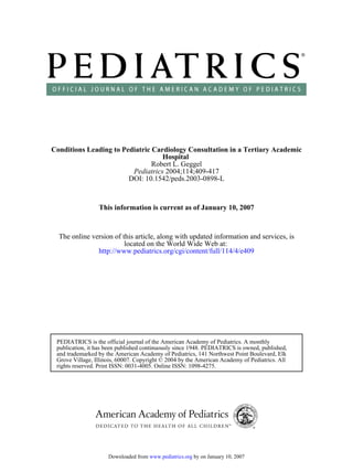
- 3. been determined. Education of medical students and residents in pediatric cardiology is challenging both because structural congenital heart disease is uncom- mon, affecting Ͻ1% of children, and because condi- tions potentially having a cardiac basis frequently have other causes.1–5 To establish the appropriate emphasis for clinical instruction in pediatric resi- dency training programs, I determined the basis for cardiac consultations in a tertiary academic pediatric hospital. METHODS Study Population The activities of the cardiology consultation service were tab- ulated from July 1, 2001, to June 30, 2002. Patients were evaluated in the emergency department, neonatal and pediatric intensive care units, inpatient wards, and occasional outpatient settings at Children’s Hospital, Boston, and in the neonatal intensive care units and nurseries at Brigham and Women’s Hospital and Beth Israel-Deaconess Hospital. There is a general cardiology ward and a cardiac intensive care unit at Children’s Hospital, Boston, which are not staffed by the consultation service. Patients were identified from 4 sources, ie, a monthly log of patient encounters maintained by the consultation service, encounter forms submitted to the billing office, consultation notes maintained in a central file, and a departmental list of echocardiographic studies. Although cardiol- ogy consultation is generally required for echocardiography, our departmental policy has permitted independent ordering of echo- cardiographic tests by the oncology service, to evaluate cardiac function associated with chemotherapy protocols, and by the neo- natology service, to screen for patent ductus arteriosus among premature infants. Patients who required clearance for noncardiac surgical proce- dures were generally evaluated in the cardiology clinic and not by the consultation service. The study was approved by the Chil- dren’s Hospital Committee on Clinical Investigation. Data Tabulation Patient data were obtained from consultation and echocardiog- raphy reports and Children’s Hospital computer-based records for discharge summaries for inpatient admissions, emergency depart- ment encounter summaries, and laboratory reports. Tabulated data included patient age, date, site, and basis of consultation, whether the patient was new or was known to the cardiology service, performance of echocardiography, electrocardiography, or chest radiography, and final diagnosis. For each patient, con- sultations were tabulated as separate encounters if they occurred on different days in the emergency department, during separate admissions, or for a different clinical concern during a single admission. Patient ages were tabulated as age at the first encoun- ter for a given diagnosis. Chest pain was classified with previously reported criteria.2 Statistical Analyses Patient ages are expressed as mean Ϯ SD. Analyses of the occurrence of heart disease among patients with supraventricular tachycardia or atrial fibrillation/atrial flutter and among patients with syncope/dizziness associated with exertion were performed with Fisher’s exact test. Comparisons of the age at first encounter between patients with supraventricular tachycardia and those with atrial fibrillation/atrial flutter and between patients with supraventricular tachycardia with or without congenital heart disease were made with the Wilcoxon rank sum test. RESULTS Number of Consultations During the study period, 2071 consultations were performed for 1724 patients. The age at the time of consultation was 6.6 Ϯ 9.3 years (median: 1.2 years; range: 1 day to 60.6 years). A single consultation was performed for 1507 patients. Two hundred seventeen patients (12.6%) had multiple encounters, ranging from 2 to 9, accounting for 564 consultations (27.2%). The monthly total of consultations ranged from 154 to 201 (average: 173). Patients were evaluated most often in the neonatal intensive care unit, inpatient wards, or the emergency department, for known car- diac patients (Fig 1). Reasons for Consultations Murmurs Evaluation of a murmur represented 18.5% of en- counters (Tables 1 and 2). The median age was 6 days (range: 1 day to 18.5 years). Echocardiography was used for all patients except for ϳ50% of patients with pulmonary branch murmurs of infancy or other in- nocent murmurs and for 10% of patients with ven- tricular septal defects, each of which was judged to be small. For patients evaluated for a murmur, the most common diagnosis or clinical concern in the neonatal intensive care unit was patent ductus arte- riosus (68%), in the well-child nursery was ventricu- lar septal defect (64%), and on the medical ward was innocent murmur (62%). Fig 1. Numbers of consultations performed on a monthly basis at different sites. Numbers in parentheses indicate the total numbers of patients evaluated. “Other” sites included the preoperative clinic, post-anesthesia care unit, radiology department, and infusion center. ER indicates emergency department; NICU, neonatal intensive care unit; PICU, pediatric intensive care unit. e410 CARDIOLOGY CONSULTATION IN AN ACADEMIC HOSPITAL by on January 10, 2007www.pediatrics.orgDownloaded from
- 4. Function Cardiac function evaluation accounted for 12.7% of consultations (Table 1). The patient age was 8.7 Ϯ 8.5 years (median: 6.4 years). Common conditions prompting evaluation of cardiac function included oncologic disease (40.5%), congenital heart disease (13.6%), sepsis (10.6%), postoperative general surgi- cal procedures (7.2%), neuromuscular disease (6.8%), arteriovenous malformations (3.8%), and hemato- logic disease (sickle cell disease or -thalassemia) TABLE 1. Sites and Bases for Consultations Basis for consultation No. of Cases ED, Known ED, New NICU Well Nursery PICU Medical Ward Other Total Murmur 17 254 73 3 34 3 384 Function 35 4 20 53 150 2 264 Arrhythmia 106 43 20 8 15 62 8 262 Intercurrent illness 210 2 21 233 Cyanosis (postnatal diagnosis) 13 12 47 5 8 1 86 Syndrome 97 2 4 15 118 Chest pain 57 35 13 2 107 Syncope/dizziness 25 45 22 1 93 Subacute bacterial endocarditis 5 2 9 17 58 91 Fetal diagnosis follow-up Arrhythmia 6 1 7 Cyanotic heart disease 45 45 Acyanotic heart disease 25 25 Normal postnatal status 8 5 13 Kawasaki disease 1 3 1 65 70 PAH/cor pulmonale 11 37 14 62 Recent cardiac surgery 25 25 Recent catheterization 8 8 CVA/TIA 8 3 6 7 24 Other conditions Surgical clearance 2 15 11 28 Cardiomegaly 8 4 2 6 20 Abnormal ECG,-nonarrhythmic 4 2 3 8 1 18 Pericardial effusion 4 3 10 17 ALTE 2 1 12 15 Thrombus 3 1 8 12 Systemic hypertension 1 1 1 6 1 10 Vascular ring 1 1 1 6 9 Rheumatic fever 1 6 7 Miscellaneous 9* 4† 2‡ 1§ 2¶ 18 ALTE indicates apparent life-threatening episode; CVA/TIA, cerebrovascular accident/transient ischemic attack; ED, emergency depart- ment; NICU, neonatal intensive care unit; PAH, pulmonary artery hypertension; PICU, pediatric intensive care unit; ECG, electrocar- diogram. * Five central venous line malfunctions (for prostacyclin or antibiotic administration), 2 hemoptysis in patients with pulmonary vascular obstructive disease, hypercalcemia, and 1 femoral artery pseudoaneurysm (catheterization performed 1 year previously). † Two thoracopagus conjoined twins, 1 sibling with congenital heart disease, and 1 rule out coarctation. ‡ Two siblings with congenital heart disease. § Rule out pulmonary embolism. ¶ Tachypnea. TABLE 2. Consultations for Evaluation of a Murmur Diagnosis No. of Cases ED, New NICU Well Nursery PICU Medical Ward Other Total Echocardiography Aortic stenosis 1 1 2 4 4 Atrial myxoma 1 1 1 Atrial septal defect, secundum 1 6 1 1 9 9 Atrioventricular canal, transitional 1 1 1 Innocent or flow murmur 2 19 5 21 1 48 26 Mitral regurgitation 1 1 1 Mitral stenosis 1 1 1 Patent ductus arteriosus 135 5 1 1 1 143 143 Rule out patent ductus arteriosus, not present 37 37 37 Peripheral pulmonary stenosis 4 27 5 1 8 1 46 24 Pulmonary stenosis 3 1 4 4 Truncus arteriosus 1 1 1 Ventricular septal defect 5 21 47 1 2 76 68 Ventricular septal defect/coarctation 1 1 1 Resolved before consultation 7 4 11 0 Abbreviations as in Table 1. www.pediatrics.org/cgi/doi/10.1542/peds.2003-0898-L e411 by on January 10, 2007www.pediatrics.orgDownloaded from
- 5. (3.4%). There were 3 new diagnoses of structural congenital heart disease among neonates who were examined initially in the emergency department or pediatric intensive care unit; 2 had severe coarctation and the other an interrupted aortic arch. Arrhythmias Evaluation of an arrhythmia accounted for 12.7% of consultations (Tables 1 and 3). Atrial arrhythmias represented 63% of consultations for the indication of arrhythmia. The most common arrhythmias requir- ing treatment were supraventricular tachycardia, atrial flutter, and atrial fibrillation. Compared with patients with atrial flutter/atrial fibrillation, patients with supraventricular tachycardia were younger (7.9 Ϯ 8.5 years; median: 5.4 years; vs 26.1 Ϯ 12.1 years; median: 22.7 years; P Ͻ .001) and had a lower incidence of congenital heart disease (8% vs 78%, P Ͻ .001). Patients with supraventricular tachycardia with congenital heart disease presented at older ages than did those without structural defects (median age: 15.1 years vs 3.1 years; P ϭ .05). Of the 17 patients evaluated for possible pro- longed QT interval, 1 neonate had congenital prolon- gation of the QT interval. The other patients had prolongation of the interval resulting from medica- tion effects (n ϭ 4; clomipramine, nortriptyline, or pentamidine) or metabolic defects (n ϭ 6; hypocal- cemia in 5 cases and hypokalemia in 1 case) or had a normal interval (n ϭ 6). An associated disease was present for 1 patient each with first-degree atrioven- tricular block (anorexia nervosa), transient second- degree atrioventricular block (Lyme disease), and neonatal complete heart block (maternal systemic lupus erythematosus). Intercurrent Illnesses Evaluation of patients with known cardiac disease presenting with an intercurrent noncardiac illness (mainly viral syndromes) accounted for 11.3% of consultations (Table 1). The patient age was 5.3 Ϯ 7.4 years (median: 2.0 years). Cyanosis Cyanosis formed the basis of consultations for 6.3% of patients (Tables 1 and 4). Three-quarters of the patients were Ͻ1 week of age. Of 54 patients with structural cyanotic heart disease, 45 (83%) were di- agnosed with fetal echocardiography. The mothers of these neonates had delivery scheduled at a nearby hospital. Intercurrent illnesses among patients with palliated cyanotic heart disease produced increasing cyanosis if there was fever for patients with system- ic-pulmonary shunts or dehydration for those with intracardiac right-to-left shunts. TABLE 3. Consultations for Evaluation of an Arrhythmia Arrhythmia No. of Cases ED, Known ED, New NICU Well Nursery PICU Medical Ward Other Total Atrial Atrial fibrillation 5 1 2 8* Atrial flutter 28 1 1 30† Atrial premature beats 4 2 4 4 1 1 16 Ectopic atrial tachycardia 2 2 1 1 6 Sinus arrhythmia 2 3 1 6 Sinus bradycardia 2 2 1 3 21 1 30 Sinus tachycardia 4 5 1 7 17‡ Supraventricular tachycardia 34 11 4 1 1 3 54§ Ventricular Ventricular premature beat 3 2 5 2 7 8 2 29 Ventricular tachycardia 3 2 2 7 Ventricular fibrillation 2 2 Junctional Junctional ectopic tachycardia 1 1 1 1 4 Junctional escape rhythm 2 1 3 Junctional premature beat 1 1 2 Heart block First-degree heart block 1 1 2 Second-degree heart block 2 1 3 Congenital complete heart block 1 1 Normal 1 1 2 2 1 7 Palpitations, no identified arrhythmia 7 8 1 16 Pacemaker ICD discharge 6 6 Dual-chamber pacemaker malfunction 3 3 Prolonged OT interval 1 2 2 11 1 17 Values include prenatal and postnatal evaluations. ICD indicates implantable cardioverter-defibrillator; other abbreviations as in Table 1. * Six different patients (congenital heart disease in 3; 1 patient each with Ebstein’s anomaly, tricuspid atresia status post Fontan, and cardiomyopathy). † Seventeen different patients (congenital heart disease in 15: single ventricle, status post Fontan, n ϭ 9; dextro-transposition of the great arteries, status post atrial switch procedure, n ϭ 2; cardiomyopathy, n ϭ 2; 1 patient each with tetralogy of Fallot and levo-transposition of the great arteries). ‡ Fifteen different patients. § Thirty-eight different patients. e412 CARDIOLOGY CONSULTATION IN AN ACADEMIC HOSPITAL by on January 10, 2007www.pediatrics.orgDownloaded from
- 6. Syndromes The evaluation of cardiac involvement in syn- dromes prompted consultation for 5.7% of patients (Tables 1 and 5). The median age was 2 days. Three syndromes represented 57% of the consultations for this indication, ie, VACTERL association (vertebral anomalies, anal atresia, congenital heart disease, tra- cheoesophageal fistula, renal abnormality, and limb anomalies), trisomy 21, and infant of a diabetic mother. Chest Pain Chest pain accounted for 5.2% of consultations, 13% of all emergency department evaluations, and 19% of emergency department consultations for new patients (Tables 1 and 6). The patient age was 16.1 Ϯ 7.4 years (median: 15.4 years). For this symptom, the 2 most common categories were musculoskeletal/ costochondritic (42%) and idiopathic (22%) condi- tions. There was a cardiac or pericardial basis in 11% of cases; these patients either had a known cardiac history or had pericarditis presenting with systemic symptoms and abnormal cardiac auscultatory or electrocardiographic findings (6). Chest pain with exertion occurred for 2 patients, each with known heart disease. One patient with palliated cyanotic heart disease and normocytic erythrocytes experi- enced resolution of chest pain with reduction of he- matocrit values from 71% to 63%. A chest radiograph was obtained in 92 episodes (86%) and contributed to the diagnosis for the 13 patients with pulmonary abnormalities and the 5 patients with pericarditis. An electrocardiogram was obtained in 106 episodes (99%) and differed from baseline tracings or was abnormal for 4 patients with pericarditis. Concern about possible pulmonary embolism was raised for 6 patients, but the results of imaging studies were normal. Syncope/Dizziness Evaluation for syncope or dizziness accounted for 4.5% of consultations, 10% of all emergency depart- ment evaluations, and 25% of emergency department evaluations for new patients. The patient age was 14.4 Ϯ 5.4 years (median: 14.9 years). The most com- mon causes were vasovagal (47 episodes, 50.5%) or orthostatic (23 episodes, 24.7%). For 5 patients, or- thostasis was related to medication (clonidine, quetiapine fumarate, or furosemide). Six episodes (6.5%) had an idiopathic basis and 2 had other causes (hyperventilation or menorrhagia). Five patients (5.4%) each demonstrated a cardiac, neurologic, or psychiatric basis. Cardiac diseases included 2 new patients with congenital prolonged QT interval and 1 known patient each with primary pulmonary hyper- tension, severe subaortic stenosis, and bradycardia secondary to pacemaker dysfunction. Neurologic conditions included a seizure disorder, breath-hold- ing spell, and migraine headache. Patients with a cardiac basis were more likely to have symptoms associated with exercise (P ϭ .001). Of 13 patients who experienced syncope (n ϭ 8) or dizziness (n ϭ 1) during exercise or syncope shortly after completion of exercise (n ϭ 4), 7 demonstrated a vasovagal basis, 4 had a cardiac condition, and 1 each exhibited a psychiatric or idiopathic cause. Subacute Endocarditis Evaluation for subacute endocarditis accounted for 4.4% of consultations (Tables 1 and 7). The patient age was 7.7 Ϯ 7.3 years (median: 5.2 years). Of 91 consultations for this concern, there were 3 cases of subacute endocarditis (3.3%) (Table 7). Two events occurred among known cardiac patients, presenting to the emergency department with either fever and bacteremia or an indolent course of weight loss and anorexia. For each patient, the blood cultures yielded Streptococcus viridans. A premature neonate had an umbilical venous line, Staphylococcus aureus bactere- mia, and right-sided intracardiac vegetations. Two patients had candidemia and intravascular thrombus associated with a central venous line. Transthoracic echocardiograms were obtained in 79 consultations (87%) and transesophageal echocar- TABLE 4. Consultations for Evaluation of Cyanosis Diagnosis No. of Cases ED, Known ED, New NICU PICU Medical Ward Other Total Acrocyanosis 3 6 2 11 Apnea-bradycardia 1 2 1 4 Cyanotic CHD, Follow-up fetal diagnosis 45* 45 Cyanotic CHD, intercurrent illness 9 1 10 Cyanotic CHD, new diagnosis 2† 7‡ 9 Cyanosis, crying only, neonate 2 1 3 Obstructive sleep apnea 1 1 PPHN 26 3 29 Pulmonary disease§ 1 8 2 5 1 17 Rule out tetralogy spell 1 1 Seizure 1 1 CHD indicates congenital heart disease; PPHN, persistent pulmonary hypertension of newborn; other abbreviations as in Table 1. * Double-outlet right ventricle, 1; hypoplastic left heart syndrome, 17; pulmonary atresia, intact ventricular septum, 5; single ventricle, 4; tetralogy of Fallot, 11; dextro-transposition of the great arteries, 5; tricuspid atresia, 2. † Total anomalous pulmonary venous connection, 2 (1 infradiaphragmatic, 1 supracardiac to innominate vein). ‡ Pulmonary atresia, intact ventricular septum, 1; right ventricular endocardial fibroelastosis, 1; tetralogy of Fallot, 1; total anomalous pulmonary venous connection, 1; dextrotransposition of the great arteries, 3. § Pulmonary conditions included pneumonia associated with gastroesophageal reflux disease and aspiration, bronchiolitis, pulmonary hypoplasia or dysplasia, laryngomalacia, and postoperative bronchospasm. www.pediatrics.org/cgi/doi/10.1542/peds.2003-0898-L e413 by on January 10, 2007www.pediatrics.orgDownloaded from
- 7. diograms in 6 (6.7%). Thrombus or vegetation was noted only for the patients cited above. Echocardiog- raphy was deferred for 9 patients who had sterile blood cultures and for 3 others who had sterile cul- tures after removal of a central venous catheter and antibiotic therapy for bacteremia. The most common clinical background was septi- cemia with an indwelling central venous catheter (36 episodes, 40%). A new murmur was present for 11 patients, 6 of whom had positive blood cultures. Echocardiograms were obtained for 10 of these pa- tients. Each murmur was innocent, ie, 6 Still’s mur- murs, 4 flow murmurs associated with anemia, and 1 innocent pulmonary branch murmur of infancy. Follow-up Evaluations of Fetal Diagnoses Ninety neonates had consultations prompted by fetal echocardiographic diagnoses, representing 4.3% of consultations and 14.1% of consultations per- formed in the neonatal intensive care unit and nurs- eries (Table 1). Kawasaki Disease This clinical concern represented 3.4% of all con- sultations (Tables 1 and 2). The patient age was 4.3 Ϯ 3.3 years (median: 3.7 years). Of the 70 encounters, 51 patients were diagnosed as having Kawasaki dis- ease. The other 19 patients were diagnosed as having viral syndrome (15 patients), urinary tract infection or pyelonephritis (1 patient, 2 episodes), hypersensi- tivity reaction to dilantin, or necrotizing fasciitis. Pulmonary Artery Hypertension/Cor Pulmonale Concern about pulmonary artery hypertension ac- counted for 3.0% of consultations. The patient age was 3.7 Ϯ 6.9 years (median: 92 days; range: 1 day to TABLE 5. Consultations for Syndromes or Other Congenital Anomalies Syndrome No. % CHD CHD Lesions VACTERL 28 43 VSD, 6; ASD, 2; bicuspid AoV, 2; TOF, 1; RAA-aberrant LSCA, 3* Trisomy 21 22 45 VSD, 3; CAVC, 2; PDA, 2; TOF, 2 Infant of diabetic mother 17 65 VSD, 1; PS, 1; septal hypertrophy, 9 (2 with LVOT obstruction) Omphalocele 6 17 VSD and PDA, 1 Cleft palate 6 33 VSD, 1; RAA-aberrant LSCA, 1 Twin-twin transfusion 4 25 VSD, 1 Dandy-Walker syndrome 3 67 VSD, 1; bicuspid AoV, 1 Jejunal atresia 3 33 PS Rule out DiGeorge syndrome† 3 0 Rule out Marfan syndrome‡ 3 0 Rule out tuberous sclerosis§ 3 50 Intracardiac rhabdomyoma, 1 Miscellaneous¶ 20 45 VSD, 4; VSD and ASD, 1; ASD, 2; bicuspid AoV, 1; MR, 1 AoV indicates aortic valve; ASD, atrial septal defect; CAVC, complete atrioventricular canal defect; LSCA, left subclavian artery; LVOT, left ventricular outflow tract; MR, mitral regurgitation; PDA, patent ductus arteriosus; PS, pulmonary stenosis; RAA, right aortic arch; TOF, tetralogy of Fallot; VSD, ventricular septal defect. * Two patients with RAA-aberrant LSCA also had either VSD or ASD. † Evaluated for hypocalcemia; 1 patient had DiGeorge syndrome. ‡ Evaluated for pneumothorax, joint dislocation, or superior mesenteric artery aneurysm; none diagnosed with Marfan syndrome. § Each with seizure disorder; 2 patients diagnosed with tuberous sclerosis. ¶ Chromosome abnormality other than trisomy 21 (3; partial trisomy 9q, 14q duplication, and partial deletion of 18q); choanal atresia or stenosis 2, Klippel-Trenaunay-Weber 2; 1 patient each with Beal’s syndrome, cloacal extrophy, coloboma, extrahepatic biliary atresia, gastroschisis, Holt-Oram syndrome, Kartagener’s syndrome, lobar emphysema, Marden-Walker syndrome, nemoline rod myopathy, Russel-Silver syndrome, Smith-Lemli-Opitz syndrome, and tracheal stenosis. TABLE 6. Consultations for Evaluation of Chest Pain Diagnosis No. of Cases ED, Known ED, New Medical Ward Other Total Anxiety 4 1 5 Cardiac disease 7* 3† 2‡ 12 Gastroesophageal reflux 1 1 Idiopathic 9 9 4 2 24 Musculoskeletal/costochondritic 27 14 4 45 Polycythemia 1 1 Pulmonary Pleural effusion/pleuritis 1 2 3 Pneumonia 2 1 2 5 Pneumomediastinum 2 2 Pneumothorax 2 1 3 Skin infection at cvl site 1 1 Toxin, cannabis 2 3 5 cvl indicates central venous line; other abbreviations as in Table 1. * Known patients with pulmonary vascular obstructive disease, 2; recurrent pericarditis, 2; tetralogy of Fallot with systemic level right ventricular pressure produced by an obstructed right ventricular- pulmonary artery conduit, 1; Kawasaki disease with known giant aneurysm of left anterior descend- ing coronary artery with echocardiographic confirmation of new clot in this artery, 1; subclavicular pacemaker pocket infection, 1. † Pericarditis associated with fever and pericardial friction rub in each patient. ‡ Known patients with Takayasu’s aortitis or severe cardiomyopathy secondary to -thalassemia. e414 CARDIOLOGY CONSULTATION IN AN ACADEMIC HOSPITAL by on January 10, 2007www.pediatrics.orgDownloaded from
- 8. 25.6 years). The most common condition was con- genital diaphragmatic hernia, which was found for 34% of patients evaluated for this concern. Other conditions prompting evaluation of pulmonary ar- tery pressure included chronic lung disease associ- ated with premature birth, pneumonitis or reactive airway disease, cystic fibrosis, pulmonary hypopla- sia associated with chest wall deformity or cystic adenomatoid malformation, obstructive sleep apnea, pulmonary hemosiderosis, neurologic conditions as- sociated with hypoventilation, and orthotopic lung transplantation. Recent Cardiac Surgery or Cardiac Catheterization Eight of 25 patients evaluated within 2 months after surgery had postpericardiotomy syndrome. Other issues addressed included surgical wound evaluation, pleural effusion, intervening viral illness, pain control, and bacterial pericarditis. Evaluations after catheterization, with 1 exception, involved interventional procedures. One patient had transient fever after coil occlusion of collateral ves- sels, whereas the others had vascular access prob- lems consisting of pain (local hematoma, infected hematoma, or arterial pseudoaneurysm) or coolness of an extremity. Cerebrovascular Accidents/Transient Ischemic Attacks Evaluation of a cardiac source for loss of neuro- logic function accounted for 1.2% of consultations (Table 1). A cardiac basis was identified for 5 of the 24 patients evaluated for this concern, ie, intracardiac thrombus for 2 patients with a single ventricle and bidirectional flow across a patent foramen ovale for 3 patients, including a neonate with a thrombus at the tip of a central venous line and 2 adolescents. An additional adult patient had pulmonary vascular ob- structive disease, polycythemia (hematocrit: 62%), and microcytic anemia (mean corpuscular volume: 69 FL). The other 18 patients had a variety of condi- tions, ie, protein C, protein S, or antithrombin III deficiencies, carotid artery dissection, moyamoya disease, sickle cell disease, encephalitis, coagulopa- thy associated with intraventricular hemorrhage, se- vere hyponatremia caused by psychogenic polydip- sia, or idiopathic causes. Other Conditions A variety of other conditions were evaluated (Ta- ble 1). Eight of 20 patients (40%) who underwent consultations because of cardiomegaly on chest ra- diographs had heart disease. The cardiologist and radiologist judged the cardiac silhouette to be nor- mal in an equal number, 5 of whom had a large thymus. For 4 other patients, echocardiograms showed normal cardiac dimensions. Infants with an apparent life-threatening event had normal QT inter- vals and no other identifiable cardiac disorders; 5 patients demonstrated an idiopathic basis, whereas the others had apnea, periodic breathing, breath- holding, gastroesophageal reflux, or seizure disor- der. Systemic hypertension was associated with re- nal disease for 4 patients, agitation, headache, or postoperative pain for 5, and medication (fludrocor- tisone and midodrine) for 1. Evaluation for a vascu- lar ring was prompted mainly by the symptom of stridor, which was caused for 2 patients each by tracheomalacia and laryngomalacia and for 1 patient each by left main bronchus stenosis, subglottic hem- angioma, innominate artery compression, and uni- lateral vocal cord paresis; 1 patient with an incidental finding of a right aortic arch had a loose vascular ring with an aberrant left subclavian artery. Of 7 patients evaluated for rheumatic fever, 2 with chorea and mitral regurgitation were diagnosed with this condition. Two other patients with movement disor- ders had either Guillain-Barre´ or antiphospholipid syndrome. The other 3 patients did not fulfill Jones TABLE 7. Consultations to Rule Out Subacute Bacterial Endocarditis No. of Cases ED, Known ED, New NICU PICU Medical Ward Total Basis for consultation Positive BC 1* 1 7* 13† 35 57‡ Fever 5* 1 3 16† 42 24 New murmur 1 1 1 8 11 New S3 1 1 Weight loss, anorexia 1† 1 Fatigue 1 1 Embolic event 1 1 2 Clinical conditions Repaired/palliated CHD 4* 3 7 Unrepaired CHD 1† 4 5 Oncologic diseases 3 16 19 General surgery within previous 8 wk 4 5 11 20 Indwelling central venous catheter 5* 10† 33 48 Intravenous drug abuser 1 1 Dental abscess 1 1 BC indicates blood culture; other abbreviations as in Tables 1 and 4. * One patient with each symbol had subacute bacterial endocarditis; see text for details. † Two patients with possible infected intravascular thrombus; see text for details. ‡ Three organisms accounted for 39 (67%) of the 58 episodes of septicemia (57 presenting with septicemia and 1 presenting with constitutional symptoms and subsequently found to have bacteremia). Various species of Candida were isolated in 17 cases, Staphylococcus aureus in 14 episodes, and coagulase-negative Staphylococcus in 8. There were 5 cases of Streptococcus viridans and 2 cases each of Pseudomonas aeruginosa and group A Streptococcus. Ten other bacteria were isolated only once, for different patients. www.pediatrics.org/cgi/doi/10.1542/peds.2003-0898-L e415 by on January 10, 2007www.pediatrics.orgDownloaded from
- 9. criteria, including 2 with fever and an innocent mur- mur. DISCUSSION This review documents for the first time the issues prompting cardiac consultation in an academic pe- diatric hospital setting. Although a variety of condi- tions were assessed, some diagnoses were encoun- tered more frequently and should be given emphasis in future curriculum development for cardiac train- ing for pediatric residents. Evaluation of a murmur was the most frequent basis for consultation. Cardiac auscultation skills in various training programs, including pediatrics, have been documented to be poor.6–8 Four condi- tions (patent ductus arteriosus, ventricular septal de- fect, innocent murmur, and pulmonary branch mur- mur of infancy) accounted for 91% of diagnoses for patients with murmurs. Initially developing stetho- scope skills among pediatric residents for the detec- tion of these conditions should fulfill clinical needs frequently. Developing skills for the diagnosis of an innocent murmur should have additional benefits in reducing parental morbidity if confirmation by a cardiologist is subsequently sought, because the abil- ity of the pediatrician to provide greater reassurance has been shown to reduce parental anxiety associ- ated with referral.9 Assessment of function was frequently requested for a variety of clinical conditions. Newly developed dysfunction among infants after discharge from the birth hospital can represent duct-dependent lesions that elude detection while the ductus is widely patent.10 The majority of arrhythmias were atrial in origin. The most common arrhythmia was supraventricular tachycardia, which usually was associated with a structurally normal heart.11 Atrial fibrillation/atrial flutter occurred among older patients, who typically had congenital heart disease; the chronic effects of pressure or volume overload or surgical scars con- tribute to the development of these 2 arrhythmias,12 which are commonly seen among patients, as in our study group, who have undergone atrial baffle repair of dextrotransposition of the great arteries (Mustard or Senning procedures), modified Fontan procedures because of a functional single ventricle, or repair of tetralogy of Fallot.12,13 Young adults with repaired congenital heart disease are becoming a larger pa- tient group, because of improved survival rates, and often are cared for at pediatric hospitals; therefore, familiarity with these arrhythmias is important. Ven- tricular arrhythmias were infrequent and mainly consisted of ventricular premature beats. Prolonged QT syndrome usually was associated with a meta- bolic abnormality or medication-related side effect,14 rather than with congenital prolongation. Heart block was uncommon and was associated with an- orexia nervosa,15 Lyme disease,16 or maternal sys- temic lupus erythematosus.17 Diagnoses made with fetal echocardiography were a frequent basis for neonatal consultation. Cyanotic lesions were over-represented among the prenatally diagnosed patients, in comparison with the occur- rence in the general population,1 because delivery of these infants was arranged at a nearby hospital. Such a policy can decrease morbidity and mortality rates.18 Patients with palliated cyanotic heart disease experienced increased cyanosis with intercurrent ill- ness in the presence of dehydration, because of in- creased oxygen extraction and lower venous oxygen saturation, in conditions with right-to-left intracar- diac shunting or in the presence of fever, because of decreased shunt perfusion produced by lower sys- temic vascular resistance, in conditions with system- ic-pulmonary artery shunting. The oxygen dissocia- tion curve is steep for arterial oxygen pressure values of Ͻ50 mm Hg, so that a small decrease in oxygen tension produces a large decrease in oxygen satura- tion.19 Chest pain and syncope were frequently evaluated in the emergency department,3 and each was typi- cally associated with a benign condition.2,3,20 A seri- ous organic cause for chest pain was unusual with- out associated symptoms of illness, positive physical examination findings related to the cardiac or respi- ratory systems, or symptoms during exertion.21 The occurrence of syncope during or shortly after exer- tion also was commonly observed among patients with cardiac conditions.3 Syndromes have been reported for 8.5% of patients with congenital heart disease.22 Recognition of syn- dromes permits thorough patient evaluation, includ- ing early detection of heart disease, which may not produce clinical symptoms as long as pulmonary vascular resistance is elevated in the early neonatal period. The 3 syndromes evaluated most frequently are associated commonly with congenital heart dis- ease.23–25 Endocarditis was uncommon and occurred among patients with known congenital heart disease or in- dwelling venous catheters.26 Risk factors for trans- thoracic echocardiographic evidence of endocarditis that were identified in each of 2 pediatric reports included positive blood cultures, physical findings of new or changing heart murmur, and congestive heart failure.27,28 Indications in neonates may be dif- ferent.26,27 Our experience shows that the feature of a new murmur should be defined as a new regurgitant murmur,29 because a hyperdynamic condition asso- ciated with febrile illness can produce innocent mur- murs. Kawasaki disease was the most common acquired disease leading to consultation. Approximately one- quarter of patients evaluated for Kawasaki disease were determined to have other conditions. Consid- ering the diagnosis of Kawasaki disease for children with some features of the illness is warranted, be- cause atypical presentation has been associated with coronary involvement.30 Various conditions were encountered less fre- quently. Evaluation of pulmonary hypertension mainly involved conditions associated with pulmo- nary disease. Doppler echocardiography can be used to estimate the level of pulmonary systolic pressure and to monitor the hemodynamic effects and dosing of nitric oxide or supplemental oxygen in these con- ditions.31,32 Consultation after cardiac surgery e416 CARDIOLOGY CONSULTATION IN AN ACADEMIC HOSPITAL by on January 10, 2007www.pediatrics.orgDownloaded from
- 10. chiefly involved evaluation for postpericardiotomy syndrome,33 whereas assessment after catheteriza- tion mainly involved vascular access concerns among patients who had undergone interventional procedures (a risk factor for complications).34 Al- though neurologic deficits usually had a noncardiac cause,5 our patient group confirmed risks associated with cyanotic heart disease,35 Fontan procedure,15 thrombosis related to the use of an indwelling central venous catheter,36 patent foramen ovale,37 and poly- cythemia associated with microcytosis.35 Cardiomeg- aly was not present for 60% of patients evaluated for this condition.38 Failure to distinguish a large over- lying thymus gland from true cardiac enlargement occurred relatively frequently. Systemic hyperten- sion was associated with renal disease, the most common cause in pediatrics.39 Stridor, which can be produced by vascular compression of the airways,40 was associated mainly with airway disease among our patients. CONCLUSIONS Although numerous conditions were evaluated by the cardiac consultation service, some were encoun- tered more frequently and should be given emphasis in curricula developed for pediatric residency train- ing programs. Future investigations will be needed to determine the effectiveness of acquiring skills for the diagnosis of cardiac symptoms and disease dur- ing pediatric residency. ACKNOWLEDGMENT I thank the fellows and attending physicians who helped staff the cardiology consultation service during the study period. REFERENCES 1. Hoffman JIE. Incidence of congenital heart disease: part I: postnatal incidence. Pediatr Cardiol. 1995;16:103–113 2. Selbst SM, Ruddy RM, Clark BJ, Henretig FM, Santulli T Jr. Pediatric chest pain: a prospective study. Pediatrics. 1988;82:319–323 3. Driscoll DJ, Jacobsen SJ, Porter CJ, Wolland PC. Syncope in children and adolescents. J Am Coll Cardiol. 1997;29:1039–1045 4. McCrindle BW, Shaffer KM, Kan JS, Zahka KG, Rowe SA, Kidd L. Cardinal clinical signs in the differentiation of heart murmurs in chil- dren. Arch Pediatr Adolesc Med. 1996;150:169–174 5. Carvalho KS, Garg BP. Arterial strokes in children. Neurol Clin. 2002; 20:1079–1100 6. Gaskin PRA, Owens SE, Talner NS, Sanders SP, Li JS. Clinical auscul- tation skills in pediatric residents. Pediatrics. 2000;105:1184–1187 7. Mangione S, Nieman LZ. Cardiac auscultation skills of internal medi- cine and family practice trainees: a comparison of diagnostic profi- ciency. JAMA. 1997;278:717–722 8. St Clair EW, Oddone EZ, Waugh RA, Corey GR, Feussner JR. Assessing housestaff diagnostic skills using a cardiology patient simulator. Ann Intern Med. 1992;117:751–756 9. Geggel RL, Horowitz LM, Brown EA, Parsons M, Wang PS, Fulton DR. Parental anxiety associated with referral of a child to a pediatric cardi- ologist for evaluation of a Still’s murmur. J Pediatr. 2002;140:747–752 10. Abu-Harb M, Hey E, Wren C. Death in infancy from unrecognized congenital heart disease. Arch Dis Child. 1994;71:3–7 11. Garson A Jr. Supraventricular tachycardia. In: Gillette PC, Garson A Jr, eds. Pediatric Cardiac Dysrhythmias. New York, NY: Grune & Stratton; 1981:177–253 12. Triedman JK. Arrhythmias in adults with congenital heart disease. Heart. 2002;87:383–389 13. Geggel RL. Update on the modified Fontan procedure. Curr Opin Car- diol. 1997;12:51–62 14. Viskin S. Long QT syndromes and torsade de pointes. Lancet. 1999;354: 1625–1633 15. Palla B, Litt IF. Medical complications of eating disorders in adoles- cents. Pediatrics. 1988;81:613–623 16. Steere AC. Lyme disease. N Engl J Med. 345:115–125, 2001 17. Chameides L, Truex RC, Vetter V, Rashkind WJ, Galioto FM Jr, Noonan JA. Association of maternal systemic lupus erythematosus with congen- ital complete heart block. N Engl J Med. 1977;297:1204–1207 18. Allan L. Antenatal diagnosis of heart disease. Heart. 2000;83:367–370 19. Rudolph A. Congenital Diseases of the Heart. Chicago, IL: Year Book Medical Publishers; 1974:102 20. Kapoor WN. Current evaluation and management of syncope. Circula- tion. 2002;106:1606–1609 21. Geggel RL. Evaluation of pediatric chest pain. In: Rose BD, ed. UpTo- Date. Wellesley, MA: UpToDate; 2003 22. Greenwood RD, Rosenthal A, Parisi L, Fyler DC, Nadas AS. Extracar- diac anomalies in children with congenital heart disease. Pediatrics. 1975;55:485–492 23. Zahka KG. Associated abnormalities in children with congenital heart disease. In: Emmanouilides GC, Riemenschneider TA, Allen HD, Gut- gesell HP, eds. Heart Disease in Infants, Children, and Adolescents. 5th ed. Baltimore, MD: Williams & Wilkins; 1995;1:614–628 24. Lin AE. Congenital heart defects in chromosome abnormality syn- dromes. In: Emmanouilides GC, Riemenschneider TA, Allen HD, Gut- gesell HP, eds. Heart Disease in Infants, Children, and Adolescents. 5th ed. Baltimore, MD: Williams & Wilkins; 1995;1:633–643 25. Caddell JL. Metabolic and nutritional diseases. In: Emmanouilides GC, Riemenschneider TA, Allen HD, Gutgesell HP, eds. Heart Disease in Infants, Children, and Adolescents. 5th ed. Baltimore, MD: Williams & Wilkins; 1995;1:1453–1486 26. Ferrieri P, Gewitz MH, Gerber MA, et al. Unique features of infective endocarditis in childhood. Circulation. 2002;105:2115–2127 27. Sable CA, Rome JJ, Martin GR, Patel KM, Karr SS. Indications for echocardiography in the diagnosis of infective endocarditis in children. Am J Cardiol. 1995;75:801–804 28. Aly AM, Simpson PM, Humes RA. The role of transthoracic echocar- diography in the diagnosis of infective endocarditis in children. Arch Pediatr Adolesc Med. 1999;153:950–954 29. Lindner JR, Case A, Dent JM, Abbott RD, Scheld WM, Kaul S. Diagnos- tic value of echocardiography in suspected endocarditis. Circulation. 1996;93:730–736 30. Newburger JW, Burns JC. Kawasaki disease. Vasc Med. 1999;4:187–202 31. Fraser KL, Tullis E, Sasson Z, Hyland RH, Thornley KS, Hanly PJ. Pulmonary hypertension and cardiac function in adult cystic fibrosis. Chest. 1999;115:1321–1328 32. Lonnqvist PA. Inhaled nitric oxide in newborn and paediatric patients with pulmonary hypertension and moderate to severe impaired oxygenation: effects of doses of 3–100 parts per million. Intensive Care Med. 1997;23:773–779 33. Engle MA, O’Loughlin JE. Complications of cardiac surgery in children. Pediatr Rev. 1987;9:147–154 34. Vitiello R, McCrindle BW, Nykanen D, Freedom RM, Benson LN. Complications associated with pediatric cardiac catheterization. J Am Coll Cardiol. 1998;32:1433–1440 35. Ammash N, Warnes CA. Cerebrovascular events in adult patients with cyanotic congenital heart disease. J Am Coll Cardiol. 1996;28:768–772 36. McGee DC, Gould MK. Preventing complications of central venous catheterization. N Engl J Med. 2003;348:1123–1133 37. Windecker S, Wahl A, Chatterjee T, et al. Percutaneous closure of patent foramen ovale in patients with paradoxical embolism. Circulation. 2000; 101:893–898 38. Satou GM, Lacro RV, Chung T, Gauvreau K, Jenkins KJ. Heart size on chest x-ray as a predictor of cardiac enlargement by echocardiography in children. Pediatr Cardiol. 2001;22:218–222 39. Sinaiko AR. Hypertension in children. N Engl J Med. 1996;335:1968–1973 40. Anand R, Dooley KJ, Williams WH, Vincent RN. Follow-up of surgical correction of vascular anomalies causing tracheobronchial compression. Pediatr Cardiol. 1994;15:58–61 www.pediatrics.org/cgi/doi/10.1542/peds.2003-0898-L e417 by on January 10, 2007www.pediatrics.orgDownloaded from
- 11. DOI: 10.1542/peds.2003-0898-L 2004;114;409-417Pediatrics Robert L. Geggel Hospital Conditions Leading to Pediatric Cardiology Consultation in a Tertiary Academic This information is current as of January 10, 2007 & Services Updated Information http://www.pediatrics.org/cgi/content/full/114/4/e409 including high-resolution figures, can be found at: References http://www.pediatrics.org/cgi/content/full/114/4/e409#BIBL at: This article cites 33 articles, 20 of which you can access for free Subspecialty Collections s http://www.pediatrics.org/cgi/collection/heart_and_blood_vessel Heart & Blood Vessels following collection(s): This article, along with others on similar topics, appears in the Permissions & Licensing http://www.pediatrics.org/misc/Permissions.shtml tables) or in its entirety can be found online at: Information about reproducing this article in parts (figures, Reprints http://www.pediatrics.org/misc/reprints.shtml Information about ordering reprints can be found online: by on January 10, 2007www.pediatrics.orgDownloaded from