
Recomendados
Mais conteúdo relacionado
Semelhante a hemoglobinestimation2022-220717075634-6da2d22b (1).pptx
Semelhante a hemoglobinestimation2022-220717075634-6da2d22b (1).pptx (20)
Mais de sandeep singh
Mais de sandeep singh (20)
Último
Último (20)
hemoglobinestimation2022-220717075634-6da2d22b (1).pptx
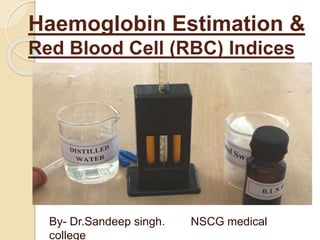
- 1. Haemoglobin Estimation & Red Blood Cell (RBC) Indices By- Dr.Sandeep singh. NSCG medical college
- 3. Red Blood Cell (RBC) Indices Red blood cell (RBC) indices measure the size, shape, and quality of your red blood cells. Red blood cells, also known as erythrocytes, carry oxygen from your lungs to every cell in your body. Your cells need oxygen to grow, reproduce, and stay healthy.
- 7. What are they used for? Red blood cell (RBC) indices are part of a complete blood count . The results of RBC indices are used to diagnose different types of anemia. There are several types of anemia, and each type has a different effect on the size, shape and/or quality of red blood cells
- 8. Following red blood cell indices: Mean corpuscular volume (MCV), which measures the average size of your red blood cells Mean corpuscular hemoglobin (MCH), which measures the average amount of hemoglobin in a single red blood cell. Hemoglobin in red blood cells that carries oxygen. Mean corpuscular hemoglobin concentration (MCHC), which also measures hemoglobin in red blood cells. In addition, it includes a calculation of the size and volume of your red blood cells. Red cell distribution width (RDW) which measures differences in the volume and size of your red blood cells.
- 9. Mean corpuscular volume (MCV) If your red blood cells - smaller than normal, microcytic it may mean you have: Iron deficiency anemia, thalassemia If your red blood cells are larger than normal, Macrocytic it may mean you have: vitamin B deficiency ,Liver disease
- 10. Mean corpuscular hemoglobin (MCH) If the amount of hemoglobin is lower than normal, it may mean you have: Iron deficiency anemia If the amount of hemoglobin is higher than normal, it may mean you have: High level of cholesterol in the blood, vitamin B deficiency
- 11. Mean corpuscular hemoglobin concentration (MCHC) If the average amount of hemoglobin is lower than normal it may mean you have: Iron deficiency anemia Thalassemia If the average amount of hemoglobin is higher than normal, it may mean you have: Hemolytic anemia, a type of anemia that happens when red blood cells are broken up Hereditary spherocytosis, a rare genetic
- 12. Samples used for Hb estimation Capillary blood from finger prick. Intravenous sample It should be taken in anticoagulated tubes preferably in EDTA.
- 13. Normal Values of Hb Men 13.0 to 16.0 g/dl Women 11.0 to 15.0 g/dl Infants 16.0 to 19.0 g/dl Critical Values- Less than 5gm/dl- Severe Anemia More than 20 gm/dl- Polycythemia.
- 14. METHODS FOR ESTIMATION OF HAEMOGLOBIN The measurement of HB concentration in the blood is known as HEMOGLOBINOMETRY. Methods- Colorimetric method Gasometric method. Specific gravity method Chemical method.
- 15. Colorimetric method: Colorimetric method is based on intensity of color developed on addition of some substance to the blood. values are measured by comparing with known standard. Include the following: A. Visual Methods B. Photoelectric Methods
- 16. Visual Methods ◦ Sahli’s method (Acid haematin method) ◦ Colour comparison ◦ Photoelectric Method
- 17. Commonly used methods Sahli’s Method – A Color Based Method Visual method. Principle : ◦ Hb is converted into acid hematin with the action of dilute hydrochloric acid (N/10 HCl). ◦ The acid hematin is brown in color and its intensity is matched with a standard brown glass comparator in a visual colorimeter called Sahli’s colorimeter.
- 18. Equipment: ◦ Sahli’s Hemaglobinometer consisting of : Graduated hemoglobin tube Comparator box with a brown glass standard ◦ Hb Pipette ◦ Stirrer ◦ Dropper (dropping pipette) REAGENTS: a. N/10 HCl b. Distilled Water Sample: EDTA anticoagulated venous blood. Blood obtained by skin puncture
- 19. Sahli’s Method Procedure Fill Sahli’s Hb tube upto mark 2 with N/10 HCl. Deliver 20 μl (0.02 ml) of blood from a Hb pipette into it.Stir with a stirrer and wait for 10 minutes. Add distilled water drop by drop and stir till color matches with the comparator. Take the reading of meniscus from graduated tube in grams
- 22. ADVANTAGES: ◦ Simple bedside test no colorimeter is required. ◦ Reagents and apparatus are cheap. DISADVANTAGES ◦ There can be visual error. ◦ Carboxy, met and sulfhaemoglobins cannot be converted to acid hematin. ◦ Comparator can fade over the years. ◦ Color appearing of acid hematin takes long time and also fades quickly. ◦ Source of light (day light or artificial) influences the color comparison.
- 23. Hemoglobin Color Scale This is rapid, simple, inexpensive and reliable. Procedure: A drop of blood is placed on strip of chromatography paper and the color developed is matched visually against the printed color
- 24. B. Photoelectric Method Cyanmethemoglobin method: This is method of choice for estimation of Hb, recommended by ICSH (International committee for standardisation in haematology) .
- 25. Principle – Blood is diluted in a solution called drabkin’s fluid containing Pot. Ferricyanide converts Hb to methamoglobin. Methamoglobin reacts with Pot. Cyanide to form cyanmetHb. All forms are converted except SULF-Hb.
- 26. Sample: Blood from Skin Puncture or EDTA anti-coagulated blood ◦ Take 5 ml of Drabkin solution in two test tubes each. ◦ Add 20 micro liter of blood and mix well ◦ Allow the tube to stand for at least 5 min. or more. ◦ Read in Spectrometer at 540 nm.
- 27. Colorimeter
- 28. Advantage- Less error all forms of hemoglobin estimated except sulphaemoglobin. Disadvantage- Hyperbilirubinemia affect value. Turbidity effect.
- 29. Specific Gravity Method This method gives approximate value of Hb. Method A drop of blood is allowed to fall in copper If drop sinks within this time its specific gravity is higher. If it floats- specific gravity is low hence low Hb. • Normal specific gravity of blood ranges from 1.048-1.066.
- 30. Now days Analyzers are commonly used for Blood components & parameters includes – Hb, WBC, Blood indices , Platelets