SlideShare uma empresa Scribd logo
1 de 72
STRUCTURE AND FUNCTION OF
PLASMA MEMBRANE
Cell membrane /
Bio membrane/Plasmalemma
By
Prof (Dr.) Ichha Purak
Department of Botany
Ranchi Women’s College,Ranchi
CONTENTS
DEFINITION : STRUCTURE OF PLASMA MEMBRANE
COMPONENTS OF PLASMA MEMBRANE ( (BIOCHEMICAL PROPERTIES)
LIPID BILAYER
PROTEINS
CARBOHYDRATES
CHOLESTEROL
MODELS EXPLAINING STRUCTURE OF BIO MEMBRANE
FLUID MOSAIC MODEL
MOBILITY OF MEMBRANE
GLYCOCALYX : GLYCOPROTEINS AND GLYCOLIPIDS
TRANSPORT OF IONS AND MOLECULES ACROSS PLASMA MEMBRANE
FUNCTIONS OF PLASMA MEMBRANE
DIVERSITY OF CELL MEMBRANES
SITE OF ATPase ION CARRIER CHANNELS AND PUMPS-RECEPTORS
Plasma membrane is the biological membrane which is present both in
prokaryotic and eukaryotic cells. It acts as a barrier between outer and inner
surface of a cell. Plasma membrane is present as outer most layer or envelope
in animal cell but is present beneath the cell wall in plant cell as well as most of
prokaryotic cells.
The plasma membrane, which is also referred to as the cytoplasmic
membrane, is a biological membrane that encloses the contents of the cell
(protoplasm) and separates it from the outer environment
The cell or bio membrane has extracellular face(facing outer environment)
and intracellular face (facing cytoplasm) .
Plasma membranes appears trilaminar ( Protein-phospholipid bilayer-
protein ) when viewed under electron microscope.
Protoplasm of all type of cells is surrounded by plasma membrane, which in
eukaryotic cells extends into the interior of the cell to divide the protoplasm into
a number of compartments to form cell organelles as Endoplasmic reticulum,
mitochondria, plastids, golgi apparatus etc. for various cellular functions.
In prokaryotic cells also the plasma membrane extends into the interior of the
cell as mesosomes for different biochemical processes but does not form
separate compartments.
In animal cells this is the outer most covering of the Cell. In plant cells, plasma
membrane is surrounded by cell wall which is comparatively rigid in texture and
provides a definite shape to plant cell.
I P
Prokaryotic cells as Bacteria and Cyanobacteria also have cell wall which
surrounds the cell membrane or plasma membrane. The Red blood cells of
human beings are enucleated and are surrounded by plasma membrane
consisting of lipid bilayer with embedded proteins.
Plasma membrane is thin (4-10nm) ,delicate ,permeable , semi-porous barrier to
outside environment. The membrane acts as a boundary, holding the cell
constituents together and keeping other substances from entering. Membranes
are impermeable to most polar or charged solutes
but permeable to non polar compounds.
Permeability of cell membrane depends on physiological state of cell and size
and nature of molecules.
CHEMICAL COMPOSITION OF PLASMA MEMBRANE
It is composed of lipids (phosphatidylcholine, phosphatidyleserine,
phosphatidylethanolamine,sphingomyelin, cardiolipin, etc ), proteins and small
amount of carbohydrates ( as glycolipids or glycoproteins ) and cholesterol
The ratio of protein to lipids vary from 80:20 (Bacteria) ,20:80 (nerve cells) and
50:50 in most of the cells.
Proteins - major component provide 60% mechanical strength and help in
transportation.
Lipids 28-70% vary with different cells, provide continuity to the membrane
and facilitate the fusion and splitting of membrane.
Lipid molecules constitute about 50% of the mass of most animal cell
membranes, nearly all of the remainder being protein
Component Location
Phospholipids Main fabric (core) of plasma membrane
Integral proteins
Embedded in the phospholipid bilayer; may or may not
extend through both layers
Peripheral proteins
On the inner or outer surface of the phospholipid bilayer,
but not embedded in its hydrophobic core
Carbohydrates
Attached to proteins or lipids on the extracellular side of
the membrane (forming glycoproteins and glycolipids)
Together form Glycocalyx
Cholesterol
Distributed randomly among hydrophobic tails of the
membrane phospholipids
Table-The components of the plasma membrane
Carbohydrates or sugars (2-10% of Membrane) are sometimes found attached
to proteins and lipids as glycoproteins and glycolipids on the outside of a cell
membrane. Glycolipids help the cell to recognize other cells of the body
Sugars are only found on the extracellular side of a cell membrane . These
carbohydrates together form the glycocalyx.
Common sugars present in glycoproteins and glycolipids are D-Glucose, D-
mannose and D-Galactose, N-acetyle glucosamine, N-acetyleneuramic acid and
sialic acid
Cholesterol is another lipid component of animal cell membrane, is selectively
dispersed between phospholipid bilayer, prevent phospholipids from being too
closely packed . Cholesterol is not found in the membranes of plant cells .
Various models have been proposed from time to time to
explain the arrangement of molecules in plasma membrane
Some of which are as follows :
Langmuir –lipid monolayer Model
It is made up of phospholipids of one molecule thick (because of amphipathic
nature, phospholipids become arranged in the form of a layer-with heads
(hydrophillic) towards aqueous phase and tails (hydrophobic) away from
aqueous phase.
Langmuir lipid monolayer Model
Danielli and Davson (1935-52) proposed another model to explain the structure
of the cell membrane- Sandwich Model
According to this Model Lipid bilayer is sandwiched between two dense protein
layers.
Outer ends of lipid molecules are hydrophilic and polar. Proteins are attached at
the outer ends of lipid layer by ionic exchanges and hydrostatic forces.
Inner ends of lipid molecules are hydrophobic and non polar, membranes are held
together by electrostatic attraction between lipid layers.
They also proposed the presence of protein coated pores that make the membrane
a seive like structure.
This model fails to explain all the functions of the membrane.
Robertson (1950) -Unit membrane
Model
The cell membrane generally appears to have three layers –two dense layers of
2nm (protein) and a clear middle layer of about 3.5nm (lipid bilayer). Clear layer
corresponded to the hydrocarbon chains of lipids and dense layers to the proteins.
On this basis Robertson called it unit membrane Model( a unit of these three
layers) Proteins found in plasma membrane are generally globular proteins.It does
not explain how some molecules pass through it.
Schematic diagram of the Robertson model of
membrane structure. Lipid layer is defined as
bimolecular and the protein is extended on two
faces of the membrane
Fluid Mosaic Model (Singer and Nicholson,1972)
According to this Model
The main component of plasma membrane is the phospholipid bilayer with
hydrophilic (water attracting) polar heads of lipids oriented outwards and
hydrophobic (water repelling) tails towards inside forming the core of plasma
membrane.
Proteins are arranged in two ways
1) Extrinsic or peripheral proteins are associated with the surface of lipid bilayer.
They can be easily removed as ATPase in Mitochondria and spectrin in Red
Blood Cells (RBC)
2) Intrinsic or Integral proteins penetrate the lipid bilayer wholly (from one end to
other end ) or partially . The portion of the polypeptide chain that extend through
lipid bilayer typically occur as α helix composed of hydrophobic amino acids.
Lipids and intrinsic proteins form a mosaic pattern ( proteins are embedded
randomly in the lipid bilayer ).
The lipids, many of intrinsic proteins and glycoproteins of the membrane are
amphipathic molecules constitute liquid crystalline aggregate in which the polar
groups are directed towards the outer phase and the non polar groups are situated
inside the bilayer.
Transmembranous proteins which pass throughout the lipid bilayer (Enzymes and
glycoproteins ) possess their active side towards outer phase and hydrophobic part
towards interior and are of appropriate size to fit in lipid bilayer.
The distribution of phospholipids in the outer and inner surface is highly
asymmetrical. Outer layer consists mainly of lecithin and sphingomyelin while inner
is composed mainly of phosphatidylethanolamine and phosphatidylserine.
Glycolipids are mainly present in outer half of the bilayer.
Figure-11-3 Fluid mosaic model for membrane structure
Courtsey: Principles of Biochemistry, Lehninger,Nelson and Cox
Chapter 11 : Biological membranes and Transport Page : 372
.
1.The fatty acyl chains in the interior of the membrane form a fluid, hydrophobic
region.
2.Integral proteins float in this sea of lipid, held by hydrophobic interactions with
their nonpolar amino acid side chains.
3.Both proteins and lipids are free to move laterally in the plane of the bilayer,
but movement of either from one face of the bilayer to the other is restricted.
4.The carbohydrate moieties attached to some proteins and lipids of the plasma
membrane are exposed on the extracellular surface of the membrane
Description of Figure-11-3 Fluid Mosaic Model for membrane structure
Lehninger
Figure 12.3 Fluid mosaic model of the plasma membrane
Integral membrane proteins are inserted into the lipid bilayer, whereas peripheral
proteins are bound to the external face of the membrane
Most integral proteins are transmembranous, portions of these proteins are
glycosylated.
Courtsey: The cell: A molecular
Approach 2nd edition Sunderland (MA):
Sinauer Associates;2000
Lipid bilayer
With the help of electron microscopy and x-ray diffraction studies ,it has been
established that lipid bilayer is the basic structural element of plasma membrane.
Membrane lipids are amphipathic(amphiphillic) molecules having a
hydrophilic(water-loving,or polar) end and a hydrophobic(water-hating, or nonpolar
) end, most of which spontaneously form bilayers.
These have a polar head group and two hydrophobic hydrocarbon tails.
The tails are usually fatty acids, and they can differ in length (14-24 carbon atoms)
,one of fatty acid tail is unsaturated having double bond whereas other is saturated
fatty acid.
The hydrophobic fatty acid portions face each other within the interior of the
membrane itself.
Figure-Two-dimensional and three –dimensional view of plasma
membrane
Electron micrograph of
cross section of human
RBC plasma membrane
General structural formula of a
glycerophospholipid . (Encyclopaedia
Britannica)
Phosphate is linked to one side by
Alcoholic group and Glycerol on other
side. R1 and R2 are two fatty acids
attached to glycerol
Glycerol
Phosphate
Choline
Phospholipid bilayer
Phospholipid
R1
R2
Kink
Double bond
Figure- A-PhosphoLipid bilayer,B- a phospholipid, C & D-Parts of Phosphatidyl Choline
(structural formula) R1 & R2 –Fatty acids
Phospholipids are asymmetrically distributed between the two halves of the
membrane layer. Cholesterol is present in both leaflets.
The outer leaflet of plasma membrane consists mainly of
phosphatidylcholine,sphingomyelin and glycolipids,whereas the inner leaflet
contains phosphatidylethanolamine,phosphatidyl Serine and phosphatidylinositol
(Figure-12.2)
The head groups of both phosphatidylserine and phosphatidyl inositol are
negatively charged,that results in a net negative charge on the cytosolic face of
plasma membrane
Figure: 12.2 Lipid components of the animal plasma membrane
Courtsey: The cell: A
molecular Approach 2nd
edition Sunderland
(MA): Sinauer
Associates;2000
Figure Phospholipid Structure. A
phospholipid molecule consists of a polar
phosphate “head,” which is hydrophilic
and a non-polar lipid “tail,” which is
hydrophobic. Unsaturated fatty acids
result in kinks in the hydrophobic tails.
Figure Phospolipid Bilayer. The phospholipid
bilayer consists of two adjacent sheets of
phospholipids, arranged tail to tail. The
hydrophobic tails associate with one another,
forming the interior of the membrane. The polar
heads contact the fluid inside and outside of the
cell.
Courtsey :Chapter 3. The Cellular Level of Organization
The Cell Membrane :Anatomy and Physiology files
Fluidity of the membrane
ESR(Electron Spin Resonance) technique has been used to establish fluidity of
the membrane which shows that lipid molecules of the biological membrane are
neither in a fixed state as in crystal,nor like completely mobile molecule (liquid)
.
Hence the intermediate state of the membrane is referred as liquid crystal
state.
lipid molecules can move laterally within the bilayer keeping the orientation
intact (polar head exposed towards membranous surface and tails (nonpolar)
towards interior .
This difference in length and saturation of the fatty acid tails influence the ability
of phospholipid molecules to pack against one another and also affect fluidity of
the membrane.
The fluidity of the membrane depends on degree of saturation of fatty acids
(hydrocarbon chain ), chain length and the temperature
The temperature again depends on composition of lipids.
A shorter chain length reduces the tendency of the hydrocarbon tails to interact
with one another, and double bonds of unsaturated fatty acids produce kinks
(bends) in the hydrocarbon chains that make them more difficult to pack together
and cause molecules to spread out, so that the membrane remains fluid at lower
temperatures .
Phospholipid molecules move side ways at a rate about 2 µ/sec (length of a
prokaryotic cell).
The transition of the lipid layer from non fluid (gel) condition to a liquid crystalline
(fluid) state depends on the temperature of the cell.
The Lipid bilayer is a two-dimensional fluid, precise fluidity of cell membranes is
biologically important.
The mobility (fluidity) of membrane is controlled by different factors .
The increase in membrane fluidity is related with the increase of unsaturated
fatty acids and decrease in fatty acid chain length and cholesterol content.
Again the increase in membrane fluidity is inversely proportional to transition
temperature.
This alteration in the physical state of the membrane has an important role in
the function of membrane .
The membrane proteins are also capable of rapid lateral migration in fluid lipid
layer.
Thus the random distribution of membrane proteins depend on the membrane
fluidity.
In mitochondria and chloroplast membranes 50% of fatty acids are unsaturated and
help in oxidation reduction through electron transport chain (ETC) .
The lipid bilayer of some cell membranes is not exclusively composed of
phospholipids but also contain glycolipids.(chloroplast)
Eukaryotic plasma membrane contains large amount of cholesterol (animal cell )
which enhances the permeability barrier properties of the lipid bilayer.
Eukaryotic plasma membrane contains four major phospholipids viz.
phosphatidylcholine, sphingomyelin, phosphatidylserine, and
phosphatidylethanolamine of these phosphatidylserine carries a net negative
charge.
Other phospholipids, such as the inositol phospholipids, are present in smaller
quantities but are functionally very important having a crucial role in cell signaling.
The lipid bilayer is asymmetrical
The lipid composition of two halves of the lipid bilayer differ strikingly in some
membranes
In the human red blood cell (erythrocytes) membrane , phosphatidylcholine and
sphingomyelin are present in the outer half of lipid bilayer whereas
phosphatidylethanolamine , phosphatidylserine and phosphatidylinositols are
present in the inner half (cytoplasmic side )of the membrane
Because the negatively charged phosphatidylserine is located in the inner
monolayer, there is a significant difference in charge between the two halves of the
bilayer.
Phospholipid asymmetry in lipid bilayer can be functionally important.
The enzyme protein kinase is activated in response to various extracellular signals,
it binds to the cytoplasmic face of plasma membrane where phosphatidylserine is
concentrated , its negative charge is required for its activity.
Membrane Proteins
Although the basic structure of biological membranes is provided by the lipid
bilayer, most of the specific functions are carried out by proteins.
The amount of proteins in a membrane are highly variable, in nerve cell
membrane (25%), in mitochondria and chloroplast membrane (75%) and usual
plasma membrane it is about 50%.
Because lipid molecules are small in comparison to protein molecules, there are
always many more lipid molecules in number than protein molecules in
membranes
on the basis of orientation in lipid bilayer-
proteins can be integral (intrinsic) embedded (can be transmembranous, which
extends through both sides of cell membrane or unilateral which reach only
partway across the membrane and
peripheral (extrinsive) attached to the membrane surface only by weak ionic
bonds.
Integral proteins are attached to the membrane by hydrophobic interactions with
fatty acid tails and are buried inside (difficult to extract)
Detergent sodium dodecyl sulphate can help extraction .
Such proteins have both hydrophilic amino acid and hydrophobic amino acids.
Polar amino acids are exposed at the surface of membranes. For this reason
interaction with ions, hormones, antigens can occur on the membrane surface
Some of the proteins in the membrane act as enzymes (ATPase), others function
as transporters or signal receptors and ion channels.
Peripheral proteins are bound to the hydrophilic head groups of the lipid and they
do not form a continuous layer, but are randomly distributed .
The random distribution of integral and peripheral proteins in the fluid mosaic model
satisfies many of the problems raised against the previous model.
Different membrane proteins are associated with the membrane in different ways
Many membrane proteins extend through the lipid bilayer having their part on either
side, these are transmembranous proteins ,are amphipathic having both
hydrophobic and hydrophilic regions.
Their hydrophobic regions pass through membrane and interact with the
hydrophobic tails of the lipid molecules in the interior of the bilayer. The hydrophilic
regions are exposed to water on one or both sides of membrane
Other membrane proteins are located in the cytosol, associated with bilayer only by
means of one or more covalently attached fatty acid chains.
Other membrane proteins are entirely exposed at the external cell surface, being
attached to the bilayer only by a covalent linkage (via a specific oligo-saccharide)
to phosphatidylinositol in the outer lipid monolayer of the plasma membrane.
Figure- Different ways in which membrane proteins associate with lipid bilayer
Most transmembranous proteins extend across the bilayer as a single α helix (1) or as multiple α Helices
(2) Some of these are single pass and multi pass proteins having covalently attached fatty acid chains
inserted in the cytoplasmic monolayer . Other membrane proteins are attached to the bilayer by covalently
attached lipid in the cytoplasmic monolayer (3) or via an oligosaccharide to a minor
phoshpolipid,phosphatidylinositol in the non cytoplasmic monolayer (4) Some proteins are attached to the
membrane only by noncolent interaction with other membrane proteins (5) and (6 )
Membrane proteins can be associated with the lipid bilayer in various ways
In most transmembrane proteins the polypeptide chain is thought to cross the lipid
bilayer in an α-helical conformation
Spectrin Is a cytoskeletal protein noncovalently associated with the cytoplasmic side
of the red blood cell membrane
Glycophorin extends through the red blood cell lipid bilayer as a single α Helix
Glycophorin transmembranous α helix protein having 131 amino acids, glycosylated.
Band 3 has multiple transmembranous α helices (929 amino acids ,cross the
membrane 14 times. ((Figure- 12.6)
Bacteriorhodopsin is a proton pump that traverses the lipid bilayer as seven α helices
Porins are pore-forming transmembrane proteins that cross the lipid bilayer as a β
barrel membrane
Figure 12.6 Integral membrane proteins of red blood cells
Courtsey: The cell: A
molecular Approach 2nd
edition Sunderland (MA):
Sinauer Associates;2000
The plasma membrane of some bacteria is surrounded by a cell wal and a distinct
outer membrane.
Figure:12.8 Bacterial outer membranes
Courtsey: The cell: A
molecular Approach 2nd
edition Sunderland (MA):
Sinauer Associates;2000
In bacterial cell ,the outer membrane contains porins ,which form open
aqueous channels allowing the free passage of ions and small molecules.
GLYCOCALYX
In the plasma membrane of all eucaryotic cells most of the proteins exposed on the
cell surface and some of the lipid molecules have oligosaccharide chains covalently
attached to them.
Sugar groups of glycolipids and glycoproteins exposed at the cell surface together
form glycocalyx, play a role in interaction of cell with its surroundings.
This sugar coating helps to protect the cell surface from mechanical and chemical
damage, and some of the oligosaccharide chains are recognized by cell-surface
carbohydrate-binding proteins (lectins) that mediate specific, transient, cell-cell
adhesion events.
Being of a hydrophilic nature, the glycocalyx attracts large amounts of water to the
surface of the cell, which in turn facilitates the interaction between the cell and its
aqueous environment.
Figure- Simplified diagram of the cell coat
(glycocalyx). The cell coat is made up of
the oligosaccharide side chains of
glycolipids and integral membrane
glycoproteins and the polysaccharide
chains on integral membrane
proteoglycans.
Structure of glycolipids
Two hydrocarbon chains are joined to
a polar head group formed from serine
containing a carbohydrate
( e.g. glucose)
Glycoolipids help the cell to recognize
other cells of the body
The hydroxyl group can form hydrogen bond with carbonyl oxygen of phospholipid
or sphingolipid head groups.
Cholesterol is absent in plant ,prokaryotic and intracellular membranes.
Cholesterol Cholesterol is a lipid consisting of four
fused hydrocarbon rings forming the
bulky steroid structure. A hydrocarbon
tail is linked to one end of steroid . It
is amphipathic molecule because of a
polar hydroxyl group linked to other
end of steroid. Cholesterol helps to
minimize the effects of temperature on
fluidity.
C27H46O
Cholesterol is selectively
dispersed in both leaf lets of
animal cell membranes, prevent
phospholipids from being too
closely packed.
At low temperature,cholesterol increases fluidity while at high temperature,it reduces
fluidity.
Principles of membrane transport
Transport of ions and molecules across plasma membrane
The lipid bilayer (core) of plasma membrane serves as a barrier to the passage of
most polar molecules because of its hydrophobic nature.
This barrier function allows the cell to maintain concentration of solute in its cytosol
,which differs from extracellular fluid as well as membrane bound compartments or
cell organelles.
Transport of inorganic ions and small water soluble organic molecules across the lipid
bilayer is achieved by specialized transmembrane proteins, each of which is
responsible for the transfer of a specific ion or molecule or a group of closely related
ions or molecules.
lipid bilayers are highly impermeable to charged molecules (ions), charge prevents
small ions as Na+ or K+ from entering the hydrocarbon phase of the bilayer
Cell membranes allow water and nonpolar molecules to permeate by simple
diffusion.
Cells can also transfer macromolecules and larger particles across plasma
membrane by different mechanism from those used for transferring small
molecules.
Some membrane transport proteins are multi pass transmembrane proteins - that
is, their polypeptide chains traverse the lipid bilayer multiple times
Cell membrane also allow various polar molecules,such as ions,sugars,amino
acids ,nucleotides and many cell metabolites to pass through it .
Special membrane proteins are responsible for transferring such solutes across
cell membranes.
These proteins, referred to as membrane transport proteins, occur in many forms
and are present in all types of biological membranes
Each protein transports a particular class of molecule (such as ions, sugars, or
amino acids) and often only certain molecular species of the class.
There are two classes of membrane proteins –
Carrier membrane proteins and Channel membrane proteins
Carrier proteins (also called carriers, permeases, or transporters) bind the specific
solute to be transported and undergo a series of conformational changes in order to
transfer the bound solute across the membrane
Channel proteins, on the other hand, need not bind the solute. Instead, they form
hydrophilic pores that extend across the lipid bilayer; when these pores are open,
they allow specific solutes (usually inorganic ions of appropriate size and charge) to
pass through them and thereby cross the membrane
Transport through channel proteins occurs at a very much faster rate than
transport mediated by carrier proteins.
Active transport is mediated by carrier proteins coupled to an energy source
All channel proteins and many carrier proteins allow solutes to cross the
membrane only passively(downhill) ,a process called passive transport or
facilitated diffusion
If the transported molecule is uncharged, it is simply the difference in its
concentration on the two sides of the membrane (concentration gradient) that
determines the direction and drives passive transport.
If the solute carries a net charge then both its concentration gradient and the
electrical potential difference across the membrane influence its transport.
Almost all plasma membranes have an electrical potential difference (voltage
gradient) across them, with the inside usually negative with respect to the
outside.
This potential difference favours the entry of positively charged ions into the cell
but opposes the entry of negatively charged ions.
In active transport the pumping activity of the carrier protein is directional because it
is tightly coupled to a source of metabolic energy,such as ATP hydrolysis or an ion
gradient
Thus transport by carrier proteins can be either active or passive, whereas transport
by channel proteins is always passive
Cells also require transport proteins that can actively pump certain solutes across
the membrane against their electrochemical gradient (uphill) , this process is
known as active transport,is always mediated by carrier proteins
Figure- A schematic view of the two classes of membrane transport proteins.
A carrier protein is thought to alternate between two confirmations ,so that the solute
binding site is sequentially accessible on one side of the bilayer and then on the other.
In contrast, a channel protein is thought to form a water filled pore across the bilayer
through which specific ions can diffuse
Figure –Comparision of passive transport down an electrochemical gradient with active
transport against an electrochemical gradient.
Simple diffusion and passive transport by membrane transport proteins (facilitated
diffusion) occur spontaneously, active transport requires an input of metabolic energy.
Only carrier proteins can carry out active transport,both carrier proteins and channel
proteins mediate facilitated diffusion.
Transport of small molecules (such as glucose, amino acids, water, mineral ions
etc).
(i) Diffusion : molecules of substances move from their region of higher
concentration to their region of lower concentration. This does not require energy.
Example : absorption of glucose in a cell
(ii) Osmosis : movement of water molecules from the region of their higher
concentration to the region of their lower concentration through a semipermeable
membrane. There is no expenditure of energy in osmosis. This kind of movement
is along concentration gradient.
(iii) Active Transport : When the direction of movement of a certain molecules is
opposite that of diffusion i.e. from region of their lower concentration towards the
region of their higher concentration, it would require an “active effort” by the cell for
which energy is needed. This energy is provided by ATP (adenosine triphosphate).
The active transport may also be through a carrier molecule
phagocytosis pinocytosis
1. intake of solid
particles
2. membrane folds
out going round
the particle,
forming a cavity
and thus
engulfing the
particle
1. intake of fluid
droplets
2. membrane
folds in and
forms a cup like
structure sucks
in the droplets
Endocytosis
Cell membrane regulates movement of substance into and out of the cell. If the
cell membrane fails to function normally the cell dies.
Diagrammatic representation of
(a) phagocytosis; (b) pinocytosis
Transport of large molecules (bulk transport)
During bulk transport the membrane changes its form and shape.
It occurs in two ways: (i) endocytosis (taking the substance in)
(ii) exocytosis (passing the substance out)
Endocytosis is of two types :
FUNCTIONS OF PLASMA MEMBRANE
The most important function of plasma membrane is to provide passage for
various substances, into and out of the cell and regulates flow of water and
inorganic molecules through it, as lipid molecules are only closely placed to each
other but are not joined to each other.
 Plasma membrane is selectively permeable, allows some solute particles
(1-15A⁰) to pass through it readily along with solvents.
 It acts as a protective layer, from the uptake of some harmful molecules.
. Cell membranes often include receptor sites for interaction with specific
biochemicals such as certain hormones, neurotransmitters and immune proteins.
It helps in conversion of signals conveyed by some extra cellular agents
 In this way the cell can recognize and process some signals received from the
extracellular environment.
For Active transport chemical energy is required because molecules move against
the normal diffusion gradient.
Passive transport- Movement of molecules from the region of higher concentration
to lower concentration.
Diffusion of different gases takes place through plasma membrane
Oligosaccharide molecules (in the form of Glycolipid/Glycoprotein) of the cell
membrane help in cell to cell recognition/recognizing self from non self.
Plasma membrane is responsible for the transportation of materials, molecules or
ions through it, by various ways
Exocytosis- Molecules are taken out of the cell
Endocytosis- Ingestion of molecules either in solid form (phagocytosis) cell eating
or in liquid form (pinocytosis) cell drinking.
Plasma membrane separates the components of the cell from its outside
environment
It allows only selected substances into the cell and keeps others out.
Plasma membrane has a major role in protecting the integrity of the interior of the
cell.
•Plasma membrane serves as a base of attachment for the cytoskeleton in some
organisms and cell walls in other organisms
Plasma membrane provides cell shape (in animal cells) e.g. the characteristic
shape of red blood cells, nerve cells, bone cells, etc
Plasma membrane allows transport of certain substances into and out of the cell
but not all substance, so it is termed selectively permeable.
•The cell membrane maintains the physical integrity of the cell.
It’s most obvious in the cases of animal cells (because they don’t have cell
walls) that the cell membrane holds the cell together by enclosing the cytoplasm
and organelles within it.
The cell membrane forms a barrier between the inside of the cell and the
environment outside the cell – enclosing cytoplasm and any organelles within the
cell, and enabling different chemical environments to exist on each side of the
cell membrane.
The cell membrane physically separates the intracellular components (e.g.
organelles in eukaryotic cells) from the extracellular environment.
The cell membrane protects the cell from some harmful chemicals in its external
environment.
The cell membrane also protects the cell from loss of useful biological
macromolecules held within the cell
Endocytosis is the process in which cells absorb molecules by engulfing them.
The plasma membrane creates a small deformation inward, called an
invagination, in which the substance to be transported is captured.
Endocytosis is a pathway for internalizing solid particles (“cell eating” or
phagocytosis), small molecules and ions (“cell drinking” or pinocytosis), and
macromolecules.
Endocytosis requires energy and is thus a form of active transport.
Proteins and lipids make up the composition of a cell membrane. There are three
different types of proteins found within a cell membrane: structural protein,
transport protein and glycoprotein.
These support cell structure and shape, move molecules through the membrane
and transmit signals between cells.
Diversity of cell membranes/Organelle membranes
In prokaryotic cells the plasma only encases the entire cell.
membrane
At some places plasma membranes gives invagination which provide sites for
various chemical processes, are called as mesosomes .
In eukaryotic cells ,the extensions of plasma membrane surrounds various
organelles .
In such cells, the plasma membrane is part of an extensive endomembrane
system that includes the Endoplasmic Reticulum (ER), Nuclear membrane,Golgi
apparatus and Lysosomes.
The components are exchanged throughout the endomembranous system in
an organized fashion.
Mitochondria and chloroplasts are surrounded by two (double) membranes
instead of just one.
The outer membrane of these organelles has pores that allow molecules to
pass easily.
The inner membrane is loaded with proteins that make up the electron
transport chain and help to generate energy through ATP formation.
The membranes of the different organelles vary in molecular composition and
are well suited for the functions they perform.
Organelle membranes play important role for several vital cell functions such as
cellular respiration, photosynthesis, protein synthesis and lipid metabolism
The lipid bilayer of biological membranes is impermeable to ions and polar
molecules. Membrane proteins may act as pumps or channels,which induces
permeability.
Pumps use a source of free energy as ATP or light to drive uphill transport of ions or
molecules. It is active transport.
Channels in contrast , enable ions to flow rapidly through membranes in a downhill
direction. It is passive transport or facilitated diffusion.
SITE OF ATPase ION CARRIER CHANNELS AND PUMPS-RECEPTORS
Pumps are energy transducers ,they convert one form of free energy into
another.
Two types of ATP driven pumps (P-type ATPases and the ATP-binding cassette
pumps, undergo conformational changes on ATP binding and hydrolysis that
cause a bound ion to be transported across the membrane.
Phosphorylation and dephosphorylation of both the Ca++K+ ATPase and Na +
K+ATPase pumps are reprentative of P-type ATPase, are coupled to changes
in orientation and affinity of their ion –binding sites .
P-type ATPases are the largest and most diverse of the ATP-dependent ion
transporters involved in transporting many different ions ,metals and other
substances .
Multiple Choice Questions
1. Lipid bi layer is
a) hydrophilic
b) hydrophobic
c) hydrophilic and hydrophobic
d) depends on the surrounding medium
2. Which of the following membrane has the largest amount of proteins
a) erythrocyte membrane
b) myelin sheath membrane
c) inner mitochondrial membrane
d) outer mitochondrial membrane
3. High lipid content is a characteristic of
a) erythrocyte membrane
b) myelin sheath membrane
c) inner mitochondrial membrane
d) outer mitochondrial membrane
4. The distribution of intrinsic proteins in the cell membrane is
a) symmetrical
b)asymetrical
c) random
d)uniform
5. In cell membrane, carbohydrates in glycoproteins or glycolipids are
oriented
a) towards outside
b)towards inside
c) towards outside and inside
d)randomly distributed
6. The plasma membrane is impermeable to all molecules except
a) Glucose
b) ATP
c)urea
d)K+
7. Which of the following transport induces conformational change in
protein
a) simple diffusion
b) active transport
c) facilitated diffusion
d) ion driven active transport
8. Na+ glucose transporter is an example of
a) facilitated diffusion
b) ATP driven active transport
c) Symport
d) antiport
9. Clathrin coated pits are associated with
a) phagocytosis
b) pinocytosis
c) receptor mediated endocytosis
d) exocytosis
10.The erythrocyte glucose transporter is an example of
a)simple diffusion
b)active transport
c) facilitated diffusion
d) ion driven active transport
11. Because they are embedded within the membrane, ion channels are
examples of ________.
a) receptor proteins
b) integral proteins
c) peripheral proteins
d) glycoproteins
12. The diffusion of substances within a solution tends to move those substances
________ their ________ gradient.
a) up; electrical
b) up; electrochemical
c) down; pressure
d) down; concentration
13. Ion pumps and phagocytosis are both examples of ________.
a) endocytosis
b) passive transport
c) active transport
d) facilitated diffusion
14. Choose the answer that best completes the following analogy: Diffusion is
to ________ as endocytosis is to ________.
a) filtration; phagocytosis
b) osmosis; pinocytosis
c) solutes; fluid
d) gradient; chemical energy
15. The average thickness of plasma membrane of eukaryotic cell is
a) 5 to 10 nm
b) 5 to 10 A⁰
c) 5 to 10 µm
d) 5 to 10 mm
16. Which of the following is NOT the function of plasma membrane
a) Intercellular interactions
b) Responding to external stimulli
c) Energy transduction
d) Assisting in chromosome segregation
17.Glycolipids in plasma membrane are usually located at
a) Outer leaflet of plasma membrane
b) Inner leaflet of plasma membrane
c) Evenly distributed in both inner and outer layers of plasma membrane
d) cannot be predicted, it varies according to cell types17.
18. Which of the following events in a biological membrane would not be
energetically favourable and therefore not occur simultaneously
a) The rotation of membrane proteins
b) The rotation of phospholipids
c) The lateral movement of phospholipids
d) The flip-flop of phospholipids to opposite leaflet
19. Which of the following statement best describes the chemical composition of
plasma membrane?
a) Plasma membrane is composed of two layers-one layer of phospholipids and
one layer of proteins.
b) Plasma membrane is composed of equal numbers of phospholipids,proteins
and carbohydrates.
c) Plasma membrane is bilayer of proteins with associated lipids and
carbohydrates.
d) Plasma membrane is bilayer of phospholipids with associated proteins and
carbohydrates
20.The main role of carbohydrates in the cell membrane is
a) Adhesion
b) Recognition
c) Locomotion
d) Reception
21. Match the following
i)Hydrophillic end a) cell wall
ii) Microfibrils b) inner ends of lipids
iii) Fluid mosaic model c) fluid droplets
iv) Hydrophobic end d) outer end of lipids
v) Pinocytosis e) Nicholson and Singer
Long Answer Questions
1. What materials can easily diffuse through the lipid bilayer, and why?
2. Why is receptor-mediated endocytosis said to be more selective than
phagocytosis or pinocytosis?
3. What do osmosis, diffusion, filtration, and the movement of ions away from like
charge all have in common? In what way do they differ?
4. Describe the molecular components that make up the cell membrane
5. Explain the major features and properties of the cell membrane
6. Differentiate between materials that can and cannot diffuse through the lipid
bilayer
7. Compare and contrast different types of passive transport with active
transport, providing examples of each
8.Define diffusion and osmosis.
9.What does active transport mean?
10.Give one point of difference between Phagocytosis and pinocytosis
Write short notes on the following
Phospholipid bilayer
Glycerophospholipid
Membrane proteins
Fluid Mosaic Model of Plasma membrane
Integrated proteins of plasma membrane
Peripheral proteins of Plasma membrane
Glycoproteins
Glycolipids
Glycocalyx
Carrier transport proteins
Channel transport protein
Active transport across plasma membrane
Passive transport across Plasma membrane
Osmosis
Diffusion
Endocytosis
Exocytosis
Phagocytosis
Pinocytosis
Transmembranous proteins
Bretscher, M.S. The molecules of the cell membrane. Sci. Am. 253(4): 100-109.
1985. (PubMed) Gennis, R.B. Biomembranes: Molecular Structure and Function.
New York: Springer-Verlag, 1989.
Jain, M.K. Introduction to Biological Membranes, 2nd ed. New York: Wiley, 1988.
Singer, S.J.; Nicolson, G.L. The fluid mosaic model of the structure of cell
membranes. Science 175: 720-731. 1972. (PubMed)
REFERENCES
Molecular Biology of the Cell, 4th edition
Bruce Alberts, Alexander Johnson, Julian Lewis, Martin Raff, Keith Roberts,
and Peter Walter. 2002.NewYork
Chapter 10. Membrane Structure
Chapter 11. Membrane Transport of Small Molecules and the Ionic Basis of
Membrane Excitabilit
BOOKS REFERRED
The cell: A molecular Approach 2nd edition 2000 Sunderland (MA): Sinauer
Associates; Cooper G M.
Principles of Biochemistry Fourth Edison Albert L Lehninger, David L Nelson
and Michael M Cox 2008 W. H. Freeman and Company
Biochemistry Third Edison 1988 Lubert Stryer W H Freeman and
Company New York
The cell membrane Encyclopaedia Britannica
Cell and Molecular biology By P K Gupta: Rastogi Publications
Cell Biology (Cytology,Biomolecules and Molecular Biology) By Verma PS and
Agarwal V K ) S Chand & Company Pvt Ltd.
Cell Biology by Dr S P Singh and Dr B S Tomar Rastogi Publication,Meerut,U
P ,India
Structure and function of plasma membrane 2

Mais conteúdo relacionado

Mais procurados

Gap junction by subhrajyoti sahoo
Gap junction by subhrajyoti sahooGap junction by subhrajyoti sahoo
Gap junction by subhrajyoti sahooSubhrajyotisahoo6
 
Structure and functions of Mitochondria
Structure and functions of MitochondriaStructure and functions of Mitochondria
Structure and functions of MitochondriaICHHA PURAK
 
Mitochondria ppt
Mitochondria pptMitochondria ppt
Mitochondria pptdevadevi666
 
Ribosomes structure & function
Ribosomes structure & functionRibosomes structure & function
Ribosomes structure & functionAbu Raihan
 
Structure and functions of endoplasmic reticulum
Structure and functions of endoplasmic reticulumStructure and functions of endoplasmic reticulum
Structure and functions of endoplasmic reticulumICHHA PURAK
 
The fluid mosaic model of membrane structure
The fluid mosaic model of membrane structureThe fluid mosaic model of membrane structure
The fluid mosaic model of membrane structureJaya Kumar
 
microtubules and microfilaments
microtubules and microfilamentsmicrotubules and microfilaments
microtubules and microfilamentskarthika05
 
TRANSPORT ACROSS CELL MEMBRANE
TRANSPORT ACROSS CELL MEMBRANETRANSPORT ACROSS CELL MEMBRANE
TRANSPORT ACROSS CELL MEMBRANEDr Nilesh Kate
 
Cilia and flagella ppt by ramana babu
Cilia and flagella ppt by ramana babuCilia and flagella ppt by ramana babu
Cilia and flagella ppt by ramana baburamana babu marri
 
THE CYTOSKELETON
THE CYTOSKELETONTHE CYTOSKELETON
THE CYTOSKELETONMariya Raju
 
Plasma/Cell Membrane
Plasma/Cell MembranePlasma/Cell Membrane
Plasma/Cell MembraneGul Muneer
 

Mais procurados (20)

Gap junction by subhrajyoti sahoo
Gap junction by subhrajyoti sahooGap junction by subhrajyoti sahoo
Gap junction by subhrajyoti sahoo
 
Structure and functions of Mitochondria
Structure and functions of MitochondriaStructure and functions of Mitochondria
Structure and functions of Mitochondria
 
Mitochondria ppt
Mitochondria pptMitochondria ppt
Mitochondria ppt
 
Nucleus
NucleusNucleus
Nucleus
 
Chloroplast
ChloroplastChloroplast
Chloroplast
 
Peroxisome
PeroxisomePeroxisome
Peroxisome
 
Ribosomes structure & function
Ribosomes structure & functionRibosomes structure & function
Ribosomes structure & function
 
Vacuole
VacuoleVacuole
Vacuole
 
Peroxisomes
PeroxisomesPeroxisomes
Peroxisomes
 
Structure and functions of endoplasmic reticulum
Structure and functions of endoplasmic reticulumStructure and functions of endoplasmic reticulum
Structure and functions of endoplasmic reticulum
 
Nucleus Structure
Nucleus StructureNucleus Structure
Nucleus Structure
 
Microtubules
MicrotubulesMicrotubules
Microtubules
 
Lysosome
LysosomeLysosome
Lysosome
 
The fluid mosaic model of membrane structure
The fluid mosaic model of membrane structureThe fluid mosaic model of membrane structure
The fluid mosaic model of membrane structure
 
Mitochondria
MitochondriaMitochondria
Mitochondria
 
microtubules and microfilaments
microtubules and microfilamentsmicrotubules and microfilaments
microtubules and microfilaments
 
TRANSPORT ACROSS CELL MEMBRANE
TRANSPORT ACROSS CELL MEMBRANETRANSPORT ACROSS CELL MEMBRANE
TRANSPORT ACROSS CELL MEMBRANE
 
Cilia and flagella ppt by ramana babu
Cilia and flagella ppt by ramana babuCilia and flagella ppt by ramana babu
Cilia and flagella ppt by ramana babu
 
THE CYTOSKELETON
THE CYTOSKELETONTHE CYTOSKELETON
THE CYTOSKELETON
 
Plasma/Cell Membrane
Plasma/Cell MembranePlasma/Cell Membrane
Plasma/Cell Membrane
 

Semelhante a Structure and function of plasma membrane 2

HMP01 02Membrane structure of excitable cell (1).pdf
HMP01 02Membrane structure of excitable cell (1).pdfHMP01 02Membrane structure of excitable cell (1).pdf
HMP01 02Membrane structure of excitable cell (1).pdfkennedymaweu032
 
chemical composition of plasma membrane
chemical composition of plasma membranechemical composition of plasma membrane
chemical composition of plasma membraneBasharat Sangal
 
The cell membrane
The cell membraneThe cell membrane
The cell membraneIIDC
 
Structure of plasma membrane
Structure of plasma membraneStructure of plasma membrane
Structure of plasma membraneshilpa nair
 
introduction physiology.pptx
introduction physiology.pptxintroduction physiology.pptx
introduction physiology.pptxNusratJahan25636
 
Components of plasma membrane
Components of plasma membraneComponents of plasma membrane
Components of plasma membranePadhmasri Krish
 
Structure, composition of plasma membrane by muhammad akmal bhikh
Structure, composition of plasma membrane by muhammad akmal bhikhStructure, composition of plasma membrane by muhammad akmal bhikh
Structure, composition of plasma membrane by muhammad akmal bhikhmuhammad akmal
 
Microbiology 5 - Microbial Structures - Cell Membrane, Ribosome.pdf
Microbiology 5 - Microbial Structures - Cell Membrane, Ribosome.pdfMicrobiology 5 - Microbial Structures - Cell Membrane, Ribosome.pdf
Microbiology 5 - Microbial Structures - Cell Membrane, Ribosome.pdfShashwatsingh500380
 
General morphology of the cell
General morphology of the cellGeneral morphology of the cell
General morphology of the cellEneutron
 
Structure and composition of plasma membrane by MUHAMMAD AKMAL BHIKH
Structure and composition of plasma membrane by MUHAMMAD AKMAL BHIKHStructure and composition of plasma membrane by MUHAMMAD AKMAL BHIKH
Structure and composition of plasma membrane by MUHAMMAD AKMAL BHIKHmuhammad akmal
 

Semelhante a Structure and function of plasma membrane 2 (20)

HMP01 02Membrane structure of excitable cell (1).pdf
HMP01 02Membrane structure of excitable cell (1).pdfHMP01 02Membrane structure of excitable cell (1).pdf
HMP01 02Membrane structure of excitable cell (1).pdf
 
Cell Membarne ppt.pptx
Cell Membarne ppt.pptxCell Membarne ppt.pptx
Cell Membarne ppt.pptx
 
Cell
CellCell
Cell
 
chemical composition of plasma membrane
chemical composition of plasma membranechemical composition of plasma membrane
chemical composition of plasma membrane
 
Plasma membrane
Plasma membranePlasma membrane
Plasma membrane
 
Copy of plasma membrane
Copy of plasma membraneCopy of plasma membrane
Copy of plasma membrane
 
Plasma membrane.pptx
Plasma membrane.pptxPlasma membrane.pptx
Plasma membrane.pptx
 
Plasma membrne
Plasma membrnePlasma membrne
Plasma membrne
 
The cell membrane
The cell membraneThe cell membrane
The cell membrane
 
Structure of plasma membrane
Structure of plasma membraneStructure of plasma membrane
Structure of plasma membrane
 
Cell membrane
Cell membraneCell membrane
Cell membrane
 
Biological membrane
Biological membraneBiological membrane
Biological membrane
 
introduction physiology.pptx
introduction physiology.pptxintroduction physiology.pptx
introduction physiology.pptx
 
Components of plasma membrane
Components of plasma membraneComponents of plasma membrane
Components of plasma membrane
 
cell membrane supriya
cell membrane supriyacell membrane supriya
cell membrane supriya
 
Structure, composition of plasma membrane by muhammad akmal bhikh
Structure, composition of plasma membrane by muhammad akmal bhikhStructure, composition of plasma membrane by muhammad akmal bhikh
Structure, composition of plasma membrane by muhammad akmal bhikh
 
Microbiology 5 - Microbial Structures - Cell Membrane, Ribosome.pdf
Microbiology 5 - Microbial Structures - Cell Membrane, Ribosome.pdfMicrobiology 5 - Microbial Structures - Cell Membrane, Ribosome.pdf
Microbiology 5 - Microbial Structures - Cell Membrane, Ribosome.pdf
 
General morphology of the cell
General morphology of the cellGeneral morphology of the cell
General morphology of the cell
 
Structure and composition of plasma membrane by MUHAMMAD AKMAL BHIKH
Structure and composition of plasma membrane by MUHAMMAD AKMAL BHIKHStructure and composition of plasma membrane by MUHAMMAD AKMAL BHIKH
Structure and composition of plasma membrane by MUHAMMAD AKMAL BHIKH
 
Membrane proteins
Membrane proteinsMembrane proteins
Membrane proteins
 

Mais de ICHHA PURAK

Cytological methods
Cytological methodsCytological methods
Cytological methodsICHHA PURAK
 
Micrometry and karyotype analysis
Micrometry and karyotype analysisMicrometry and karyotype analysis
Micrometry and karyotype analysisICHHA PURAK
 
PHOTORESPIRATION(C2 cycle)/Glycolate Cycle/PCO Cycle
PHOTORESPIRATION(C2 cycle)/Glycolate Cycle/PCO CyclePHOTORESPIRATION(C2 cycle)/Glycolate Cycle/PCO Cycle
PHOTORESPIRATION(C2 cycle)/Glycolate Cycle/PCO CycleICHHA PURAK
 
Structure and function of lysosome
Structure and function of lysosomeStructure and function of lysosome
Structure and function of lysosomeICHHA PURAK
 
Polygenic inheritance
Polygenic inheritancePolygenic inheritance
Polygenic inheritanceICHHA PURAK
 
Structure and functon of golgi apparatus
Structure and functon of golgi apparatusStructure and functon of golgi apparatus
Structure and functon of golgi apparatusICHHA PURAK
 
Cytological features of green algae
Cytological features of green algaeCytological features of green algae
Cytological features of green algaeICHHA PURAK
 
Cell as basic unit of life ppt 88 slides
Cell as basic unit of life ppt 88 slidesCell as basic unit of life ppt 88 slides
Cell as basic unit of life ppt 88 slidesICHHA PURAK
 
Regulation of gene expression in prokaryotes final
Regulation of gene expression in prokaryotes finalRegulation of gene expression in prokaryotes final
Regulation of gene expression in prokaryotes finalICHHA PURAK
 
Structure and function of Messenger RNA (mRNA )
Structure and function of Messenger RNA (mRNA )Structure and function of Messenger RNA (mRNA )
Structure and function of Messenger RNA (mRNA )ICHHA PURAK
 
DNA fingerprinting 7 jan 2015
DNA fingerprinting 7 jan 2015DNA fingerprinting 7 jan 2015
DNA fingerprinting 7 jan 2015ICHHA PURAK
 
Southern Blotting (SB) 4 jan 2015 final
Southern Blotting (SB) 4 jan 2015 finalSouthern Blotting (SB) 4 jan 2015 final
Southern Blotting (SB) 4 jan 2015 finalICHHA PURAK
 
Agrobacterium mediated gene transfer in plants.
Agrobacterium mediated gene transfer in plants.Agrobacterium mediated gene transfer in plants.
Agrobacterium mediated gene transfer in plants.ICHHA PURAK
 
Interaction of genes for slide share
Interaction of genes for slide shareInteraction of genes for slide share
Interaction of genes for slide shareICHHA PURAK
 
Transfer rna final
Transfer rna finalTransfer rna final
Transfer rna finalICHHA PURAK
 
Ribonucleic acid
Ribonucleic acidRibonucleic acid
Ribonucleic acidICHHA PURAK
 
Dna structure slide share
Dna structure slide shareDna structure slide share
Dna structure slide shareICHHA PURAK
 

Mais de ICHHA PURAK (20)

Cytological methods
Cytological methodsCytological methods
Cytological methods
 
Micrometry and karyotype analysis
Micrometry and karyotype analysisMicrometry and karyotype analysis
Micrometry and karyotype analysis
 
PHOTORESPIRATION(C2 cycle)/Glycolate Cycle/PCO Cycle
PHOTORESPIRATION(C2 cycle)/Glycolate Cycle/PCO CyclePHOTORESPIRATION(C2 cycle)/Glycolate Cycle/PCO Cycle
PHOTORESPIRATION(C2 cycle)/Glycolate Cycle/PCO Cycle
 
Structure and function of lysosome
Structure and function of lysosomeStructure and function of lysosome
Structure and function of lysosome
 
Pleiotropy
PleiotropyPleiotropy
Pleiotropy
 
Polygenic inheritance
Polygenic inheritancePolygenic inheritance
Polygenic inheritance
 
Structure and functon of golgi apparatus
Structure and functon of golgi apparatusStructure and functon of golgi apparatus
Structure and functon of golgi apparatus
 
Cytological features of green algae
Cytological features of green algaeCytological features of green algae
Cytological features of green algae
 
Cell wall
Cell wallCell wall
Cell wall
 
Cell as basic unit of life ppt 88 slides
Cell as basic unit of life ppt 88 slidesCell as basic unit of life ppt 88 slides
Cell as basic unit of life ppt 88 slides
 
Regulation of gene expression in prokaryotes final
Regulation of gene expression in prokaryotes finalRegulation of gene expression in prokaryotes final
Regulation of gene expression in prokaryotes final
 
Structure and function of Messenger RNA (mRNA )
Structure and function of Messenger RNA (mRNA )Structure and function of Messenger RNA (mRNA )
Structure and function of Messenger RNA (mRNA )
 
DNA fingerprinting 7 jan 2015
DNA fingerprinting 7 jan 2015DNA fingerprinting 7 jan 2015
DNA fingerprinting 7 jan 2015
 
Southern Blotting (SB) 4 jan 2015 final
Southern Blotting (SB) 4 jan 2015 finalSouthern Blotting (SB) 4 jan 2015 final
Southern Blotting (SB) 4 jan 2015 final
 
Agrobacterium mediated gene transfer in plants.
Agrobacterium mediated gene transfer in plants.Agrobacterium mediated gene transfer in plants.
Agrobacterium mediated gene transfer in plants.
 
Interaction of genes for slide share
Interaction of genes for slide shareInteraction of genes for slide share
Interaction of genes for slide share
 
Dihybrid cross
Dihybrid crossDihybrid cross
Dihybrid cross
 
Transfer rna final
Transfer rna finalTransfer rna final
Transfer rna final
 
Ribonucleic acid
Ribonucleic acidRibonucleic acid
Ribonucleic acid
 
Dna structure slide share
Dna structure slide shareDna structure slide share
Dna structure slide share
 

Último

Sanyam Choudhary Chemistry practical.pdf
Sanyam Choudhary Chemistry practical.pdfSanyam Choudhary Chemistry practical.pdf
Sanyam Choudhary Chemistry practical.pdfsanyamsingh5019
 
The Most Excellent Way | 1 Corinthians 13
The Most Excellent Way | 1 Corinthians 13The Most Excellent Way | 1 Corinthians 13
The Most Excellent Way | 1 Corinthians 13Steve Thomason
 
Paris 2024 Olympic Geographies - an activity
Paris 2024 Olympic Geographies - an activityParis 2024 Olympic Geographies - an activity
Paris 2024 Olympic Geographies - an activityGeoBlogs
 
Measures of Dispersion and Variability: Range, QD, AD and SD
Measures of Dispersion and Variability: Range, QD, AD and SDMeasures of Dispersion and Variability: Range, QD, AD and SD
Measures of Dispersion and Variability: Range, QD, AD and SDThiyagu K
 
Activity 01 - Artificial Culture (1).pdf
Activity 01 - Artificial Culture (1).pdfActivity 01 - Artificial Culture (1).pdf
Activity 01 - Artificial Culture (1).pdfciinovamais
 
SOCIAL AND HISTORICAL CONTEXT - LFTVD.pptx
SOCIAL AND HISTORICAL CONTEXT - LFTVD.pptxSOCIAL AND HISTORICAL CONTEXT - LFTVD.pptx
SOCIAL AND HISTORICAL CONTEXT - LFTVD.pptxiammrhaywood
 
Introduction to Nonprofit Accounting: The Basics
Introduction to Nonprofit Accounting: The BasicsIntroduction to Nonprofit Accounting: The Basics
Introduction to Nonprofit Accounting: The BasicsTechSoup
 
Key note speaker Neum_Admir Softic_ENG.pdf
Key note speaker Neum_Admir Softic_ENG.pdfKey note speaker Neum_Admir Softic_ENG.pdf
Key note speaker Neum_Admir Softic_ENG.pdfAdmir Softic
 
Software Engineering Methodologies (overview)
Software Engineering Methodologies (overview)Software Engineering Methodologies (overview)
Software Engineering Methodologies (overview)eniolaolutunde
 
1029-Danh muc Sach Giao Khoa khoi 6.pdf
1029-Danh muc Sach Giao Khoa khoi  6.pdf1029-Danh muc Sach Giao Khoa khoi  6.pdf
1029-Danh muc Sach Giao Khoa khoi 6.pdfQucHHunhnh
 
Grant Readiness 101 TechSoup and Remy Consulting
Grant Readiness 101 TechSoup and Remy ConsultingGrant Readiness 101 TechSoup and Remy Consulting
Grant Readiness 101 TechSoup and Remy ConsultingTechSoup
 
Beyond the EU: DORA and NIS 2 Directive's Global Impact
Beyond the EU: DORA and NIS 2 Directive's Global ImpactBeyond the EU: DORA and NIS 2 Directive's Global Impact
Beyond the EU: DORA and NIS 2 Directive's Global ImpactPECB
 
social pharmacy d-pharm 1st year by Pragati K. Mahajan
social pharmacy d-pharm 1st year by Pragati K. Mahajansocial pharmacy d-pharm 1st year by Pragati K. Mahajan
social pharmacy d-pharm 1st year by Pragati K. Mahajanpragatimahajan3
 
BASLIQ CURRENT LOOKBOOK LOOKBOOK(1) (1).pdf
BASLIQ CURRENT LOOKBOOK  LOOKBOOK(1) (1).pdfBASLIQ CURRENT LOOKBOOK  LOOKBOOK(1) (1).pdf
BASLIQ CURRENT LOOKBOOK LOOKBOOK(1) (1).pdfSoniaTolstoy
 
Interactive Powerpoint_How to Master effective communication
Interactive Powerpoint_How to Master effective communicationInteractive Powerpoint_How to Master effective communication
Interactive Powerpoint_How to Master effective communicationnomboosow
 
Sports & Fitness Value Added Course FY..
Sports & Fitness Value Added Course FY..Sports & Fitness Value Added Course FY..
Sports & Fitness Value Added Course FY..Disha Kariya
 
microwave assisted reaction. General introduction
microwave assisted reaction. General introductionmicrowave assisted reaction. General introduction
microwave assisted reaction. General introductionMaksud Ahmed
 

Último (20)

Sanyam Choudhary Chemistry practical.pdf
Sanyam Choudhary Chemistry practical.pdfSanyam Choudhary Chemistry practical.pdf
Sanyam Choudhary Chemistry practical.pdf
 
Código Creativo y Arte de Software | Unidad 1
Código Creativo y Arte de Software | Unidad 1Código Creativo y Arte de Software | Unidad 1
Código Creativo y Arte de Software | Unidad 1
 
The Most Excellent Way | 1 Corinthians 13
The Most Excellent Way | 1 Corinthians 13The Most Excellent Way | 1 Corinthians 13
The Most Excellent Way | 1 Corinthians 13
 
Paris 2024 Olympic Geographies - an activity
Paris 2024 Olympic Geographies - an activityParis 2024 Olympic Geographies - an activity
Paris 2024 Olympic Geographies - an activity
 
Measures of Dispersion and Variability: Range, QD, AD and SD
Measures of Dispersion and Variability: Range, QD, AD and SDMeasures of Dispersion and Variability: Range, QD, AD and SD
Measures of Dispersion and Variability: Range, QD, AD and SD
 
Activity 01 - Artificial Culture (1).pdf
Activity 01 - Artificial Culture (1).pdfActivity 01 - Artificial Culture (1).pdf
Activity 01 - Artificial Culture (1).pdf
 
SOCIAL AND HISTORICAL CONTEXT - LFTVD.pptx
SOCIAL AND HISTORICAL CONTEXT - LFTVD.pptxSOCIAL AND HISTORICAL CONTEXT - LFTVD.pptx
SOCIAL AND HISTORICAL CONTEXT - LFTVD.pptx
 
Introduction to Nonprofit Accounting: The Basics
Introduction to Nonprofit Accounting: The BasicsIntroduction to Nonprofit Accounting: The Basics
Introduction to Nonprofit Accounting: The Basics
 
Key note speaker Neum_Admir Softic_ENG.pdf
Key note speaker Neum_Admir Softic_ENG.pdfKey note speaker Neum_Admir Softic_ENG.pdf
Key note speaker Neum_Admir Softic_ENG.pdf
 
Advance Mobile Application Development class 07
Advance Mobile Application Development class 07Advance Mobile Application Development class 07
Advance Mobile Application Development class 07
 
Software Engineering Methodologies (overview)
Software Engineering Methodologies (overview)Software Engineering Methodologies (overview)
Software Engineering Methodologies (overview)
 
1029-Danh muc Sach Giao Khoa khoi 6.pdf
1029-Danh muc Sach Giao Khoa khoi  6.pdf1029-Danh muc Sach Giao Khoa khoi  6.pdf
1029-Danh muc Sach Giao Khoa khoi 6.pdf
 
Grant Readiness 101 TechSoup and Remy Consulting
Grant Readiness 101 TechSoup and Remy ConsultingGrant Readiness 101 TechSoup and Remy Consulting
Grant Readiness 101 TechSoup and Remy Consulting
 
Beyond the EU: DORA and NIS 2 Directive's Global Impact
Beyond the EU: DORA and NIS 2 Directive's Global ImpactBeyond the EU: DORA and NIS 2 Directive's Global Impact
Beyond the EU: DORA and NIS 2 Directive's Global Impact
 
social pharmacy d-pharm 1st year by Pragati K. Mahajan
social pharmacy d-pharm 1st year by Pragati K. Mahajansocial pharmacy d-pharm 1st year by Pragati K. Mahajan
social pharmacy d-pharm 1st year by Pragati K. Mahajan
 
BASLIQ CURRENT LOOKBOOK LOOKBOOK(1) (1).pdf
BASLIQ CURRENT LOOKBOOK  LOOKBOOK(1) (1).pdfBASLIQ CURRENT LOOKBOOK  LOOKBOOK(1) (1).pdf
BASLIQ CURRENT LOOKBOOK LOOKBOOK(1) (1).pdf
 
Interactive Powerpoint_How to Master effective communication
Interactive Powerpoint_How to Master effective communicationInteractive Powerpoint_How to Master effective communication
Interactive Powerpoint_How to Master effective communication
 
Sports & Fitness Value Added Course FY..
Sports & Fitness Value Added Course FY..Sports & Fitness Value Added Course FY..
Sports & Fitness Value Added Course FY..
 
Mattingly "AI & Prompt Design: The Basics of Prompt Design"
Mattingly "AI & Prompt Design: The Basics of Prompt Design"Mattingly "AI & Prompt Design: The Basics of Prompt Design"
Mattingly "AI & Prompt Design: The Basics of Prompt Design"
 
microwave assisted reaction. General introduction
microwave assisted reaction. General introductionmicrowave assisted reaction. General introduction
microwave assisted reaction. General introduction
 

Structure and function of plasma membrane 2

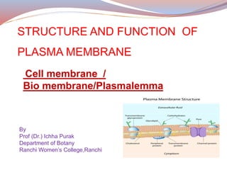
  • 1. STRUCTURE AND FUNCTION OF PLASMA MEMBRANE Cell membrane / Bio membrane/Plasmalemma By Prof (Dr.) Ichha Purak Department of Botany Ranchi Women’s College,Ranchi
  • 2. CONTENTS DEFINITION : STRUCTURE OF PLASMA MEMBRANE COMPONENTS OF PLASMA MEMBRANE ( (BIOCHEMICAL PROPERTIES) LIPID BILAYER PROTEINS CARBOHYDRATES CHOLESTEROL MODELS EXPLAINING STRUCTURE OF BIO MEMBRANE FLUID MOSAIC MODEL MOBILITY OF MEMBRANE GLYCOCALYX : GLYCOPROTEINS AND GLYCOLIPIDS TRANSPORT OF IONS AND MOLECULES ACROSS PLASMA MEMBRANE FUNCTIONS OF PLASMA MEMBRANE DIVERSITY OF CELL MEMBRANES SITE OF ATPase ION CARRIER CHANNELS AND PUMPS-RECEPTORS
  • 3. Plasma membrane is the biological membrane which is present both in prokaryotic and eukaryotic cells. It acts as a barrier between outer and inner surface of a cell. Plasma membrane is present as outer most layer or envelope in animal cell but is present beneath the cell wall in plant cell as well as most of prokaryotic cells. The plasma membrane, which is also referred to as the cytoplasmic membrane, is a biological membrane that encloses the contents of the cell (protoplasm) and separates it from the outer environment The cell or bio membrane has extracellular face(facing outer environment) and intracellular face (facing cytoplasm) . Plasma membranes appears trilaminar ( Protein-phospholipid bilayer- protein ) when viewed under electron microscope.
  • 4. Protoplasm of all type of cells is surrounded by plasma membrane, which in eukaryotic cells extends into the interior of the cell to divide the protoplasm into a number of compartments to form cell organelles as Endoplasmic reticulum, mitochondria, plastids, golgi apparatus etc. for various cellular functions. In prokaryotic cells also the plasma membrane extends into the interior of the cell as mesosomes for different biochemical processes but does not form separate compartments. In animal cells this is the outer most covering of the Cell. In plant cells, plasma membrane is surrounded by cell wall which is comparatively rigid in texture and provides a definite shape to plant cell. I P
  • 5. Prokaryotic cells as Bacteria and Cyanobacteria also have cell wall which surrounds the cell membrane or plasma membrane. The Red blood cells of human beings are enucleated and are surrounded by plasma membrane consisting of lipid bilayer with embedded proteins. Plasma membrane is thin (4-10nm) ,delicate ,permeable , semi-porous barrier to outside environment. The membrane acts as a boundary, holding the cell constituents together and keeping other substances from entering. Membranes are impermeable to most polar or charged solutes but permeable to non polar compounds. Permeability of cell membrane depends on physiological state of cell and size and nature of molecules.
  • 6. CHEMICAL COMPOSITION OF PLASMA MEMBRANE It is composed of lipids (phosphatidylcholine, phosphatidyleserine, phosphatidylethanolamine,sphingomyelin, cardiolipin, etc ), proteins and small amount of carbohydrates ( as glycolipids or glycoproteins ) and cholesterol The ratio of protein to lipids vary from 80:20 (Bacteria) ,20:80 (nerve cells) and 50:50 in most of the cells. Proteins - major component provide 60% mechanical strength and help in transportation. Lipids 28-70% vary with different cells, provide continuity to the membrane and facilitate the fusion and splitting of membrane. Lipid molecules constitute about 50% of the mass of most animal cell membranes, nearly all of the remainder being protein
  • 7. Component Location Phospholipids Main fabric (core) of plasma membrane Integral proteins Embedded in the phospholipid bilayer; may or may not extend through both layers Peripheral proteins On the inner or outer surface of the phospholipid bilayer, but not embedded in its hydrophobic core Carbohydrates Attached to proteins or lipids on the extracellular side of the membrane (forming glycoproteins and glycolipids) Together form Glycocalyx Cholesterol Distributed randomly among hydrophobic tails of the membrane phospholipids Table-The components of the plasma membrane
  • 8. Carbohydrates or sugars (2-10% of Membrane) are sometimes found attached to proteins and lipids as glycoproteins and glycolipids on the outside of a cell membrane. Glycolipids help the cell to recognize other cells of the body Sugars are only found on the extracellular side of a cell membrane . These carbohydrates together form the glycocalyx. Common sugars present in glycoproteins and glycolipids are D-Glucose, D- mannose and D-Galactose, N-acetyle glucosamine, N-acetyleneuramic acid and sialic acid Cholesterol is another lipid component of animal cell membrane, is selectively dispersed between phospholipid bilayer, prevent phospholipids from being too closely packed . Cholesterol is not found in the membranes of plant cells .
  • 9. Various models have been proposed from time to time to explain the arrangement of molecules in plasma membrane Some of which are as follows : Langmuir –lipid monolayer Model It is made up of phospholipids of one molecule thick (because of amphipathic nature, phospholipids become arranged in the form of a layer-with heads (hydrophillic) towards aqueous phase and tails (hydrophobic) away from aqueous phase. Langmuir lipid monolayer Model
  • 10. Danielli and Davson (1935-52) proposed another model to explain the structure of the cell membrane- Sandwich Model
  • 11. According to this Model Lipid bilayer is sandwiched between two dense protein layers. Outer ends of lipid molecules are hydrophilic and polar. Proteins are attached at the outer ends of lipid layer by ionic exchanges and hydrostatic forces. Inner ends of lipid molecules are hydrophobic and non polar, membranes are held together by electrostatic attraction between lipid layers. They also proposed the presence of protein coated pores that make the membrane a seive like structure. This model fails to explain all the functions of the membrane.
  • 12. Robertson (1950) -Unit membrane Model The cell membrane generally appears to have three layers –two dense layers of 2nm (protein) and a clear middle layer of about 3.5nm (lipid bilayer). Clear layer corresponded to the hydrocarbon chains of lipids and dense layers to the proteins. On this basis Robertson called it unit membrane Model( a unit of these three layers) Proteins found in plasma membrane are generally globular proteins.It does not explain how some molecules pass through it. Schematic diagram of the Robertson model of membrane structure. Lipid layer is defined as bimolecular and the protein is extended on two faces of the membrane
  • 13. Fluid Mosaic Model (Singer and Nicholson,1972) According to this Model The main component of plasma membrane is the phospholipid bilayer with hydrophilic (water attracting) polar heads of lipids oriented outwards and hydrophobic (water repelling) tails towards inside forming the core of plasma membrane. Proteins are arranged in two ways 1) Extrinsic or peripheral proteins are associated with the surface of lipid bilayer. They can be easily removed as ATPase in Mitochondria and spectrin in Red Blood Cells (RBC) 2) Intrinsic or Integral proteins penetrate the lipid bilayer wholly (from one end to other end ) or partially . The portion of the polypeptide chain that extend through lipid bilayer typically occur as α helix composed of hydrophobic amino acids.
  • 14. Lipids and intrinsic proteins form a mosaic pattern ( proteins are embedded randomly in the lipid bilayer ). The lipids, many of intrinsic proteins and glycoproteins of the membrane are amphipathic molecules constitute liquid crystalline aggregate in which the polar groups are directed towards the outer phase and the non polar groups are situated inside the bilayer. Transmembranous proteins which pass throughout the lipid bilayer (Enzymes and glycoproteins ) possess their active side towards outer phase and hydrophobic part towards interior and are of appropriate size to fit in lipid bilayer. The distribution of phospholipids in the outer and inner surface is highly asymmetrical. Outer layer consists mainly of lecithin and sphingomyelin while inner is composed mainly of phosphatidylethanolamine and phosphatidylserine. Glycolipids are mainly present in outer half of the bilayer.
  • 15. Figure-11-3 Fluid mosaic model for membrane structure Courtsey: Principles of Biochemistry, Lehninger,Nelson and Cox Chapter 11 : Biological membranes and Transport Page : 372
  • 16. . 1.The fatty acyl chains in the interior of the membrane form a fluid, hydrophobic region. 2.Integral proteins float in this sea of lipid, held by hydrophobic interactions with their nonpolar amino acid side chains. 3.Both proteins and lipids are free to move laterally in the plane of the bilayer, but movement of either from one face of the bilayer to the other is restricted. 4.The carbohydrate moieties attached to some proteins and lipids of the plasma membrane are exposed on the extracellular surface of the membrane Description of Figure-11-3 Fluid Mosaic Model for membrane structure Lehninger
  • 17. Figure 12.3 Fluid mosaic model of the plasma membrane Integral membrane proteins are inserted into the lipid bilayer, whereas peripheral proteins are bound to the external face of the membrane Most integral proteins are transmembranous, portions of these proteins are glycosylated. Courtsey: The cell: A molecular Approach 2nd edition Sunderland (MA): Sinauer Associates;2000
  • 18.
  • 19. Lipid bilayer With the help of electron microscopy and x-ray diffraction studies ,it has been established that lipid bilayer is the basic structural element of plasma membrane. Membrane lipids are amphipathic(amphiphillic) molecules having a hydrophilic(water-loving,or polar) end and a hydrophobic(water-hating, or nonpolar ) end, most of which spontaneously form bilayers. These have a polar head group and two hydrophobic hydrocarbon tails. The tails are usually fatty acids, and they can differ in length (14-24 carbon atoms) ,one of fatty acid tail is unsaturated having double bond whereas other is saturated fatty acid. The hydrophobic fatty acid portions face each other within the interior of the membrane itself.
  • 20. Figure-Two-dimensional and three –dimensional view of plasma membrane Electron micrograph of cross section of human RBC plasma membrane General structural formula of a glycerophospholipid . (Encyclopaedia Britannica) Phosphate is linked to one side by Alcoholic group and Glycerol on other side. R1 and R2 are two fatty acids attached to glycerol
  • 21. Glycerol Phosphate Choline Phospholipid bilayer Phospholipid R1 R2 Kink Double bond Figure- A-PhosphoLipid bilayer,B- a phospholipid, C & D-Parts of Phosphatidyl Choline (structural formula) R1 & R2 –Fatty acids
  • 22. Phospholipids are asymmetrically distributed between the two halves of the membrane layer. Cholesterol is present in both leaflets. The outer leaflet of plasma membrane consists mainly of phosphatidylcholine,sphingomyelin and glycolipids,whereas the inner leaflet contains phosphatidylethanolamine,phosphatidyl Serine and phosphatidylinositol (Figure-12.2) The head groups of both phosphatidylserine and phosphatidyl inositol are negatively charged,that results in a net negative charge on the cytosolic face of plasma membrane Figure: 12.2 Lipid components of the animal plasma membrane Courtsey: The cell: A molecular Approach 2nd edition Sunderland (MA): Sinauer Associates;2000
  • 23. Figure Phospholipid Structure. A phospholipid molecule consists of a polar phosphate “head,” which is hydrophilic and a non-polar lipid “tail,” which is hydrophobic. Unsaturated fatty acids result in kinks in the hydrophobic tails. Figure Phospolipid Bilayer. The phospholipid bilayer consists of two adjacent sheets of phospholipids, arranged tail to tail. The hydrophobic tails associate with one another, forming the interior of the membrane. The polar heads contact the fluid inside and outside of the cell. Courtsey :Chapter 3. The Cellular Level of Organization The Cell Membrane :Anatomy and Physiology files
  • 24. Fluidity of the membrane ESR(Electron Spin Resonance) technique has been used to establish fluidity of the membrane which shows that lipid molecules of the biological membrane are neither in a fixed state as in crystal,nor like completely mobile molecule (liquid) . Hence the intermediate state of the membrane is referred as liquid crystal state. lipid molecules can move laterally within the bilayer keeping the orientation intact (polar head exposed towards membranous surface and tails (nonpolar) towards interior . This difference in length and saturation of the fatty acid tails influence the ability of phospholipid molecules to pack against one another and also affect fluidity of the membrane.
  • 25. The fluidity of the membrane depends on degree of saturation of fatty acids (hydrocarbon chain ), chain length and the temperature The temperature again depends on composition of lipids. A shorter chain length reduces the tendency of the hydrocarbon tails to interact with one another, and double bonds of unsaturated fatty acids produce kinks (bends) in the hydrocarbon chains that make them more difficult to pack together and cause molecules to spread out, so that the membrane remains fluid at lower temperatures . Phospholipid molecules move side ways at a rate about 2 µ/sec (length of a prokaryotic cell). The transition of the lipid layer from non fluid (gel) condition to a liquid crystalline (fluid) state depends on the temperature of the cell. The Lipid bilayer is a two-dimensional fluid, precise fluidity of cell membranes is biologically important.
  • 26. The mobility (fluidity) of membrane is controlled by different factors . The increase in membrane fluidity is related with the increase of unsaturated fatty acids and decrease in fatty acid chain length and cholesterol content. Again the increase in membrane fluidity is inversely proportional to transition temperature. This alteration in the physical state of the membrane has an important role in the function of membrane . The membrane proteins are also capable of rapid lateral migration in fluid lipid layer. Thus the random distribution of membrane proteins depend on the membrane fluidity. In mitochondria and chloroplast membranes 50% of fatty acids are unsaturated and help in oxidation reduction through electron transport chain (ETC) .
  • 27. The lipid bilayer of some cell membranes is not exclusively composed of phospholipids but also contain glycolipids.(chloroplast) Eukaryotic plasma membrane contains large amount of cholesterol (animal cell ) which enhances the permeability barrier properties of the lipid bilayer. Eukaryotic plasma membrane contains four major phospholipids viz. phosphatidylcholine, sphingomyelin, phosphatidylserine, and phosphatidylethanolamine of these phosphatidylserine carries a net negative charge. Other phospholipids, such as the inositol phospholipids, are present in smaller quantities but are functionally very important having a crucial role in cell signaling.
  • 28. The lipid bilayer is asymmetrical The lipid composition of two halves of the lipid bilayer differ strikingly in some membranes In the human red blood cell (erythrocytes) membrane , phosphatidylcholine and sphingomyelin are present in the outer half of lipid bilayer whereas phosphatidylethanolamine , phosphatidylserine and phosphatidylinositols are present in the inner half (cytoplasmic side )of the membrane Because the negatively charged phosphatidylserine is located in the inner monolayer, there is a significant difference in charge between the two halves of the bilayer. Phospholipid asymmetry in lipid bilayer can be functionally important. The enzyme protein kinase is activated in response to various extracellular signals, it binds to the cytoplasmic face of plasma membrane where phosphatidylserine is concentrated , its negative charge is required for its activity.
  • 29. Membrane Proteins Although the basic structure of biological membranes is provided by the lipid bilayer, most of the specific functions are carried out by proteins. The amount of proteins in a membrane are highly variable, in nerve cell membrane (25%), in mitochondria and chloroplast membrane (75%) and usual plasma membrane it is about 50%. Because lipid molecules are small in comparison to protein molecules, there are always many more lipid molecules in number than protein molecules in membranes on the basis of orientation in lipid bilayer- proteins can be integral (intrinsic) embedded (can be transmembranous, which extends through both sides of cell membrane or unilateral which reach only partway across the membrane and peripheral (extrinsive) attached to the membrane surface only by weak ionic bonds.
  • 30. Integral proteins are attached to the membrane by hydrophobic interactions with fatty acid tails and are buried inside (difficult to extract) Detergent sodium dodecyl sulphate can help extraction . Such proteins have both hydrophilic amino acid and hydrophobic amino acids. Polar amino acids are exposed at the surface of membranes. For this reason interaction with ions, hormones, antigens can occur on the membrane surface Some of the proteins in the membrane act as enzymes (ATPase), others function as transporters or signal receptors and ion channels. Peripheral proteins are bound to the hydrophilic head groups of the lipid and they do not form a continuous layer, but are randomly distributed . The random distribution of integral and peripheral proteins in the fluid mosaic model satisfies many of the problems raised against the previous model.
  • 31. Different membrane proteins are associated with the membrane in different ways Many membrane proteins extend through the lipid bilayer having their part on either side, these are transmembranous proteins ,are amphipathic having both hydrophobic and hydrophilic regions. Their hydrophobic regions pass through membrane and interact with the hydrophobic tails of the lipid molecules in the interior of the bilayer. The hydrophilic regions are exposed to water on one or both sides of membrane Other membrane proteins are located in the cytosol, associated with bilayer only by means of one or more covalently attached fatty acid chains. Other membrane proteins are entirely exposed at the external cell surface, being attached to the bilayer only by a covalent linkage (via a specific oligo-saccharide) to phosphatidylinositol in the outer lipid monolayer of the plasma membrane.
  • 32. Figure- Different ways in which membrane proteins associate with lipid bilayer Most transmembranous proteins extend across the bilayer as a single α helix (1) or as multiple α Helices (2) Some of these are single pass and multi pass proteins having covalently attached fatty acid chains inserted in the cytoplasmic monolayer . Other membrane proteins are attached to the bilayer by covalently attached lipid in the cytoplasmic monolayer (3) or via an oligosaccharide to a minor phoshpolipid,phosphatidylinositol in the non cytoplasmic monolayer (4) Some proteins are attached to the membrane only by noncolent interaction with other membrane proteins (5) and (6 )
  • 33. Membrane proteins can be associated with the lipid bilayer in various ways In most transmembrane proteins the polypeptide chain is thought to cross the lipid bilayer in an α-helical conformation Spectrin Is a cytoskeletal protein noncovalently associated with the cytoplasmic side of the red blood cell membrane Glycophorin extends through the red blood cell lipid bilayer as a single α Helix Glycophorin transmembranous α helix protein having 131 amino acids, glycosylated. Band 3 has multiple transmembranous α helices (929 amino acids ,cross the membrane 14 times. ((Figure- 12.6) Bacteriorhodopsin is a proton pump that traverses the lipid bilayer as seven α helices Porins are pore-forming transmembrane proteins that cross the lipid bilayer as a β barrel membrane
  • 34. Figure 12.6 Integral membrane proteins of red blood cells Courtsey: The cell: A molecular Approach 2nd edition Sunderland (MA): Sinauer Associates;2000
  • 35. The plasma membrane of some bacteria is surrounded by a cell wal and a distinct outer membrane. Figure:12.8 Bacterial outer membranes Courtsey: The cell: A molecular Approach 2nd edition Sunderland (MA): Sinauer Associates;2000 In bacterial cell ,the outer membrane contains porins ,which form open aqueous channels allowing the free passage of ions and small molecules.
  • 36. GLYCOCALYX In the plasma membrane of all eucaryotic cells most of the proteins exposed on the cell surface and some of the lipid molecules have oligosaccharide chains covalently attached to them. Sugar groups of glycolipids and glycoproteins exposed at the cell surface together form glycocalyx, play a role in interaction of cell with its surroundings. This sugar coating helps to protect the cell surface from mechanical and chemical damage, and some of the oligosaccharide chains are recognized by cell-surface carbohydrate-binding proteins (lectins) that mediate specific, transient, cell-cell adhesion events. Being of a hydrophilic nature, the glycocalyx attracts large amounts of water to the surface of the cell, which in turn facilitates the interaction between the cell and its aqueous environment.
  • 37. Figure- Simplified diagram of the cell coat (glycocalyx). The cell coat is made up of the oligosaccharide side chains of glycolipids and integral membrane glycoproteins and the polysaccharide chains on integral membrane proteoglycans. Structure of glycolipids Two hydrocarbon chains are joined to a polar head group formed from serine containing a carbohydrate ( e.g. glucose) Glycoolipids help the cell to recognize other cells of the body
  • 38. The hydroxyl group can form hydrogen bond with carbonyl oxygen of phospholipid or sphingolipid head groups. Cholesterol is absent in plant ,prokaryotic and intracellular membranes. Cholesterol Cholesterol is a lipid consisting of four fused hydrocarbon rings forming the bulky steroid structure. A hydrocarbon tail is linked to one end of steroid . It is amphipathic molecule because of a polar hydroxyl group linked to other end of steroid. Cholesterol helps to minimize the effects of temperature on fluidity. C27H46O Cholesterol is selectively dispersed in both leaf lets of animal cell membranes, prevent phospholipids from being too closely packed. At low temperature,cholesterol increases fluidity while at high temperature,it reduces fluidity.
  • 39. Principles of membrane transport Transport of ions and molecules across plasma membrane The lipid bilayer (core) of plasma membrane serves as a barrier to the passage of most polar molecules because of its hydrophobic nature. This barrier function allows the cell to maintain concentration of solute in its cytosol ,which differs from extracellular fluid as well as membrane bound compartments or cell organelles. Transport of inorganic ions and small water soluble organic molecules across the lipid bilayer is achieved by specialized transmembrane proteins, each of which is responsible for the transfer of a specific ion or molecule or a group of closely related ions or molecules.
  • 40. lipid bilayers are highly impermeable to charged molecules (ions), charge prevents small ions as Na+ or K+ from entering the hydrocarbon phase of the bilayer Cell membranes allow water and nonpolar molecules to permeate by simple diffusion. Cells can also transfer macromolecules and larger particles across plasma membrane by different mechanism from those used for transferring small molecules. Some membrane transport proteins are multi pass transmembrane proteins - that is, their polypeptide chains traverse the lipid bilayer multiple times
  • 41. Cell membrane also allow various polar molecules,such as ions,sugars,amino acids ,nucleotides and many cell metabolites to pass through it . Special membrane proteins are responsible for transferring such solutes across cell membranes. These proteins, referred to as membrane transport proteins, occur in many forms and are present in all types of biological membranes Each protein transports a particular class of molecule (such as ions, sugars, or amino acids) and often only certain molecular species of the class. There are two classes of membrane proteins – Carrier membrane proteins and Channel membrane proteins
  • 42. Carrier proteins (also called carriers, permeases, or transporters) bind the specific solute to be transported and undergo a series of conformational changes in order to transfer the bound solute across the membrane Channel proteins, on the other hand, need not bind the solute. Instead, they form hydrophilic pores that extend across the lipid bilayer; when these pores are open, they allow specific solutes (usually inorganic ions of appropriate size and charge) to pass through them and thereby cross the membrane Transport through channel proteins occurs at a very much faster rate than transport mediated by carrier proteins.
  • 43. Active transport is mediated by carrier proteins coupled to an energy source All channel proteins and many carrier proteins allow solutes to cross the membrane only passively(downhill) ,a process called passive transport or facilitated diffusion If the transported molecule is uncharged, it is simply the difference in its concentration on the two sides of the membrane (concentration gradient) that determines the direction and drives passive transport. If the solute carries a net charge then both its concentration gradient and the electrical potential difference across the membrane influence its transport.
  • 44. Almost all plasma membranes have an electrical potential difference (voltage gradient) across them, with the inside usually negative with respect to the outside. This potential difference favours the entry of positively charged ions into the cell but opposes the entry of negatively charged ions. In active transport the pumping activity of the carrier protein is directional because it is tightly coupled to a source of metabolic energy,such as ATP hydrolysis or an ion gradient Thus transport by carrier proteins can be either active or passive, whereas transport by channel proteins is always passive Cells also require transport proteins that can actively pump certain solutes across the membrane against their electrochemical gradient (uphill) , this process is known as active transport,is always mediated by carrier proteins
  • 45. Figure- A schematic view of the two classes of membrane transport proteins. A carrier protein is thought to alternate between two confirmations ,so that the solute binding site is sequentially accessible on one side of the bilayer and then on the other. In contrast, a channel protein is thought to form a water filled pore across the bilayer through which specific ions can diffuse
  • 46. Figure –Comparision of passive transport down an electrochemical gradient with active transport against an electrochemical gradient. Simple diffusion and passive transport by membrane transport proteins (facilitated diffusion) occur spontaneously, active transport requires an input of metabolic energy. Only carrier proteins can carry out active transport,both carrier proteins and channel proteins mediate facilitated diffusion.
  • 47.
  • 48. Transport of small molecules (such as glucose, amino acids, water, mineral ions etc). (i) Diffusion : molecules of substances move from their region of higher concentration to their region of lower concentration. This does not require energy. Example : absorption of glucose in a cell (ii) Osmosis : movement of water molecules from the region of their higher concentration to the region of their lower concentration through a semipermeable membrane. There is no expenditure of energy in osmosis. This kind of movement is along concentration gradient. (iii) Active Transport : When the direction of movement of a certain molecules is opposite that of diffusion i.e. from region of their lower concentration towards the region of their higher concentration, it would require an “active effort” by the cell for which energy is needed. This energy is provided by ATP (adenosine triphosphate). The active transport may also be through a carrier molecule
  • 49. phagocytosis pinocytosis 1. intake of solid particles 2. membrane folds out going round the particle, forming a cavity and thus engulfing the particle 1. intake of fluid droplets 2. membrane folds in and forms a cup like structure sucks in the droplets Endocytosis Cell membrane regulates movement of substance into and out of the cell. If the cell membrane fails to function normally the cell dies. Diagrammatic representation of (a) phagocytosis; (b) pinocytosis Transport of large molecules (bulk transport) During bulk transport the membrane changes its form and shape. It occurs in two ways: (i) endocytosis (taking the substance in) (ii) exocytosis (passing the substance out) Endocytosis is of two types :
  • 50. FUNCTIONS OF PLASMA MEMBRANE The most important function of plasma membrane is to provide passage for various substances, into and out of the cell and regulates flow of water and inorganic molecules through it, as lipid molecules are only closely placed to each other but are not joined to each other.  Plasma membrane is selectively permeable, allows some solute particles (1-15A⁰) to pass through it readily along with solvents.  It acts as a protective layer, from the uptake of some harmful molecules. . Cell membranes often include receptor sites for interaction with specific biochemicals such as certain hormones, neurotransmitters and immune proteins. It helps in conversion of signals conveyed by some extra cellular agents  In this way the cell can recognize and process some signals received from the extracellular environment.
  • 51. For Active transport chemical energy is required because molecules move against the normal diffusion gradient. Passive transport- Movement of molecules from the region of higher concentration to lower concentration. Diffusion of different gases takes place through plasma membrane Oligosaccharide molecules (in the form of Glycolipid/Glycoprotein) of the cell membrane help in cell to cell recognition/recognizing self from non self. Plasma membrane is responsible for the transportation of materials, molecules or ions through it, by various ways Exocytosis- Molecules are taken out of the cell Endocytosis- Ingestion of molecules either in solid form (phagocytosis) cell eating or in liquid form (pinocytosis) cell drinking.
  • 52. Plasma membrane separates the components of the cell from its outside environment It allows only selected substances into the cell and keeps others out. Plasma membrane has a major role in protecting the integrity of the interior of the cell. •Plasma membrane serves as a base of attachment for the cytoskeleton in some organisms and cell walls in other organisms Plasma membrane provides cell shape (in animal cells) e.g. the characteristic shape of red blood cells, nerve cells, bone cells, etc Plasma membrane allows transport of certain substances into and out of the cell but not all substance, so it is termed selectively permeable.
  • 53. •The cell membrane maintains the physical integrity of the cell. It’s most obvious in the cases of animal cells (because they don’t have cell walls) that the cell membrane holds the cell together by enclosing the cytoplasm and organelles within it. The cell membrane forms a barrier between the inside of the cell and the environment outside the cell – enclosing cytoplasm and any organelles within the cell, and enabling different chemical environments to exist on each side of the cell membrane. The cell membrane physically separates the intracellular components (e.g. organelles in eukaryotic cells) from the extracellular environment. The cell membrane protects the cell from some harmful chemicals in its external environment. The cell membrane also protects the cell from loss of useful biological macromolecules held within the cell
  • 54. Endocytosis is the process in which cells absorb molecules by engulfing them. The plasma membrane creates a small deformation inward, called an invagination, in which the substance to be transported is captured. Endocytosis is a pathway for internalizing solid particles (“cell eating” or phagocytosis), small molecules and ions (“cell drinking” or pinocytosis), and macromolecules. Endocytosis requires energy and is thus a form of active transport. Proteins and lipids make up the composition of a cell membrane. There are three different types of proteins found within a cell membrane: structural protein, transport protein and glycoprotein. These support cell structure and shape, move molecules through the membrane and transmit signals between cells.
  • 55. Diversity of cell membranes/Organelle membranes In prokaryotic cells the plasma only encases the entire cell. membrane At some places plasma membranes gives invagination which provide sites for various chemical processes, are called as mesosomes . In eukaryotic cells ,the extensions of plasma membrane surrounds various organelles . In such cells, the plasma membrane is part of an extensive endomembrane system that includes the Endoplasmic Reticulum (ER), Nuclear membrane,Golgi apparatus and Lysosomes.
  • 56. The components are exchanged throughout the endomembranous system in an organized fashion. Mitochondria and chloroplasts are surrounded by two (double) membranes instead of just one. The outer membrane of these organelles has pores that allow molecules to pass easily. The inner membrane is loaded with proteins that make up the electron transport chain and help to generate energy through ATP formation. The membranes of the different organelles vary in molecular composition and are well suited for the functions they perform. Organelle membranes play important role for several vital cell functions such as cellular respiration, photosynthesis, protein synthesis and lipid metabolism
  • 57. The lipid bilayer of biological membranes is impermeable to ions and polar molecules. Membrane proteins may act as pumps or channels,which induces permeability. Pumps use a source of free energy as ATP or light to drive uphill transport of ions or molecules. It is active transport. Channels in contrast , enable ions to flow rapidly through membranes in a downhill direction. It is passive transport or facilitated diffusion. SITE OF ATPase ION CARRIER CHANNELS AND PUMPS-RECEPTORS Pumps are energy transducers ,they convert one form of free energy into another. Two types of ATP driven pumps (P-type ATPases and the ATP-binding cassette pumps, undergo conformational changes on ATP binding and hydrolysis that cause a bound ion to be transported across the membrane.
  • 58. Phosphorylation and dephosphorylation of both the Ca++K+ ATPase and Na + K+ATPase pumps are reprentative of P-type ATPase, are coupled to changes in orientation and affinity of their ion –binding sites . P-type ATPases are the largest and most diverse of the ATP-dependent ion transporters involved in transporting many different ions ,metals and other substances .
  • 59. Multiple Choice Questions 1. Lipid bi layer is a) hydrophilic b) hydrophobic c) hydrophilic and hydrophobic d) depends on the surrounding medium 2. Which of the following membrane has the largest amount of proteins a) erythrocyte membrane b) myelin sheath membrane c) inner mitochondrial membrane d) outer mitochondrial membrane 3. High lipid content is a characteristic of a) erythrocyte membrane b) myelin sheath membrane c) inner mitochondrial membrane d) outer mitochondrial membrane
  • 60. 4. The distribution of intrinsic proteins in the cell membrane is a) symmetrical b)asymetrical c) random d)uniform 5. In cell membrane, carbohydrates in glycoproteins or glycolipids are oriented a) towards outside b)towards inside c) towards outside and inside d)randomly distributed 6. The plasma membrane is impermeable to all molecules except a) Glucose b) ATP c)urea d)K+
  • 61. 7. Which of the following transport induces conformational change in protein a) simple diffusion b) active transport c) facilitated diffusion d) ion driven active transport 8. Na+ glucose transporter is an example of a) facilitated diffusion b) ATP driven active transport c) Symport d) antiport 9. Clathrin coated pits are associated with a) phagocytosis b) pinocytosis c) receptor mediated endocytosis d) exocytosis
  • 62. 10.The erythrocyte glucose transporter is an example of a)simple diffusion b)active transport c) facilitated diffusion d) ion driven active transport 11. Because they are embedded within the membrane, ion channels are examples of ________. a) receptor proteins b) integral proteins c) peripheral proteins d) glycoproteins 12. The diffusion of substances within a solution tends to move those substances ________ their ________ gradient. a) up; electrical b) up; electrochemical c) down; pressure d) down; concentration
  • 63. 13. Ion pumps and phagocytosis are both examples of ________. a) endocytosis b) passive transport c) active transport d) facilitated diffusion 14. Choose the answer that best completes the following analogy: Diffusion is to ________ as endocytosis is to ________. a) filtration; phagocytosis b) osmosis; pinocytosis c) solutes; fluid d) gradient; chemical energy 15. The average thickness of plasma membrane of eukaryotic cell is a) 5 to 10 nm b) 5 to 10 A⁰ c) 5 to 10 µm d) 5 to 10 mm
  • 64. 16. Which of the following is NOT the function of plasma membrane a) Intercellular interactions b) Responding to external stimulli c) Energy transduction d) Assisting in chromosome segregation 17.Glycolipids in plasma membrane are usually located at a) Outer leaflet of plasma membrane b) Inner leaflet of plasma membrane c) Evenly distributed in both inner and outer layers of plasma membrane d) cannot be predicted, it varies according to cell types17. 18. Which of the following events in a biological membrane would not be energetically favourable and therefore not occur simultaneously a) The rotation of membrane proteins b) The rotation of phospholipids c) The lateral movement of phospholipids d) The flip-flop of phospholipids to opposite leaflet
  • 65. 19. Which of the following statement best describes the chemical composition of plasma membrane? a) Plasma membrane is composed of two layers-one layer of phospholipids and one layer of proteins. b) Plasma membrane is composed of equal numbers of phospholipids,proteins and carbohydrates. c) Plasma membrane is bilayer of proteins with associated lipids and carbohydrates. d) Plasma membrane is bilayer of phospholipids with associated proteins and carbohydrates 20.The main role of carbohydrates in the cell membrane is a) Adhesion b) Recognition c) Locomotion d) Reception
  • 66. 21. Match the following i)Hydrophillic end a) cell wall ii) Microfibrils b) inner ends of lipids iii) Fluid mosaic model c) fluid droplets iv) Hydrophobic end d) outer end of lipids v) Pinocytosis e) Nicholson and Singer
  • 67. Long Answer Questions 1. What materials can easily diffuse through the lipid bilayer, and why? 2. Why is receptor-mediated endocytosis said to be more selective than phagocytosis or pinocytosis? 3. What do osmosis, diffusion, filtration, and the movement of ions away from like charge all have in common? In what way do they differ? 4. Describe the molecular components that make up the cell membrane 5. Explain the major features and properties of the cell membrane 6. Differentiate between materials that can and cannot diffuse through the lipid bilayer 7. Compare and contrast different types of passive transport with active transport, providing examples of each 8.Define diffusion and osmosis. 9.What does active transport mean? 10.Give one point of difference between Phagocytosis and pinocytosis
  • 68. Write short notes on the following Phospholipid bilayer Glycerophospholipid Membrane proteins Fluid Mosaic Model of Plasma membrane Integrated proteins of plasma membrane Peripheral proteins of Plasma membrane Glycoproteins Glycolipids Glycocalyx Carrier transport proteins Channel transport protein Active transport across plasma membrane Passive transport across Plasma membrane Osmosis Diffusion Endocytosis Exocytosis Phagocytosis Pinocytosis Transmembranous proteins
  • 69. Bretscher, M.S. The molecules of the cell membrane. Sci. Am. 253(4): 100-109. 1985. (PubMed) Gennis, R.B. Biomembranes: Molecular Structure and Function. New York: Springer-Verlag, 1989. Jain, M.K. Introduction to Biological Membranes, 2nd ed. New York: Wiley, 1988. Singer, S.J.; Nicolson, G.L. The fluid mosaic model of the structure of cell membranes. Science 175: 720-731. 1972. (PubMed) REFERENCES
  • 70. Molecular Biology of the Cell, 4th edition Bruce Alberts, Alexander Johnson, Julian Lewis, Martin Raff, Keith Roberts, and Peter Walter. 2002.NewYork Chapter 10. Membrane Structure Chapter 11. Membrane Transport of Small Molecules and the Ionic Basis of Membrane Excitabilit BOOKS REFERRED The cell: A molecular Approach 2nd edition 2000 Sunderland (MA): Sinauer Associates; Cooper G M. Principles of Biochemistry Fourth Edison Albert L Lehninger, David L Nelson and Michael M Cox 2008 W. H. Freeman and Company Biochemistry Third Edison 1988 Lubert Stryer W H Freeman and Company New York The cell membrane Encyclopaedia Britannica
  • 71. Cell and Molecular biology By P K Gupta: Rastogi Publications Cell Biology (Cytology,Biomolecules and Molecular Biology) By Verma PS and Agarwal V K ) S Chand & Company Pvt Ltd. Cell Biology by Dr S P Singh and Dr B S Tomar Rastogi Publication,Meerut,U P ,India