microscopes- a brief introduction
•Transferir como PPTX, PDF•
12 gostaram•9,438 visualizações

a brief introduction about microscopes

Recomendados
Mais conteúdo relacionado
Mais procurados
Mais procurados (20)
Destaque
Destaque (19)
Semelhante a microscopes- a brief introduction
Semelhante a microscopes- a brief introduction (20)
Mais de Malathi Murugesan
Mais de Malathi Murugesan (20)
Último
Último (20)
microscopes- a brief introduction
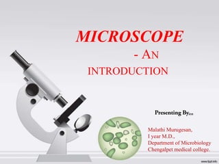
- 1. MICROSCOPE - AN INTRODUCTION Malathi Murugesan, I year M.D., Department of Microbiology Chengalpet medical college.
- 4. History of the Micro(organism)scope • 1590 –first compound microscope Discovery of Microorganisms. Anton van Leeuwenhoek (16321723) – first person to observe and describe micro-organisms accurately The way how he found micro-organism’s….
- 5. Classical Microscope A. B. C. D. E. F. G. H. I. J. K. L. M. Ocular Body tube Stage clip Revolving nose piece Objective Arm Stage Diaphragm Lever to move stage clip Course adjustment Fine adjustment Light source Base
- 6. Parts of Microscope,,, • Eyepiece Lens: the lens at the top that you look through. They are usually 10X or 15X power. • Tube: Connects the eyepiece to the objective lenses. • Arm: Supports the tube and connects it to the base • Base: The bottom of the microscope, used for support • Illuminator: A steady light source (110 volts) used in place of a mirror. If your microscope has a mirror, it is used to reflect light from an external light source up through the bottom of the stage.
- 7. Cont,,, • Stage: The flat platform where you place your slides. Stage clips hold the slides in place. If your microscope has a mechanical stage, you will be able to move the slide around by turning two knobs. One moves it left and right, the other moves it up and down. • Revolving Nosepiece or Turret: This is the part that holds two or more objective lenses and can be rotated to easily change power. • Rack Stop: This is an adjustment that determines how close the objective lens can get to the slide. • Diaphragm or Iris: This diaphragm has different sized holes and is used to vary the intensity and size of the cone of light that is projected upward into the slide.
- 8. Lens Objective lens Condenser Lens Usually you will find 3 or 4 objective lenses on a microscope The purpose of the condenser lens is to focus the light onto the specimen It consist of 4X, 10X, 40X and 100X powers. Condenser lenses are most useful at the highest powers (400X and above). When coupled with a 10X (most common) eyepiece lens, we get total magnifications of 40X (4X times 10X), 100X , 400X and 1000X Microscopes with in stage condenser lenses render a sharper image than those with no lens (at 400X) If the microscope has a maximum power of 400X, you will get the maximum benefit by using a condenser lenses rated at 0.65 NA or greater
- 9. Microscope Vocabulary • Magnification: increase of an object’s apparent size • Resolution: power to show details clearly Both are needed to see a clear image
- 10. Types of Microscopy • • • • • • • TEM Compound Light Microscope Phase contrast microscope Dissection or stereoscope Electron Microscope Transmission Electron Microscope Scanning Electron Microscope Flourescence microscope SEM
- 11. Light mechanism • What is Light??? Visible light (commonly referred to simply as light) is an electromagnetic radiation that is visible to the human eye, and is responsible for the sense of sight. Visible light has a wavelength in the range of about 380 nanometres (nm), or 380 10−9 m, to about 740 nanometres. The natural agent that stimulates sight and makes things visible. Used for illuminations…..
- 12. Angle of Incidence • Angle of incidence is a measure of deviation of something from "straight on―
- 13. Snell’s Law Snel l 's l aw(al so know as t he Snel l –D n escar t es l awand t he l awof r ef r act i on) i s a f or m a used t o descr i be t he r el at i onshi p ul bet w een t he angl es of i nci dence and r ef r act i on, w hen r ef er r i ng t o l i ght or ot her w aves passi ng t hr ough a boundar y bet w een t w o di f f er ent i sot r opi c m a, such as w er , gl ass and ai r . edi at
- 14. Cont,,, Refraction of light at the interface between two media of different refractive indices, with n2 > n1. Since the velocity is lower in the second medium (v2 < v1), the angle of refraction θ2 is less than the angle of incidence θ1; that is, the ray in the higher-index medium is closer to the normal.
- 15. Do light waves have amplitude??? • Of course it has, • Yes… Waves in general have three properties Frequency (related to wavelength), Amplitude, and Speed Freq- No of cycles. • • • Frequency tells us how many waves are passing a point per second, the inverse of time. Wavelength tells us the length of those waves in metres, almost like adisplacement. If we multiply these two together, we are really multiplying 1/s and m… which gives us m/s, the velocity of the wave!
- 16. Cont,,,
- 17. Lenses and the Bending of Light • Light is refracted (bent) when passing from one medium to another • Refractive index – a measure of how greatly a substance slows the velocity of light , where c is the speed of light in vacuum and v is the speed of light in the substance • Direction and magnitude of bending is Determined by the refractive indexes of the two media forming the interface . 17
- 18. Focal point and Focal length • Focus light rays at a specific place called the focal point • Distance between center of lens and focal point is the focal length • Strength of lens related to focal length • short focal length magnification more 18
- 19. The Light Microscope • Many types – – – – bright-field microscope dark-field microscope phase-contrast microscope fluorescence microscopes • are compound microscopes – image formed by action of 2 lenses 19
- 20. Mechanism of Light Microscopes
- 21. The Bright-Field Microscope • Produces a dark image against a brighter background • Has several objective lenses – parfocal microscopes remain in focus when objectives are changed • Total magnification – product of the magnifications of the ocular lens and the objective lens 21
- 22. Microscope Resolution • Ability of a lens to separate or distinguish small objects that are close together • Wavelength of light used is major factor in resolution shorter wavelength greater resolution 22
- 23. Working Distance •working distance — Distance between the front surface of lens and surface of cover glass or specimen
- 24. The Dark-Field Microscope • Produces a bright image of the object against a dark background • Used to observe living, unstained preparations 24
- 25. The Phase-Contrast Microscope • Enhances the contrast between intracellular structures having slight differences in refractive index • Excellent way to observe living cells Working Principle,,, 25
- 26. Cont,,,
- 27. The Differential Interference Contrast Microscope • Creates image by detecting differences in refractive indices and thickness of different parts of specimen • Excellent way to observe living cells 27
- 28. Thanking you
Notas do Editor
- MalathiMurugesan Presenting By,,,Department of Microbiology
- Usually you will find 3 or 4 objective lenses on a microscope
- TEMSEM
- Working Principle,,,
- Thanking you