SlideShare uma empresa Scribd logo
1 de 6
Baixar para ler offline
General Histology and Histotechnique (1st semester; 2012-2013) by K-Dumlao


INTEGUMENTARY SYSTEM

Functions:
1. Body temperature regulation.
2. Reservoir for blood
3. Protection from external environment
4. Cutaneous sensations
5. Excretion and Absorption
6. Vitamin D synthesis

Composition:
- Skin
- Accessory structures:
  o Hair
  o Nails                                                   1. Keratinocytes: keratin & lamellar granules
  o Various glands                                          - 90% of the epidermal cells
                                                            - Produced keratin (protein)
Skin                                                                   Tough fibrous protein that help the skin
- Largest organ of the body                                              from heat, microorganisms, underlying
                                                                         tissues of skin and chemicals.
Two principal parts:                                        - Lamellar granules
                                                                       Stratum granulosum
1. Epidermis                                                           Functions: Releases a water and proofing
- Superficial portion                                                    sealant
- With 5 layers

2. Dermis
- Inner portion
- With 2 regions


                                                            2. Melanocytes: melanin
                                                            - 8% of the epiderma cells
                                                            - Produce pigment – melanin  skin color


Epidermis
- A keratinized stratified squamous epithelium
- Consists of 4 principal types of cells:                   3.   Langerhans cells
                                                            -    Arise from the bone marrow
                                                            -    Migrate to epidermis
                                                            -    Participate in immune responses
General Histology and Histotechnique (1st semester; 2012-2013) by K-Dumlao


                                                            -    Shed off when you take a bath
                                                                Continuous Friction
                                                            -    Callous  an abnormal thickening of epidermis due
                                                                 to friction

                                                            2.   Stratum Lucidum
                                                            -    “lucid” – “clear”
                                                            -    Present in the skin of finger tips, palms, and soles.
                                                            -    3 – 5 clear layers of flat dead keratinocytes with
                                                                 intermediate filaments and thickened plasma
4. Merkel cells                                                  membrane.
- It is the least numerous of epidermal cells
- Located at the deepest part of stratum                    3. Stratum Ganulosum
   basale/geminativum.                                      - 3 – 5 layers of flattened keratinocytes
- In contact with tactile disc or merkel disc               - Consists of keratohyaline and lamellar granules
Function:                                                                 o A distinctive feature of the layer
- Both participate in sensation of touch                                  o Darkly staining granules of the
                                                                               stratum
                                                            Function: Organized intermediate filaments into thicker
                                                            bundles.

                                                            4. Stratum Spinosum
                                                            -  The superficial of basale
                                                            -  8 – 10 layers of polyhedral keratinocytes
                                                            -  Fit closely together
                                                            -  Thorn – like spines bundles of intermediate
                                                               filaments of the cytoskeleton joins the cell tightly to
                                                               one another to provide strength and flexibility of
Layers of the Epidermis                                        the skin.
                                                            5. Stratum Basale/Geminativum
                                                            - Active in cell division
                                                            - Compose of one layer of simple cuboidal/columnar
                                                               keratinocytes.
                                                                     Some considered them as stem cells
                                                                     Continue cell divisions to produce new
                                                                        keratinocytes
                                                            - Merker cell
                                                            - Melanocytes

                                                            Dermis
                                                            - Second deeper part of the skin
                                                            - Composed of connective tissue containing collagen
                                                               and elastic fibers
1.   Stratum Corneum                                               o Functions: for extensibility (stretch; obesity)
-    25 – 30 layers of dead flat keratinocytes.                        and flexibility (Return to its original form)
-    Contains the dead flat intermediate filaments          - Include cells such as fibroblasts, macrophages, and
-    Consists of keratohyaline                                 some adipocytes
                 o Function: Water repellant barrier        - Blood vessels, nerves, glands, and hair follicles are
-    Lamellar granules                                         embedded in dermal tissue
-    Lipid from lamellar granules (barrier in between)
General Histology and Histotechnique (1st semester; 2012-2013) by K-Dumlao


Two regions of Dermis:                                      3.   Meissner’s corpuscle
                                                            -    Encapsulated
1. Papillary region                                         -    Palmar and planthar
- The superficial portion of the dermis                     -    Fingertips
- Consists of areolar connective tissue with tissue
   with elastic fibers                                      4. Ruffini corpuscles
- Contains dermal papillae                                  - Type II cutaneous mechanoreceptor
              Finger-like projections that indent the
                  epidermis to the dermis.                  5. Pacinian corpuscle
Contains the following:                                     - Located deeper part of the dermis
                                                            - Encapsulated with multicellular ovoid structures
a. Meissner’s corpuscle                                     Function: Respond to deeper pressure.
-  For touch sensation
b. Free nerve endings
-  Contains the dendrites that initiates signals that are
   felt such as warm, coolness, pain, tickling and
   itching.
*Papillary layer – small fine collagen

2. Reticular region
- Deeper portion of dermis
- Consists of dense irregular connective tissue
    containing bundles of collagen and some elastic
    fibers.
*Reticular layer – more coarsely-textured collagen
fibers.

                                                            Functions of the connective tissue of the dermis:

                                                            1. Tough collagen fibers and resilient elastic fibers
                                                               provide mechanical strength for skin. Lines of
                                                               tension in the dermis called Langer’s lines, affect
                                                               healing after surgical incision
                                                            2. The ground substances of the dermis serve as the
                                                               substrate for diffusion of nutrients and wastes to
                                                               and from various other tissue components.
                                                            3. Mast cells, lymphocytes and macrophages in the
                                                               connective tissue carry our surveillance for the
                                                               immune system.
                                                            4. Finally, the dermis together with its associated
                                                               blood vessels and nerves is capable of active
                                                               responses to injury, yielding the defensive reaction
Sensory receptors / free nerve endings of the reticular
                                                               of inflammation, followed by the healing processes
layer:
                                                               of growth and repair
1.   Nociceptotrs – pain receptor
                                                            Subcutaneous layer (Hypodermis)
2. Tactile/Merkel disc
                                                            -    The hypodermis (hypo – “under” + dermis – “skin”)
- Type one cutaneous mechanoreceptor
                                                                 or subcutaneous (sub – “below” + cutaneous) layer
- For sensation touch
General Histology and Histotechnique (1st semester; 2012-2013) by K-Dumlao


     lies below the skin and is made up of loose             -    Scatters light from the dermis without altering its
     connective and adipose tissues.                              color
-    The hypodermis binds the skin to underlying organs      -    Whiteness of white skin
     while allowing the skin to move somewhat                -    Reflection of collagen
     independently of underlying structures.
-    Adipose tissue in the hypodermis provides padding       3. Blood
     and shock-absorption that helps to protect              - Hemoglobin and RBC
     underlying tissues from damage; it is also important    - Scatters red light
     in insulating against loss of body heat.                - Responsible for the pinkness in pigmented skin
-    Because the subcutaneous layer contains numerous        ***The amount of pigment, the thickness of dermis,
     blood vessels but no vital organs, it is a near-ideal   and the degree of perfusion in dermal capillaries vary.
     place to inject drugs. This is why so many drugs are
     administered through subcutaneous injection by a        Skin texture
     hypodermic (hypo-“under”+ dermic – “skin”)              - Affect the thickness and smoothness of the
     needle.                                                     epidermis, by the quality of fibers in the dermis, and
                                                                 by the amount of fluid in dermal connective tissue.
Types of skin
                                                             Note:
1. Thin skin                                                 Both edema (accumulation of excess fluid in connective
- Covers all parts of the body except palms, palmar          tissue) and dehydration can dramatically alter the
   surfaces of the digits, and soles                         appearance of skin
- Epidermis is thin
- Stratum lucidum lacking; stratum spinosum and              Accessory structures of the skin
   stratum Corneum are thin                                  1. Hairs
- Has fewer dermal papilla, lacks epidermal ridges           2. Glands
- Has hair follicles, arrector pili muscle and oil glands    3. Nails
   but fewer sweat glands than thick
- Sparser distribution of sensory receptors.                     Hairs
                                                             -   Composed of columns of dead, keratinized cells
2.   Thick skin                                                  bounded together by extracellular proteins
-    Covers palms, palmar surfaces of digits and soles       Parts:
-    Thick epidermis                                          1. Shaft – superficial portion
-    With stratum lucidum and thicker stratum                 2. Roots – penetrates into the dermis
     spinosum and stratum corneum.                               Concentric layers of shaft and root:
-    More numerous dermal papillae thus has epidermal
     ridges                                                  a. Medulla
-    Lack hair follicles, arrector pili muscle, sebaceous    - Inner portion
     gland.                                                  - 2 or 3 rows of polyhedral-shaped cells containing
-    Has more sweat glands that thin skin                       pigment granules and hair spaces.
-    Sensory receptors are more densely clustered.
                                                             b.   Cortex
Skin color                                                   -    Middle portion
                                                             -    It forms the major part of the shaft
1.   Melanin                                                 -    Contains pigments granules of the hair
-    Produced by melanocytes                                 -    White/gray hair – air bubbles
-    Yellowish-brown color of the epidermis
-    Lesser melanin – Light will penetrate easily into the   c. Cuticle
     skin --- prone to cancer.                               - Outermost layer
                                                             - Single layer of flat cells that are heavily keratinized
2. Collagen
General Histology and Histotechnique (1st semester; 2012-2013) by K-Dumlao


 3. Follicle – tubular invagination lined by stratified
     squamous epithelium similar to epidermis;                  6. Hair root plexuses
     surrounds the root of the hair; made up of:            -      Dendrites of neurons sensitive to touch
      a. External root sheath                               -      Generate nerve impulses in hair shaft is moved.
- Downward continuation of epidermis
- Contains the stratum basale                                     Glands
      b. Internal root sheath
- It forms the cellular tubular sheath between the
    external root and the hair.
Hair follicles are associated with:

a. Sebaceous glands – secrete oil into the hair follicle
   as well as nerve ending and smooth muscle to form
   the pilosebaceous apparatus.
b. Nerve endings – detects deflection of the hair shaft
   and also controls piloerection.
c. Smooth muscle – arrector pili ; affect piloerection

 4. Bulb – the base of hair follicle; onion-shaped
     structure; houses
   a. Papilla of the hair
- Nipple-shaped indentation
- Contains areolar connective tissue
- A lot of Blood vessels that nourish the growing hair
    follicle
   b. Matrix
- Germinal layer of the cell w/c rise from stratum
    basale.
Function:
- Responsible for growth of existing hairs
- Produce new hairs when old hairs are shed.


                                                             Sebaceous glands
 5. Arrector pili muscle – smooth muscle cells              - Associated with hair follicles
- Stimulated by autonomic nerve endings to contract         - Holocrine glands
    pulling the hair shaft perpendicular to the skin                    The whole cell is secreted
    surface during cold or fright.
General Histology and Histotechnique (1st semester; 2012-2013) by K-Dumlao


Function:
- It coats the surface of the hair and help keep them
   from drying and becoming brittle
- It prevents excessive evaporation of water from the
   skin.
- It keeps the skin soft and pliable
- It inhibits growth of certain bacteria

 Sweat glands
- 3-4 millions producing their secretion
- Exocytosis – release secretion
Two types:

a. Ecrine                                                       1. Nail body – visible portion of the nail
- Simple coiled tubular gland                                   2. Free edge – part of that may end pass the distal
- Common sweat gland                                               ends of digits
Function:                                                       3. Nail root – proximal edge of the nail plate that
- Regulates body temperature                                       is buried in the skin.
- Waste removal                                                 4. Lunula – whitish crescent shaped covered by
                                                                   portion of the nail bed.
b. Apocrine                                                     5. Hyponichium – nail bed; skin of nail bed;
- Simple coiled tubular gland                                      provide support
- Stimulated during emotional stress and sexual                 6. Eponychium/cuticle – a horny epidermal
   excitement                                                      extension of the tip of the nail folds.
- Secretion of cold sweat

    Nails
-   Plates of tightly packed, hard, keratinized epidermal
    cells.
-   Cells form a clear, solid covering over the dorsal
    surfaces of the distal portions of the digits
-   Parts:

Mais conteúdo relacionado

Destaque

GHH Nervous Tissue
GHH Nervous TissueGHH Nervous Tissue
GHH Nervous TissueKzen D.
 
GHH (prelim handout)
GHH (prelim handout)GHH (prelim handout)
GHH (prelim handout)Kzen D.
 
Tugas III evaluasi pembelajaran pkn lampiran i(nina)
Tugas III evaluasi pembelajaran pkn lampiran i(nina)Tugas III evaluasi pembelajaran pkn lampiran i(nina)
Tugas III evaluasi pembelajaran pkn lampiran i(nina)Nina Ruspina
 
Tugas v evaluasi nina ruspina
Tugas v evaluasi nina ruspinaTugas v evaluasi nina ruspina
Tugas v evaluasi nina ruspinaNina Ruspina
 
GHH Muscle Tissues
GHH Muscle TissuesGHH Muscle Tissues
GHH Muscle TissuesKzen D.
 
32 Ways a Digital Marketing Consultant Can Help Grow Your Business
32 Ways a Digital Marketing Consultant Can Help Grow Your Business32 Ways a Digital Marketing Consultant Can Help Grow Your Business
32 Ways a Digital Marketing Consultant Can Help Grow Your BusinessBarry Feldman
 

Destaque (12)

GHH Nervous Tissue
GHH Nervous TissueGHH Nervous Tissue
GHH Nervous Tissue
 
GHH (prelim handout)
GHH (prelim handout)GHH (prelim handout)
GHH (prelim handout)
 
Vol.01 Iss.22
Vol.01 Iss.22Vol.01 Iss.22
Vol.01 Iss.22
 
Vol. 01 Iss. 10
Vol. 01 Iss. 10Vol. 01 Iss. 10
Vol. 01 Iss. 10
 
Vol. 01 Iss. 08
Vol. 01 Iss. 08Vol. 01 Iss. 08
Vol. 01 Iss. 08
 
Tugas III evaluasi pembelajaran pkn lampiran i(nina)
Tugas III evaluasi pembelajaran pkn lampiran i(nina)Tugas III evaluasi pembelajaran pkn lampiran i(nina)
Tugas III evaluasi pembelajaran pkn lampiran i(nina)
 
Vol. 01 Iss. 13
Vol. 01 Iss. 13Vol. 01 Iss. 13
Vol. 01 Iss. 13
 
Tugas v evaluasi nina ruspina
Tugas v evaluasi nina ruspinaTugas v evaluasi nina ruspina
Tugas v evaluasi nina ruspina
 
Vol. 01 Iss. 23
Vol. 01 Iss. 23Vol. 01 Iss. 23
Vol. 01 Iss. 23
 
Vol. 01 Iss. 30
Vol. 01 Iss. 30Vol. 01 Iss. 30
Vol. 01 Iss. 30
 
GHH Muscle Tissues
GHH Muscle TissuesGHH Muscle Tissues
GHH Muscle Tissues
 
32 Ways a Digital Marketing Consultant Can Help Grow Your Business
32 Ways a Digital Marketing Consultant Can Help Grow Your Business32 Ways a Digital Marketing Consultant Can Help Grow Your Business
32 Ways a Digital Marketing Consultant Can Help Grow Your Business
 

Semelhante a GHH - INTEGUMENTARY SYSTEM

Nonneoplastic skin diseases
Nonneoplastic skin diseasesNonneoplastic skin diseases
Nonneoplastic skin diseaseshazemmali
 
Lect. 12 integumentary system
Lect. 12   integumentary systemLect. 12   integumentary system
Lect. 12 integumentary systemHara O.
 
Ch4 skin, martini
Ch4  skin, martiniCh4  skin, martini
Ch4 skin, martiniTheSlaps
 
STRUCTURE AND FUNCTIONS OF THE SKIN.pptx
STRUCTURE AND FUNCTIONS OF THE SKIN.pptxSTRUCTURE AND FUNCTIONS OF THE SKIN.pptx
STRUCTURE AND FUNCTIONS OF THE SKIN.pptxPrernaYadav80
 
ANATOMY AND PHYSIOLOGY OF SKIN
ANATOMY AND PHYSIOLOGY OF SKINANATOMY AND PHYSIOLOGY OF SKIN
ANATOMY AND PHYSIOLOGY OF SKINAlmasMajeeth
 
Histology (Skin) - Part 1
Histology (Skin) - Part 1Histology (Skin) - Part 1
Histology (Skin) - Part 1Omar Moatamed
 
INTEGUMENTARY SYSTEM.pdf
INTEGUMENTARY SYSTEM.pdfINTEGUMENTARY SYSTEM.pdf
INTEGUMENTARY SYSTEM.pdfJuliusKauki1
 
Structure and function of ewpidermis
Structure and function of ewpidermisStructure and function of ewpidermis
Structure and function of ewpidermisgreeshma visak
 
Skin Tissue - Histology and Embryrology.
Skin Tissue - Histology and Embryrology.Skin Tissue - Histology and Embryrology.
Skin Tissue - Histology and Embryrology.Denizasmaz
 
structure of skin024731c569dd4c199b0afdd7dbcc47f5.pptx
structure of skin024731c569dd4c199b0afdd7dbcc47f5.pptxstructure of skin024731c569dd4c199b0afdd7dbcc47f5.pptx
structure of skin024731c569dd4c199b0afdd7dbcc47f5.pptxPrernaYadav80
 
Ch 4 Skin Power Point
Ch 4 Skin Power PointCh 4 Skin Power Point
Ch 4 Skin Power Pointzernwoman
 
dermatology.Skin anatomy.(dr.darseem)
dermatology.Skin anatomy.(dr.darseem)dermatology.Skin anatomy.(dr.darseem)
dermatology.Skin anatomy.(dr.darseem)student
 
(1)Skin & Appendages.ppt
(1)Skin & Appendages.ppt(1)Skin & Appendages.ppt
(1)Skin & Appendages.pptTselisoTutuoane
 
Integumentary System Outline
Integumentary System OutlineIntegumentary System Outline
Integumentary System Outlinechuckiecalsado
 
Anatomy and Histology of Skin(Dermis & Epidermis).pptx
Anatomy and Histology  of Skin(Dermis & Epidermis).pptxAnatomy and Histology  of Skin(Dermis & Epidermis).pptx
Anatomy and Histology of Skin(Dermis & Epidermis).pptxMathew Joseph
 
Skin and Appendages of skin
Skin and Appendages of skinSkin and Appendages of skin
Skin and Appendages of skinSajid Khan
 
Histology of the Integument
Histology of the Integument Histology of the Integument
Histology of the Integument Garry D. Lasaga
 

Semelhante a GHH - INTEGUMENTARY SYSTEM (20)

Nonneoplastic skin diseases
Nonneoplastic skin diseasesNonneoplastic skin diseases
Nonneoplastic skin diseases
 
Lect. 12 integumentary system
Lect. 12   integumentary systemLect. 12   integumentary system
Lect. 12 integumentary system
 
Ch4 skin, martini
Ch4  skin, martiniCh4  skin, martini
Ch4 skin, martini
 
STRUCTURE AND FUNCTIONS OF THE SKIN.pptx
STRUCTURE AND FUNCTIONS OF THE SKIN.pptxSTRUCTURE AND FUNCTIONS OF THE SKIN.pptx
STRUCTURE AND FUNCTIONS OF THE SKIN.pptx
 
Skin and its appendages
Skin and its appendagesSkin and its appendages
Skin and its appendages
 
ANATOMY AND PHYSIOLOGY OF SKIN
ANATOMY AND PHYSIOLOGY OF SKINANATOMY AND PHYSIOLOGY OF SKIN
ANATOMY AND PHYSIOLOGY OF SKIN
 
Skin
Skin Skin
Skin
 
Histology (Skin) - Part 1
Histology (Skin) - Part 1Histology (Skin) - Part 1
Histology (Skin) - Part 1
 
INTEGUMENTARY SYSTEM.pdf
INTEGUMENTARY SYSTEM.pdfINTEGUMENTARY SYSTEM.pdf
INTEGUMENTARY SYSTEM.pdf
 
Structure and function of ewpidermis
Structure and function of ewpidermisStructure and function of ewpidermis
Structure and function of ewpidermis
 
Skin Tissue - Histology and Embryrology.
Skin Tissue - Histology and Embryrology.Skin Tissue - Histology and Embryrology.
Skin Tissue - Histology and Embryrology.
 
Oral mucosa
Oral mucosa Oral mucosa
Oral mucosa
 
structure of skin024731c569dd4c199b0afdd7dbcc47f5.pptx
structure of skin024731c569dd4c199b0afdd7dbcc47f5.pptxstructure of skin024731c569dd4c199b0afdd7dbcc47f5.pptx
structure of skin024731c569dd4c199b0afdd7dbcc47f5.pptx
 
Ch 4 Skin Power Point
Ch 4 Skin Power PointCh 4 Skin Power Point
Ch 4 Skin Power Point
 
dermatology.Skin anatomy.(dr.darseem)
dermatology.Skin anatomy.(dr.darseem)dermatology.Skin anatomy.(dr.darseem)
dermatology.Skin anatomy.(dr.darseem)
 
(1)Skin & Appendages.ppt
(1)Skin & Appendages.ppt(1)Skin & Appendages.ppt
(1)Skin & Appendages.ppt
 
Integumentary System Outline
Integumentary System OutlineIntegumentary System Outline
Integumentary System Outline
 
Anatomy and Histology of Skin(Dermis & Epidermis).pptx
Anatomy and Histology  of Skin(Dermis & Epidermis).pptxAnatomy and Histology  of Skin(Dermis & Epidermis).pptx
Anatomy and Histology of Skin(Dermis & Epidermis).pptx
 
Skin and Appendages of skin
Skin and Appendages of skinSkin and Appendages of skin
Skin and Appendages of skin
 
Histology of the Integument
Histology of the Integument Histology of the Integument
Histology of the Integument
 

Último

Industrial Policy - 1948, 1956, 1973, 1977, 1980, 1991
Industrial Policy - 1948, 1956, 1973, 1977, 1980, 1991Industrial Policy - 1948, 1956, 1973, 1977, 1980, 1991
Industrial Policy - 1948, 1956, 1973, 1977, 1980, 1991RKavithamani
 
Software Engineering Methodologies (overview)
Software Engineering Methodologies (overview)Software Engineering Methodologies (overview)
Software Engineering Methodologies (overview)eniolaolutunde
 
Science 7 - LAND and SEA BREEZE and its Characteristics
Science 7 - LAND and SEA BREEZE and its CharacteristicsScience 7 - LAND and SEA BREEZE and its Characteristics
Science 7 - LAND and SEA BREEZE and its CharacteristicsKarinaGenton
 
mini mental status format.docx
mini    mental       status     format.docxmini    mental       status     format.docx
mini mental status format.docxPoojaSen20
 
Separation of Lanthanides/ Lanthanides and Actinides
Separation of Lanthanides/ Lanthanides and ActinidesSeparation of Lanthanides/ Lanthanides and Actinides
Separation of Lanthanides/ Lanthanides and ActinidesFatimaKhan178732
 
microwave assisted reaction. General introduction
microwave assisted reaction. General introductionmicrowave assisted reaction. General introduction
microwave assisted reaction. General introductionMaksud Ahmed
 
How to Make a Pirate ship Primary Education.pptx
How to Make a Pirate ship Primary Education.pptxHow to Make a Pirate ship Primary Education.pptx
How to Make a Pirate ship Primary Education.pptxmanuelaromero2013
 
The Most Excellent Way | 1 Corinthians 13
The Most Excellent Way | 1 Corinthians 13The Most Excellent Way | 1 Corinthians 13
The Most Excellent Way | 1 Corinthians 13Steve Thomason
 
Employee wellbeing at the workplace.pptx
Employee wellbeing at the workplace.pptxEmployee wellbeing at the workplace.pptx
Employee wellbeing at the workplace.pptxNirmalaLoungPoorunde1
 
Arihant handbook biology for class 11 .pdf
Arihant handbook biology for class 11 .pdfArihant handbook biology for class 11 .pdf
Arihant handbook biology for class 11 .pdfchloefrazer622
 
Incoming and Outgoing Shipments in 1 STEP Using Odoo 17
Incoming and Outgoing Shipments in 1 STEP Using Odoo 17Incoming and Outgoing Shipments in 1 STEP Using Odoo 17
Incoming and Outgoing Shipments in 1 STEP Using Odoo 17Celine George
 
Measures of Central Tendency: Mean, Median and Mode
Measures of Central Tendency: Mean, Median and ModeMeasures of Central Tendency: Mean, Median and Mode
Measures of Central Tendency: Mean, Median and ModeThiyagu K
 
Presiding Officer Training module 2024 lok sabha elections
Presiding Officer Training module 2024 lok sabha electionsPresiding Officer Training module 2024 lok sabha elections
Presiding Officer Training module 2024 lok sabha electionsanshu789521
 
_Math 4-Q4 Week 5.pptx Steps in Collecting Data
_Math 4-Q4 Week 5.pptx Steps in Collecting Data_Math 4-Q4 Week 5.pptx Steps in Collecting Data
_Math 4-Q4 Week 5.pptx Steps in Collecting DataJhengPantaleon
 
Q4-W6-Restating Informational Text Grade 3
Q4-W6-Restating Informational Text Grade 3Q4-W6-Restating Informational Text Grade 3
Q4-W6-Restating Informational Text Grade 3JemimahLaneBuaron
 
MENTAL STATUS EXAMINATION format.docx
MENTAL     STATUS EXAMINATION format.docxMENTAL     STATUS EXAMINATION format.docx
MENTAL STATUS EXAMINATION format.docxPoojaSen20
 
PSYCHIATRIC History collection FORMAT.pptx
PSYCHIATRIC   History collection FORMAT.pptxPSYCHIATRIC   History collection FORMAT.pptx
PSYCHIATRIC History collection FORMAT.pptxPoojaSen20
 
Contemporary philippine arts from the regions_PPT_Module_12 [Autosaved] (1).pptx
Contemporary philippine arts from the regions_PPT_Module_12 [Autosaved] (1).pptxContemporary philippine arts from the regions_PPT_Module_12 [Autosaved] (1).pptx
Contemporary philippine arts from the regions_PPT_Module_12 [Autosaved] (1).pptxRoyAbrique
 

Último (20)

Industrial Policy - 1948, 1956, 1973, 1977, 1980, 1991
Industrial Policy - 1948, 1956, 1973, 1977, 1980, 1991Industrial Policy - 1948, 1956, 1973, 1977, 1980, 1991
Industrial Policy - 1948, 1956, 1973, 1977, 1980, 1991
 
Software Engineering Methodologies (overview)
Software Engineering Methodologies (overview)Software Engineering Methodologies (overview)
Software Engineering Methodologies (overview)
 
Science 7 - LAND and SEA BREEZE and its Characteristics
Science 7 - LAND and SEA BREEZE and its CharacteristicsScience 7 - LAND and SEA BREEZE and its Characteristics
Science 7 - LAND and SEA BREEZE and its Characteristics
 
mini mental status format.docx
mini    mental       status     format.docxmini    mental       status     format.docx
mini mental status format.docx
 
Separation of Lanthanides/ Lanthanides and Actinides
Separation of Lanthanides/ Lanthanides and ActinidesSeparation of Lanthanides/ Lanthanides and Actinides
Separation of Lanthanides/ Lanthanides and Actinides
 
microwave assisted reaction. General introduction
microwave assisted reaction. General introductionmicrowave assisted reaction. General introduction
microwave assisted reaction. General introduction
 
How to Make a Pirate ship Primary Education.pptx
How to Make a Pirate ship Primary Education.pptxHow to Make a Pirate ship Primary Education.pptx
How to Make a Pirate ship Primary Education.pptx
 
The Most Excellent Way | 1 Corinthians 13
The Most Excellent Way | 1 Corinthians 13The Most Excellent Way | 1 Corinthians 13
The Most Excellent Way | 1 Corinthians 13
 
Employee wellbeing at the workplace.pptx
Employee wellbeing at the workplace.pptxEmployee wellbeing at the workplace.pptx
Employee wellbeing at the workplace.pptx
 
Arihant handbook biology for class 11 .pdf
Arihant handbook biology for class 11 .pdfArihant handbook biology for class 11 .pdf
Arihant handbook biology for class 11 .pdf
 
Incoming and Outgoing Shipments in 1 STEP Using Odoo 17
Incoming and Outgoing Shipments in 1 STEP Using Odoo 17Incoming and Outgoing Shipments in 1 STEP Using Odoo 17
Incoming and Outgoing Shipments in 1 STEP Using Odoo 17
 
Measures of Central Tendency: Mean, Median and Mode
Measures of Central Tendency: Mean, Median and ModeMeasures of Central Tendency: Mean, Median and Mode
Measures of Central Tendency: Mean, Median and Mode
 
Presiding Officer Training module 2024 lok sabha elections
Presiding Officer Training module 2024 lok sabha electionsPresiding Officer Training module 2024 lok sabha elections
Presiding Officer Training module 2024 lok sabha elections
 
Staff of Color (SOC) Retention Efforts DDSD
Staff of Color (SOC) Retention Efforts DDSDStaff of Color (SOC) Retention Efforts DDSD
Staff of Color (SOC) Retention Efforts DDSD
 
_Math 4-Q4 Week 5.pptx Steps in Collecting Data
_Math 4-Q4 Week 5.pptx Steps in Collecting Data_Math 4-Q4 Week 5.pptx Steps in Collecting Data
_Math 4-Q4 Week 5.pptx Steps in Collecting Data
 
Q4-W6-Restating Informational Text Grade 3
Q4-W6-Restating Informational Text Grade 3Q4-W6-Restating Informational Text Grade 3
Q4-W6-Restating Informational Text Grade 3
 
Model Call Girl in Tilak Nagar Delhi reach out to us at 🔝9953056974🔝
Model Call Girl in Tilak Nagar Delhi reach out to us at 🔝9953056974🔝Model Call Girl in Tilak Nagar Delhi reach out to us at 🔝9953056974🔝
Model Call Girl in Tilak Nagar Delhi reach out to us at 🔝9953056974🔝
 
MENTAL STATUS EXAMINATION format.docx
MENTAL     STATUS EXAMINATION format.docxMENTAL     STATUS EXAMINATION format.docx
MENTAL STATUS EXAMINATION format.docx
 
PSYCHIATRIC History collection FORMAT.pptx
PSYCHIATRIC   History collection FORMAT.pptxPSYCHIATRIC   History collection FORMAT.pptx
PSYCHIATRIC History collection FORMAT.pptx
 
Contemporary philippine arts from the regions_PPT_Module_12 [Autosaved] (1).pptx
Contemporary philippine arts from the regions_PPT_Module_12 [Autosaved] (1).pptxContemporary philippine arts from the regions_PPT_Module_12 [Autosaved] (1).pptx
Contemporary philippine arts from the regions_PPT_Module_12 [Autosaved] (1).pptx
 

GHH - INTEGUMENTARY SYSTEM

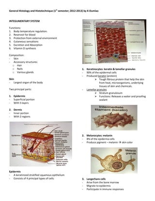
  • 1. General Histology and Histotechnique (1st semester; 2012-2013) by K-Dumlao INTEGUMENTARY SYSTEM Functions: 1. Body temperature regulation. 2. Reservoir for blood 3. Protection from external environment 4. Cutaneous sensations 5. Excretion and Absorption 6. Vitamin D synthesis Composition: - Skin - Accessory structures: o Hair o Nails 1. Keratinocytes: keratin & lamellar granules o Various glands - 90% of the epidermal cells - Produced keratin (protein) Skin  Tough fibrous protein that help the skin - Largest organ of the body from heat, microorganisms, underlying tissues of skin and chemicals. Two principal parts: - Lamellar granules  Stratum granulosum 1. Epidermis  Functions: Releases a water and proofing - Superficial portion sealant - With 5 layers 2. Dermis - Inner portion - With 2 regions 2. Melanocytes: melanin - 8% of the epiderma cells - Produce pigment – melanin  skin color Epidermis - A keratinized stratified squamous epithelium - Consists of 4 principal types of cells: 3. Langerhans cells - Arise from the bone marrow - Migrate to epidermis - Participate in immune responses
  • 2. General Histology and Histotechnique (1st semester; 2012-2013) by K-Dumlao - Shed off when you take a bath  Continuous Friction - Callous  an abnormal thickening of epidermis due to friction 2. Stratum Lucidum - “lucid” – “clear” - Present in the skin of finger tips, palms, and soles. - 3 – 5 clear layers of flat dead keratinocytes with intermediate filaments and thickened plasma 4. Merkel cells membrane. - It is the least numerous of epidermal cells - Located at the deepest part of stratum 3. Stratum Ganulosum basale/geminativum. - 3 – 5 layers of flattened keratinocytes - In contact with tactile disc or merkel disc - Consists of keratohyaline and lamellar granules Function: o A distinctive feature of the layer - Both participate in sensation of touch o Darkly staining granules of the stratum Function: Organized intermediate filaments into thicker bundles. 4. Stratum Spinosum - The superficial of basale - 8 – 10 layers of polyhedral keratinocytes - Fit closely together - Thorn – like spines bundles of intermediate filaments of the cytoskeleton joins the cell tightly to one another to provide strength and flexibility of Layers of the Epidermis the skin. 5. Stratum Basale/Geminativum - Active in cell division - Compose of one layer of simple cuboidal/columnar keratinocytes.  Some considered them as stem cells  Continue cell divisions to produce new keratinocytes - Merker cell - Melanocytes Dermis - Second deeper part of the skin - Composed of connective tissue containing collagen and elastic fibers 1. Stratum Corneum o Functions: for extensibility (stretch; obesity) - 25 – 30 layers of dead flat keratinocytes. and flexibility (Return to its original form) - Contains the dead flat intermediate filaments - Include cells such as fibroblasts, macrophages, and - Consists of keratohyaline some adipocytes o Function: Water repellant barrier - Blood vessels, nerves, glands, and hair follicles are - Lamellar granules embedded in dermal tissue - Lipid from lamellar granules (barrier in between)
  • 3. General Histology and Histotechnique (1st semester; 2012-2013) by K-Dumlao Two regions of Dermis: 3. Meissner’s corpuscle - Encapsulated 1. Papillary region - Palmar and planthar - The superficial portion of the dermis - Fingertips - Consists of areolar connective tissue with tissue with elastic fibers 4. Ruffini corpuscles - Contains dermal papillae - Type II cutaneous mechanoreceptor  Finger-like projections that indent the epidermis to the dermis. 5. Pacinian corpuscle Contains the following: - Located deeper part of the dermis - Encapsulated with multicellular ovoid structures a. Meissner’s corpuscle Function: Respond to deeper pressure. - For touch sensation b. Free nerve endings - Contains the dendrites that initiates signals that are felt such as warm, coolness, pain, tickling and itching. *Papillary layer – small fine collagen 2. Reticular region - Deeper portion of dermis - Consists of dense irregular connective tissue containing bundles of collagen and some elastic fibers. *Reticular layer – more coarsely-textured collagen fibers. Functions of the connective tissue of the dermis: 1. Tough collagen fibers and resilient elastic fibers provide mechanical strength for skin. Lines of tension in the dermis called Langer’s lines, affect healing after surgical incision 2. The ground substances of the dermis serve as the substrate for diffusion of nutrients and wastes to and from various other tissue components. 3. Mast cells, lymphocytes and macrophages in the connective tissue carry our surveillance for the immune system. 4. Finally, the dermis together with its associated blood vessels and nerves is capable of active responses to injury, yielding the defensive reaction Sensory receptors / free nerve endings of the reticular of inflammation, followed by the healing processes layer: of growth and repair 1. Nociceptotrs – pain receptor Subcutaneous layer (Hypodermis) 2. Tactile/Merkel disc - The hypodermis (hypo – “under” + dermis – “skin”) - Type one cutaneous mechanoreceptor or subcutaneous (sub – “below” + cutaneous) layer - For sensation touch
  • 4. General Histology and Histotechnique (1st semester; 2012-2013) by K-Dumlao lies below the skin and is made up of loose - Scatters light from the dermis without altering its connective and adipose tissues. color - The hypodermis binds the skin to underlying organs - Whiteness of white skin while allowing the skin to move somewhat - Reflection of collagen independently of underlying structures. - Adipose tissue in the hypodermis provides padding 3. Blood and shock-absorption that helps to protect - Hemoglobin and RBC underlying tissues from damage; it is also important - Scatters red light in insulating against loss of body heat. - Responsible for the pinkness in pigmented skin - Because the subcutaneous layer contains numerous ***The amount of pigment, the thickness of dermis, blood vessels but no vital organs, it is a near-ideal and the degree of perfusion in dermal capillaries vary. place to inject drugs. This is why so many drugs are administered through subcutaneous injection by a Skin texture hypodermic (hypo-“under”+ dermic – “skin”) - Affect the thickness and smoothness of the needle. epidermis, by the quality of fibers in the dermis, and by the amount of fluid in dermal connective tissue. Types of skin Note: 1. Thin skin Both edema (accumulation of excess fluid in connective - Covers all parts of the body except palms, palmar tissue) and dehydration can dramatically alter the surfaces of the digits, and soles appearance of skin - Epidermis is thin - Stratum lucidum lacking; stratum spinosum and Accessory structures of the skin stratum Corneum are thin 1. Hairs - Has fewer dermal papilla, lacks epidermal ridges 2. Glands - Has hair follicles, arrector pili muscle and oil glands 3. Nails but fewer sweat glands than thick - Sparser distribution of sensory receptors. Hairs - Composed of columns of dead, keratinized cells 2. Thick skin bounded together by extracellular proteins - Covers palms, palmar surfaces of digits and soles Parts: - Thick epidermis 1. Shaft – superficial portion - With stratum lucidum and thicker stratum 2. Roots – penetrates into the dermis spinosum and stratum corneum. Concentric layers of shaft and root: - More numerous dermal papillae thus has epidermal ridges a. Medulla - Lack hair follicles, arrector pili muscle, sebaceous - Inner portion gland. - 2 or 3 rows of polyhedral-shaped cells containing - Has more sweat glands that thin skin pigment granules and hair spaces. - Sensory receptors are more densely clustered. b. Cortex Skin color - Middle portion - It forms the major part of the shaft 1. Melanin - Contains pigments granules of the hair - Produced by melanocytes - White/gray hair – air bubbles - Yellowish-brown color of the epidermis - Lesser melanin – Light will penetrate easily into the c. Cuticle skin --- prone to cancer. - Outermost layer - Single layer of flat cells that are heavily keratinized 2. Collagen
  • 5. General Histology and Histotechnique (1st semester; 2012-2013) by K-Dumlao 3. Follicle – tubular invagination lined by stratified squamous epithelium similar to epidermis; 6. Hair root plexuses surrounds the root of the hair; made up of: - Dendrites of neurons sensitive to touch a. External root sheath - Generate nerve impulses in hair shaft is moved. - Downward continuation of epidermis - Contains the stratum basale Glands b. Internal root sheath - It forms the cellular tubular sheath between the external root and the hair. Hair follicles are associated with: a. Sebaceous glands – secrete oil into the hair follicle as well as nerve ending and smooth muscle to form the pilosebaceous apparatus. b. Nerve endings – detects deflection of the hair shaft and also controls piloerection. c. Smooth muscle – arrector pili ; affect piloerection 4. Bulb – the base of hair follicle; onion-shaped structure; houses a. Papilla of the hair - Nipple-shaped indentation - Contains areolar connective tissue - A lot of Blood vessels that nourish the growing hair follicle b. Matrix - Germinal layer of the cell w/c rise from stratum basale. Function: - Responsible for growth of existing hairs - Produce new hairs when old hairs are shed.  Sebaceous glands 5. Arrector pili muscle – smooth muscle cells - Associated with hair follicles - Stimulated by autonomic nerve endings to contract - Holocrine glands pulling the hair shaft perpendicular to the skin  The whole cell is secreted surface during cold or fright.
  • 6. General Histology and Histotechnique (1st semester; 2012-2013) by K-Dumlao Function: - It coats the surface of the hair and help keep them from drying and becoming brittle - It prevents excessive evaporation of water from the skin. - It keeps the skin soft and pliable - It inhibits growth of certain bacteria  Sweat glands - 3-4 millions producing their secretion - Exocytosis – release secretion Two types: a. Ecrine 1. Nail body – visible portion of the nail - Simple coiled tubular gland 2. Free edge – part of that may end pass the distal - Common sweat gland ends of digits Function: 3. Nail root – proximal edge of the nail plate that - Regulates body temperature is buried in the skin. - Waste removal 4. Lunula – whitish crescent shaped covered by portion of the nail bed. b. Apocrine 5. Hyponichium – nail bed; skin of nail bed; - Simple coiled tubular gland provide support - Stimulated during emotional stress and sexual 6. Eponychium/cuticle – a horny epidermal excitement extension of the tip of the nail folds. - Secretion of cold sweat Nails - Plates of tightly packed, hard, keratinized epidermal cells. - Cells form a clear, solid covering over the dorsal surfaces of the distal portions of the digits - Parts: