2. pollution is presently attracting more attention from
environmentalists worldwide.
Lead (II) is a heavy metal poison which forms
complexes with oxo-groups in enzymes to affect nearly
all steps in the process of hemoglobin synthesis and
porphyrin metabolism. Toxic levels of Pb (II) in man have
been associated with encephalopathy appropriations and
mental delay (Ademorati, 1996). Conventional physico-
chemical methods such as electrochemical treatment, ion
exchange, precipitation, reverse osmosis evaporation
and sorption (Kadirvelu et al., 2001, 2002) have been
used for removing heavy metals but are economically
expensive and have disadvantages. Bioremediation is a
natural process which depends on bacteria, fungi and
plants to altering pollutants as these organisms perform
their normal life functions. These organisms have the
ability of exploiting chemical contaminants as an energy
source in their metabolic processes. Therefore,
bioremediation affords alternative tool to destroy or
reduce the risky contaminants through biological activity
with an effective cost (Salem et al., 2012).
Microbial populations in metal polluted environments
become metals resistant (Prasenjit and Sumathi, 2005)
so the response of microorganisms towards toxic heavy
metals is of importance in view of the interest in the
reclamation of polluted sites (Shankar et al., 2007).
Microorganisms uptake metals either actively
(bioaccumulation) and/or passively (biosorption)
(Shumate and Strandberg, 1985; Anders and Hubert,
1992; Hussein et al., 2004). Bioaccumulation is the active
method of metal accumulation by living cells. The
capacity of living cells to remove metal ions from
environment is influenced by environmental growth
conditions, as temperature, pH and biomass
concentrations (Abd-El-Raheem et al., 2013).
TEM is a useful technique that can help to localize and
to identify metals deposited within or around microbial
cells. Identification of the site of accumulation is important
as it can give clues to the biochemical mechanisms
driving metal accumulation. Biological materials which
are largely composed of light elements such as C, N, H,
O, P, and S, do not deflect the electron beam to the same
degree. Thus, it is possible to visualize metals against the
faint image of a bacterial cell (Lloyd and Macaskie, 2002).
The present study, aims at isolating S. oneindensis
from Basra soil, south of Iraq, and evaluating metals
bioaccumulation ability, and also studying the effect of
metals initial concentration, contact times, and determine
the cellular localization of accumulated metals within this
bacterium by using Transmission electron microscope.
MATERIALS AND METHODS
Isolation of bacteria
Three soil samples (30 g each) were collected from AL-Zubair
district west of Basra city- Iraq during January 2013. The samples
were collected using a sterile plastic bag and transferred within 2 h
Jaafar et al. 371
to laboratory for analysis. One gram of air dried soil sample was
serially diluted using sterilized distilled water and spread over
nutrient agar. The plates were incubated at 30°C for 24 h.
Bacterial characterization
Properties of the bacteria included gram stain, citrate utilization,
indole production, methyl red, nitrate reduction, Voges Proskauer,
catalase, dextrose, mannitol and sucrose utilization, starch
hydrolysis, and gelatin liquefaction tests were determined according
to Sneath et al. (1986).
S16 rRNA gene based identification
The isolates were identified by sequencing of the 16S rRNA gene.
To determine the identification of bacterial isolates, the amplified
16S rRNA gene PCR products obtained from total genomic DNA
using primer set 27F (5′ AGAGTTTGATCCTGGCTCAG-3′) and
72.1492R (5′ GGTTACCTTGTTACGACTT-3′), (Lane et al., 1985)
were sequenced commercially. DNA sequences obtained were
compared to sequences available online in a GenBank database
(http://www.ncbi.nlm.nih.gov). Homology search was performed
using Bioinformatics tools available online BLASTn
www.ncbi.nlm.nih.gov/BLA (Altschul et al., 1997).
Determination of minimal inhibitory concentrations (MIC) for
Pb and Cd
The minimum inhibitory concentration (MIC) of Cd and Pb of
bacteria were determined by disc diffusion method. The
concentrations of Cd and Pb were between 40 to 2500 mgl-1
. Filter
paper discs were saturated with heavy metals for 30 min, and then
placed on nutrient agar plates and incubated for 24 h at 30°C. Pb
(NO3)2 and CdCl2 were used to prepare mother solution of these
metals in sterile distilled water and were used in various
concentrations. The lowest concentrations of Cd and Pb that
completely prevented growth of each bacterium were considered as
the MIC (Sethuraman and Kumar, 2011).
Bioaccumulation of heavy metals by bacteria
Bacteria were grown in LB broth containing 5, 10, 25 and 50 mgl-1
of lead and for cadmium 10, 20, 50 and 100 mgl-1
then incubated
for 2, 4, 6, 24 and 48 h at 30°C in a shaker incubator at 150 rpm.
Three replicates for each concentration have been done, and one
as a control. The bacterial cells were harvested by centrifugation at
6000 rpm for 15 min, and suspended in 1 ml of distilled water,
oven-dried at 80°C for 1 h and weighted. Metal concentrations were
measured by atomic absorption spectrophotometer. Control was
represented by the same microbial culture without heavy metals.
Each metals concentration is measured with two replicates
(Sprocati et al., 2006).
Transmission electron microscope
By centrifuging samples broth culture for 10 min at 3000 rpm, and
decanting the supernatant, fixing pellet with 4% gutaraldehyde for 4
h at 4°C and centrifuged again, decanting fixative and adding an
appropriate quantity animal serum to submerge sample, and
allowing serum to clot. It was washed three times with 0.1 M
Cacodylate buffer for 10 min. and Posted fix in 1% Osmium
tetrroxide for 2 h at 4°C. Also, it was washed again three times with
0.1 M Cacodylate buffer for 10 min. Dehydrating in series of
3. 372 Afr. J. Microbiol. Res.
Table 1. Biochemical characteristics of S. oneidensis isolate from
soils.
Tests Characteristics observed
Oxidase test +
Catalase test +
Indole formation -
Nitrate reduction -
Production of H2S +
Gelatin liquefaction +
Fermentation of
Sucrose +
Fructose +
D-glucose +
"+"and "-" indicate positive and negative reactions, respectively.
acetone of 35, 50, 75, 95, and 100% for 10, 10,10, 10 and 15 min
respectively. Finally, we make infiltration of the specimen with
acetone and resin:
Acetone: Resin Time
1 : 1 1 h
1 : 3 2 h
100% Overnight
100% 2 h
Embedding: Specimens were placed into beam capsule filled with
resin. Polymerization: polymerized in oven at 60°C for 24 h. Make
ultrasectioning, by choosing an area of interest, then cut for
ultrathin section, selecting the silver section, picking up a section
with a grid, then drying with filter paper. Finally staining with Uranyl
acetate for 15 min, and washed double distills water. Lead stained
for 10 min, and washed twice in distilled water. This work was done
at the Electron Microscope Laboratory Institute of Bioscience,
University Putra Malaysia.
RESULTS AND DISCUSSION
Characterization and molecular identification of
isolated bacteria
The selected bacterium was characterized and identified
by using conventional morphological, physiological and
biochemical tests (Table 1). It was presumptively
identified as Shewanella sp (Holt et al., 2005). The
sequence of 16S rRNA gene of this bacterium was
submitted to Blastn database 16S ribosomal RNA
sequences (Bacteria and Archaea) Megablast
http://www.ncbinlm.nih.gov/blast. It indicated a close
genetic relatedness of this bacterium with the rDNA
sequence of Shewanella oneidensis (Holt et al., 2005).
Minimum inhibitory concentration (MIC)
The MIC is the lowest concentration of the heavy metals
that completely inhibited bacterial growth (Froidevaux et
al., 2001) S. oneindensis showed significant resistance to
high concentrations of Pb and Cd (700 and 1000 mgl
-1
)
respectively. This may be considered new finding, that
the other studies showed different results. Chihomvu et
al. (2014) recorded MIC for Pb by Shewanella (4 Mm),
while MIC was 0 for Cd. Francis and Dodge (1988) and
Toes et al. (2008) demonstrated that, the tolerances
inhibited growth of different Shewanella strains
completely at 150 µM Co, 150 - 400 µM Zn, 75 - 150 µM
Cd, and 150 µM Cu when cultivated aerobically in 10%
LB broth. The effect of the medium on metal toxicity was
demonstrated in a study by Toes et al. (2008) where
higher tolerances of Cu by Shewanella between 75 and
750 µM in more nutrient rich media and the presence of
manganese oxides also reduce the toxicity of Cu.
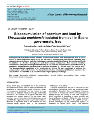
Bioaccumulation
S. oneidensis as sulfate reducing bacteria has the
potential to enhance metal retention via extracellular
binding, cellular uptake and accumulation of metals,
oxidation/reduction processes, and surface mediated
mineral precipitation (Burkhardt, 2010). From results of
the present study, S. oneindensis was able to accumulate
Cd than Pb (26.77 and 3.98 mgg
-1
) at 48 h and at
concentrations 50 and 100 mgl
-1
respectively (Figures 1
and 2). The differences in this accumulation ability for
these two metals may be related to different toxicity of
these metals to this bacterium. From the results, the
accumulation of both metals increases with increasing
the time. Varghese et al. (2012) showed that, with
increasing time, the biomass of the bacterial strains
increased. Likewise, with an increase in biomass, the
heavy metals bioaccumulation also increased. The
results of the present study showed that the high amount
of accumulation occurs with high metals concentration
(50 and 100 mgl
-1
). These results agree with the results
reported by Odokuma and Akponah (2010), where they
concluded an increasing uptake pattern observed in the
respective test isolates as the initial concentration of the
various heavy metal salts were increased. These
observations suggested that metal uptake may involve
diffusion phenomenon, whereby metal ions move from
regions of high to low concentrations.
Transmission electron microscope
Cells were evaluated by TEM to observe the locations of
precipitate of metals in relation to the S. oneidensis cells.
In order to differentiate whether extracellular or
intracellular reduction occurred, the cells were stained
with uranyl acetate. Figure 3 has shown the cells before
being exposed to the metals (a). Dark precipitate can be
seen around the inside of the cell membrane, indicating
4. Jaafar et al. 373
Figure 1. Bioaccumulation of Pb by S. oneidensis during different incubation periods and
different concentrations.
Figure 2. Bioaccumulation of Cd by S. oneidensis during different incubation periods and
different concentrations.
intracellular Cd and Pb reduction has occurred (b and c).
Also, from Figure (3b and c) there were changes in size
and shape of cells and some cells have been lysed.
These results could add to the toxicity of the substance,
and ultimately results in cell death.
The cell surface morphology considerably changed
after metals exposure. The cellular localization of the
metals bound by the cells of the bacterium was located
mainly within the cell membrane. However, some
intracellular metal accumulates were also identified in the
cytoplasm of the bacterial cells. Merroun et al. (2005)
reported that, the cellular localization of the uranium
bound by the cells of three types of Acidithiobacillus
ferrooxidans was studied using TEM. Also, El-Helow et
al. (2000) reported that, cell surfaces of cultures treated
with cadmium chloride tended to be rough, suggesting
that the cell increased its surface to improve the
interaction of toxic substances with the cell surface.
Also, Singh et al. (2013), reported cell surface
morphological changes in Cryptococcus sp. after
exposure to heavy metals, and which could be observed
by the presence of shrunken and distorted cell wall in the
presence of Cd and depressions in the presence of Pb
and Zn.
Secretion of extracellular polymeric substance by
Desulfovibrio desulfuricans during biosorption of Zn and
Cu was reported to modify its cell surface morphology
(Chen et al., 2000). Similarly, El-Meleigy et al. (2013)
reported that, high dark dense cytoplasm due to Co
2+
precipitation is partially emptied with a very thick cell
wall; changing in the morphology of vegetative cells of
Bacillus firmus and Bacillus subtilis.
0
1
2
3
4
2 4 6 24 48
Bioaccumulation(ppm)
Time(hr)
Bioaccumulation of Pb
con. 5
con. 10
con. 25
con 50
control
Time (h)
0
5
10
15
20
25
30
Bioaccumulation(ppm)
2 4 6 24 48
Time( hr)
Bioaccumulation Cd
con 10
con20
con 50
con 100
control
Time (h)
5. 374 Afr. J. Microbiol. Res.
Figure 3. Transmission electron micrographs of S. oneidensis a: control, b: treated with 50 mgl-1
of Cd for
24 h, c: Treated with 50 mgl-1
of Pb for 24 h (Scale of bar 0.5 and 2 µm).
Conflict of interests
The authors have not declared any conflict of interests.
REFERENCES
Ademorati CMA (1996). Environmental Chemistry and Toxicology.
Pollution by Heavy metals. Fludex Press Ibadan. pp. 171-172.
Altschul SF, Madden TL, Schaffer AA, Zhang J, Zhang Z, Miller W,
Lipman DJ (1997). Gapped BLAST and PSI-BLAST: A new
generation of protein database search programs. Nucl. Acids Res.
25:3389-3402.
Anders MYJH, Hubert CJ (1992). Bacterial biosorption and retention of
thorium and uranyl cations by Mycobacterium smegmatis. J.
Radioanal. Nucl. Chem. 166:431-440.
Burkhardt EM (2010). Interaction of Fe (III)- reducing bacteria with
heavy metals in contaminated soils, Ph.D. Thesis, University of
Jena, Germany. P 183.
Chen BY, Utgikar VP, Harmon SM, Tabak HH, Bishop DF, Govind R
(2000). Studies on biosorption of zinc (II) and copper (II) on
Desulfovibrio desulfuricans. Int. Biodeterior. Biodegradation 46:11-18.
Chihomvu P, Stegmann P, Pillay M (2014). Identification and
Characterization of Heavy Metal Resistant Bacteria from the Klip
River. Int. J. Biol. Vet. Agric. Food Eng. 8(11):1056-1066.
El-Helow ER, Sabry SA, Amer RM (2000). Cadmium biosorption by a
cadmium resistant strain of Bacillus thuringensis. Regulation and
optimization of cell surface affinity for metal cations. Biometals
13:273-280.
El-Meleigy MA, Abed NN, Sari IP (2013). Fate of cobalt and nickel in B.
firmus and B. subtilis. Nat. Sci. 9(8):175-189.
Francis AJ, Dodge CJ (1988). Anaerobic Microbial Dissolution of
Transition and Heavy Metal Oxides. Appl. Environ. Microbiol.
54(4):14.
Holt HM, Hansen BG, Bruun B (2005). Shewanella algae and
Shewanella putrefaciens: Clinical and microbiological characteristics
Clin. Microbiol. Infect. 11:347-352.
Hussein H, Ibrahim SF, Kandeel K, Moawad H (2004). Biosorption of
heavy metals from waste water using Pseudomonas sp. Electron. J.
Biotechnol. 7(1):30-37.
IARC (1994). Beryllium, cadmium, mercury, and exposures in the glass
manufacturing industry [M]. In: Monographs on the evaluation of
carcinogenic risks to humans. Lyon: WHO Press 58:444.
Kadirvelu K, Thamaraiselvi K, Namasivayam C (2001). Adsorption of
nickel (II) from aqueous solution onto activated carbon prepared from
coir pith. Sep. Purif. Technol. 24: 477-505.
Kadirvelu K, Senthilkumar P, Thamaraiselvi K, Subburam V (2002).
a
b
c
6. Activated carbon prepared from biomass as adsorbent: elimination of
Ni (II) from aqueous solution. Bioresour. Technol. 81:87-90.
Lloyd JR, Macaskie LE (2002). Environmental Microbe-Metal
Interactions. American Society for Microbiology Press, Washington,
DC. pp 3-30.
Merroun ML, Raff J, Rossberg A, Hennig C, Reich T, Selenska-Pobell S
(2005). Complexation of uranium by cells and S-layer sheets of
Bacillus sphaericus JG-A12. Appl. Environ. Microbiol. 71(9):5532-
5543.
Odokuma LO, Akponah E (2010). Effect of concentration and contact
time on heavy metal uptake by three bacterial isolates. J. Environ.
Chem. Ecotoxicol. 2(6):84-97.
Prasenjit B, Sumathi S (2005). Uptake of chromium by Aspergillus
foetidus. J. Mater. Cycles Waste Manage. 7:88-92.
Rathnayake IVN, Megharaj M, Bolan N, Naidu R (2010). Tolerance of
Heavy Metals by Gram Positive Soil Bacteria. Environ. Eng.
2(4):1185-1190.
Salem IB, Sghaier H, Trifi H, Héni S, Khwaldia K, Saidi M, Landoulsi A
(2012). Isolation and characterization of a novel Micrococcus strain
for bioremediation of strontium in radioactive residues. Afr. J.
Microbiol. Res. 64:851-858.
Sethuraman P, Kumar MD (2011). Bacillus subtilis on Pb2+
ion removal
from aqueous solution by Biosorption. Res. J. Pharm. Biol. Chem.
Sci. 2(4):247.
Shankar C, Sridevi D, Joonhong P, Dexilin M, Thamaraiselvi K (2007).
Biosorption of chromium and nickel by heavy metal resistant fungal
and bacterial isolates. J. Hazard. Mater. 146:270-277.
Shumate ES, Strandberg WG (1985). Accumulation of metals by
microbial cells. Compr. Biotechnol. 13:235-247.
Singh P, Raghukumar C, Parvatkar RR, Mascarenhas-Pereira MBL
(2013). Heavy metal tolerance in the psychrotolerant Cryptococcus
sp. isolated from deep-sea sediments of the Central Indian Basin.
Yeast 30(3):93-101.
Sneath PHA, Mair NS, Sharpe ME, Holt JG (1986). Bergey's manual of
systematic bacteriology. Vol. 2. Williams & Wilkins, Baltimore, U.S.A.
Jaafar et al. 375
Sprocati AR, Alisi C, Segre L, Tasso F, Galletti M, Cremisini C (2006).
Investigating heavy metal resistance, bioaccumulation and metabolic
profile of a metallophile microbial consortium native to an abandoned
mine. Sci. Total Environ. 366:649-658.
Tang XY, Zhu YG, Cui YS, Duan J, Tang L (2006). The effect of ageing
on the bioaccessibility and fractionation of cadmium in some typical
soils of China. Environ. Int. 32:682-689.
Toes ACM, Geelhoed JS, Kuenen JG, Muyzer G (2008).
Characterization of heavy metal resistance of metal-reducing
Shewanella isolates from marine sediments. Geomicrobiol. J. 25:304-
314.
Varghese R, Krishna MP, Babu A, Mohamed HA (2012). Biological
removal of lead by Bacillus sp. obtained from metal contaminated
industrial area. J. Microbiol. Biotechnol. Food Sci. 2(2):756-770.