SlideShare uma empresa Scribd logo
1 de 18
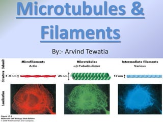
Microtubules &
Filaments
By:- Arvind Tewatia
Cytoskeleton
•The major structural elements of the
cytosketeton are:-
•microtubules,
•microfilaments
•The range of techniques to understand the
cytosketeton include fluorescence microscopy,
digital video microscopy and electron
microscopy.
Microtubules
•Microtubules were first discovered by De
Roberties and Franchi (1953)
•In the axons of medullated nerve fibres and were
named neurotubules. Porter(1955)
•Term “Microtubules” was given by
Slautterback (1963)
•Microtubules are electron-microscopic structure.
•Found only in Eukaryotic cellular structures.
•But absent in mature mammalian erythrocytes,slim moulds
and prokaryotes
•Microtubules (MTs) are largest element of the cytoskeleton.
•Cytoplasmic microtubules:- 1) maintain axons; 2) maintain
shape; 3) orient cellulose microfibrils (in plants); 4) mitotic
and meiotic spindles for chromosome movements; and 5)
vesicle movement.
•Axonemal microtubules:- highly organized, stable
microtubules in specific movement associated subcellular
structures (cilia, flagella and attachment basal bodies). The
central shaft (axoneme) consists of a highly ordered bundle of
axonemal MTs (see sperm tail axoneme).
Microtubule Structure
Microtubule Structure:- is made up
of heterodimers of alpha-tubulin and
beta-tubulin (form an alpha-beta-
heterodimer).
•An a,b-tubulin heterodimer is the
basic structural unit of microtubules.
•The heterodimer does not come
apart, once formed.
ab
GTP GTP
tubulin
heterodimer
•The alpha-tubulin and beta-tubulin molecules are 4-5
nm in diamater and 55 kDa, have almost identical
shapes but only share 40% amino acid sequence
identity.
•A microtubule is a hollow cylinder of 13
protofilaments around a lumen with an outer
diameter (25 nm), inner diameter (15 nm) and dimer
width (8 nm).
•Singlet microtubules are 13 protofilaments;
doublets are 13 plus 10 (or 11)
[in cilica or flagella] and
•Triplets are 13 plus 10 (or 11)
plus 10 (or 11)
•[in basal bodies or centrioles].
•A microtubule is a hollow cylinder,
about 24 nm in diameter.
•Along the microtubule axis, tubulin
heterodimers join end-to-end to form
protofilaments, with alternating a & b
subunits.
•Staggered assembly of 13 protofilaments
yields a helical arrangement of tubulin
heterodimers in the cylinder wall.
seam
microtubule3-starthelix
b-GDP
a-GTP
b-GTP
a-GTP
+

Note :-Electron microscopy of microtubules
decorated with motor protein heads indicate a "3-
start helix.“
Each turn of the helix spans 3 tubulin monomers
(e.g., a, b, a).
•Microtubules form by addition of tubulin dimers at the ends.
New microtubules form nucleation centres (oligomers) and
grow by addition of subunits on either end: elongation.
Critical concentration is when disassemby and assemby is
exactly balanaced: treadmilling.
The plus end: rapidly growing end; the minus end: the slower
growing end.
•Microtubule-organizing centre (MTOC)
•serves as a site for initiation and
as an anchor.
•A centrosome (animals cells) has two
centrioles surrounded by pericentriolar
material.
•The microtubule polarity can vary with cell function.
•Microtubule-associated proteins (MAPs) act as stabilizing
and bundling proteins.
Centrioles at right angles,
as in the centrosome.
cartwheel structure
at one end
Microfilaments
Microfilaments were given by PALEVIZ et.
al.(1974)
•Microfilaments (MFs) are smallest element of the
cytoskeleton.
•Thread-like protein fibers
•Found only in the cytoplasm of all eukaryotic cells
•This are present in microvilli, muscle fibres. In muscle
fibres these are called myofilaments
•They are 3-7nm in Diameter
•This are long, narrow, cylindrical,
contractile & proteinous structure
•These are absent in prokaryotes
Functions
1) As cytoskeleton:- 1) Actin filaments are interlinked by
actin-binding proteins like Fimbrin & Villin while these are
linked to the micrevillin membrane by another actin-binding
protein calmodulin
2) The arrangements of proteins make the microfilaments
make a rigid structure which provide mechanical support to
maintain the form of microvilli
2)Movement of cell membrane:- MF act as a cell
muscles & help in the cell membrane movement which also
help in endocytosis through the cell membrane
3)In movement & locomotion :- Microfilaments of the
myofibers of the muscles fibres help in movement &
locomotion during Actomyosin complexes.
These are formed bw the heads of thick myosin and actin
sites of thin actin filaments in the presence of energy released
during hydrolysis of ATP by myosin & ATPase enzyme in
presence of calcium ion released form yhe depolarized
sarcoplasmic recticulam
The head of myosin filaments undergo swivelling(rotaion)
which pull the actin filament inward which inward over the
myosin filament. This is called sliding-filament theory
Difference BW
Microtubules
• These are non-contractile
• A microtubule contain 13
protofilaments
• They are hallow tubules
• Microtubules are formed of
a&btubulin proteins.
• The diameter of
microtubule is 25 nm
Microfilaments
• These are contractile.
• These do not posses
longitudnal sub-units.
• Microfilaments are solid
structures.
• Microfilaments are mainly
made up of actin.
• The diameter of a
miocrofilaments is 5-6 nm.
•

Mais conteúdo relacionado

Mais procurados

Cytoskeleton presentation (introduction structure & function)
Cytoskeleton presentation (introduction structure & function)Cytoskeleton presentation (introduction structure & function)
Cytoskeleton presentation (introduction structure & function)Dryogeshcsv
 
Golgi apparatus ppt (introduction structure and Function)
Golgi apparatus ppt (introduction structure and Function)Golgi apparatus ppt (introduction structure and Function)
Golgi apparatus ppt (introduction structure and Function)Dryogeshcsv
 
cell adhesion molecules
cell adhesion moleculescell adhesion molecules
cell adhesion moleculesmah neem mah
 
THE CYTOSKELETON
THE CYTOSKELETONTHE CYTOSKELETON
THE CYTOSKELETONMariya Raju
 
Structure and functions of endoplasmic reticulum
Structure and functions of endoplasmic reticulumStructure and functions of endoplasmic reticulum
Structure and functions of endoplasmic reticulumICHHA PURAK
 
Structure and functions of Mitochondria
Structure and functions of MitochondriaStructure and functions of Mitochondria
Structure and functions of MitochondriaICHHA PURAK
 
Ribosomes structure & function
Ribosomes structure & functionRibosomes structure & function
Ribosomes structure & functionAbu Raihan
 
Cilia and flagella ppt by ramana babu
Cilia and flagella ppt by ramana babuCilia and flagella ppt by ramana babu
Cilia and flagella ppt by ramana baburamana babu marri
 
Cell motility cilia and flagella
Cell motility  cilia and flagellaCell motility  cilia and flagella
Cell motility cilia and flagellaALLIENU
 
Golgi complex structure and functions
Golgi complex structure and functionsGolgi complex structure and functions
Golgi complex structure and functionsbikashsingh2510
 
Intermediate filaments
Intermediate filamentsIntermediate filaments
Intermediate filamentsSarita Nanda
 
Endoplasmic reticulum- cell Organelle
Endoplasmic reticulum- cell OrganelleEndoplasmic reticulum- cell Organelle
Endoplasmic reticulum- cell OrganelleAshishNain
 

Mais procurados (20)

Cytoskeleton presentation (introduction structure & function)
Cytoskeleton presentation (introduction structure & function)Cytoskeleton presentation (introduction structure & function)
Cytoskeleton presentation (introduction structure & function)
 
Golgi apparatus ppt (introduction structure and Function)
Golgi apparatus ppt (introduction structure and Function)Golgi apparatus ppt (introduction structure and Function)
Golgi apparatus ppt (introduction structure and Function)
 
cell adhesion molecules
cell adhesion moleculescell adhesion molecules
cell adhesion molecules
 
Cytoskeleton[1]
Cytoskeleton[1]Cytoskeleton[1]
Cytoskeleton[1]
 
Microtubules
MicrotubulesMicrotubules
Microtubules
 
The Nucleus
The NucleusThe Nucleus
The Nucleus
 
Cytoskeleton
CytoskeletonCytoskeleton
Cytoskeleton
 
THE CYTOSKELETON
THE CYTOSKELETONTHE CYTOSKELETON
THE CYTOSKELETON
 
Structure and functions of endoplasmic reticulum
Structure and functions of endoplasmic reticulumStructure and functions of endoplasmic reticulum
Structure and functions of endoplasmic reticulum
 
Structure and functions of Mitochondria
Structure and functions of MitochondriaStructure and functions of Mitochondria
Structure and functions of Mitochondria
 
Ribosome ultra structure
Ribosome ultra structureRibosome ultra structure
Ribosome ultra structure
 
Micro filaments
Micro filamentsMicro filaments
Micro filaments
 
Ribosomes structure & function
Ribosomes structure & functionRibosomes structure & function
Ribosomes structure & function
 
Cilia and flagella ppt by ramana babu
Cilia and flagella ppt by ramana babuCilia and flagella ppt by ramana babu
Cilia and flagella ppt by ramana babu
 
Cytoskeletal structures
Cytoskeletal structuresCytoskeletal structures
Cytoskeletal structures
 
Cell motility cilia and flagella
Cell motility  cilia and flagellaCell motility  cilia and flagella
Cell motility cilia and flagella
 
Golgi complex structure and functions
Golgi complex structure and functionsGolgi complex structure and functions
Golgi complex structure and functions
 
Intermediate filaments
Intermediate filamentsIntermediate filaments
Intermediate filaments
 
Endoplasmic reticulum- cell Organelle
Endoplasmic reticulum- cell OrganelleEndoplasmic reticulum- cell Organelle
Endoplasmic reticulum- cell Organelle
 
Membrane proteins
Membrane proteinsMembrane proteins
Membrane proteins
 

Destaque

Microtubules in 5 minutes
Microtubules in 5 minutesMicrotubules in 5 minutes
Microtubules in 5 minutesRita Pizzi
 
Microfilaments and intermediate filaments
Microfilaments and intermediate filamentsMicrofilaments and intermediate filaments
Microfilaments and intermediate filamentsaljeirou
 
Microfilaments cytoskeleton
Microfilaments cytoskeletonMicrofilaments cytoskeleton
Microfilaments cytoskeletonSayra Medellín
 
Ch 4 structure and function of cells
Ch 4 structure and function of cellsCh 4 structure and function of cells
Ch 4 structure and function of cellsmrjones_science
 
Cell Structure And Function
Cell Structure And FunctionCell Structure And Function
Cell Structure And Functionjrt004
 
Microtubules and molecular motors
Microtubules and molecular motorsMicrotubules and molecular motors
Microtubules and molecular motorsaljeirou
 
04 lecture ppt cell structure and function
04 lecture ppt cell structure and function04 lecture ppt cell structure and function
04 lecture ppt cell structure and functionAriel. Christopher, BSW
 
Microtubules and molecular motors
Microtubules and molecular motorsMicrotubules and molecular motors
Microtubules and molecular motorsaljeirou
 
Animall Cell Powerpoint
Animall Cell PowerpointAnimall Cell Powerpoint
Animall Cell PowerpointMandy Bennett
 
Molecular Cell Biology Lodish 6th.ppt - Chapter 17 cell organization and mo...
Molecular Cell Biology Lodish 6th.ppt - Chapter 17   cell organization and mo...Molecular Cell Biology Lodish 6th.ppt - Chapter 17   cell organization and mo...
Molecular Cell Biology Lodish 6th.ppt - Chapter 17 cell organization and mo...Nattawut Huayyai
 
Cell : Structure and Function Part 01
Cell : Structure and Function Part 01Cell : Structure and Function Part 01
Cell : Structure and Function Part 01Abdul-arzeez Penai
 
The Top Skills That Can Get You Hired in 2017
The Top Skills That Can Get You Hired in 2017The Top Skills That Can Get You Hired in 2017
The Top Skills That Can Get You Hired in 2017LinkedIn
 

Destaque (19)

Microtubules in 5 minutes
Microtubules in 5 minutesMicrotubules in 5 minutes
Microtubules in 5 minutes
 
Microfilaments and intermediate filaments
Microfilaments and intermediate filamentsMicrofilaments and intermediate filaments
Microfilaments and intermediate filaments
 
Disease
DiseaseDisease
Disease
 
Microfilaments cytoskeleton
Microfilaments cytoskeletonMicrofilaments cytoskeleton
Microfilaments cytoskeleton
 
Ch 4 structure and function of cells
Ch 4 structure and function of cellsCh 4 structure and function of cells
Ch 4 structure and function of cells
 
Cell Structure And Function
Cell Structure And FunctionCell Structure And Function
Cell Structure And Function
 
12. Microtubules - cell biology
12. Microtubules - cell biology12. Microtubules - cell biology
12. Microtubules - cell biology
 
B14 eukaryotic cell structure powerpoint
 B14 eukaryotic cell structure  powerpoint B14 eukaryotic cell structure  powerpoint
B14 eukaryotic cell structure powerpoint
 
Microtubules and molecular motors
Microtubules and molecular motorsMicrotubules and molecular motors
Microtubules and molecular motors
 
Microtubules
MicrotubulesMicrotubules
Microtubules
 
04 lecture ppt cell structure and function
04 lecture ppt cell structure and function04 lecture ppt cell structure and function
04 lecture ppt cell structure and function
 
Microtubules and molecular motors
Microtubules and molecular motorsMicrotubules and molecular motors
Microtubules and molecular motors
 
Animall Cell Powerpoint
Animall Cell PowerpointAnimall Cell Powerpoint
Animall Cell Powerpoint
 
The Cytoskeleton
The CytoskeletonThe Cytoskeleton
The Cytoskeleton
 
Molecular Cell Biology Lodish 6th.ppt - Chapter 17 cell organization and mo...
Molecular Cell Biology Lodish 6th.ppt - Chapter 17   cell organization and mo...Molecular Cell Biology Lodish 6th.ppt - Chapter 17   cell organization and mo...
Molecular Cell Biology Lodish 6th.ppt - Chapter 17 cell organization and mo...
 
Cytoskeleton
CytoskeletonCytoskeleton
Cytoskeleton
 
Cytoskeleton
CytoskeletonCytoskeleton
Cytoskeleton
 
Cell : Structure and Function Part 01
Cell : Structure and Function Part 01Cell : Structure and Function Part 01
Cell : Structure and Function Part 01
 
The Top Skills That Can Get You Hired in 2017
The Top Skills That Can Get You Hired in 2017The Top Skills That Can Get You Hired in 2017
The Top Skills That Can Get You Hired in 2017
 

Semelhante a Microtubules & filaments.pptx 2

Microtubules by jimmy
Microtubules by jimmyMicrotubules by jimmy
Microtubules by jimmyAhmad Ali
 
structure and functions of cytoskeletons
structure and functions of cytoskeletonsstructure and functions of cytoskeletons
structure and functions of cytoskeletonszaferkaragozlu
 
Cytoskeleton structure. Microfilaments, microtubules and intermediate filaments.
Cytoskeleton structure. Microfilaments, microtubules and intermediate filaments.Cytoskeleton structure. Microfilaments, microtubules and intermediate filaments.
Cytoskeleton structure. Microfilaments, microtubules and intermediate filaments.GURUNath18
 
CYTOSKELETON presentation by (Aneeq) (1).pptx
CYTOSKELETON presentation  by (Aneeq) (1).pptxCYTOSKELETON presentation  by (Aneeq) (1).pptx
CYTOSKELETON presentation by (Aneeq) (1).pptxAneeqJaved
 
lecture_6_3 cells.ppt
lecture_6_3 cells.pptlecture_6_3 cells.ppt
lecture_6_3 cells.pptWoyo3
 
CYTOSKELETON STRUCTURE.ppt
CYTOSKELETON STRUCTURE.pptCYTOSKELETON STRUCTURE.ppt
CYTOSKELETON STRUCTURE.pptEstherShobhaR
 
Group 6 presentation(cytoskeleton).pptx
Group 6 presentation(cytoskeleton).pptxGroup 6 presentation(cytoskeleton).pptx
Group 6 presentation(cytoskeleton).pptxRoyBisenti
 

Semelhante a Microtubules & filaments.pptx 2 (20)

Microtubules by jimmy
Microtubules by jimmyMicrotubules by jimmy
Microtubules by jimmy
 
Cytoskeleton
Cytoskeleton  Cytoskeleton
Cytoskeleton
 
structure and functions of cytoskeletons
structure and functions of cytoskeletonsstructure and functions of cytoskeletons
structure and functions of cytoskeletons
 
Cytoskeleton structure. Microfilaments, microtubules and intermediate filaments.
Cytoskeleton structure. Microfilaments, microtubules and intermediate filaments.Cytoskeleton structure. Microfilaments, microtubules and intermediate filaments.
Cytoskeleton structure. Microfilaments, microtubules and intermediate filaments.
 
CYTOSKELETON presentation by (Aneeq) (1).pptx
CYTOSKELETON presentation  by (Aneeq) (1).pptxCYTOSKELETON presentation  by (Aneeq) (1).pptx
CYTOSKELETON presentation by (Aneeq) (1).pptx
 
Cytoskeleton
CytoskeletonCytoskeleton
Cytoskeleton
 
Cytoskeleton
CytoskeletonCytoskeleton
Cytoskeleton
 
lecture_6_3 cells.ppt
lecture_6_3 cells.pptlecture_6_3 cells.ppt
lecture_6_3 cells.ppt
 
Cytoskeleton
CytoskeletonCytoskeleton
Cytoskeleton
 
CYTOSKELETON
CYTOSKELETONCYTOSKELETON
CYTOSKELETON
 
Cytoskeleton
CytoskeletonCytoskeleton
Cytoskeleton
 
Cytoskeleton
CytoskeletonCytoskeleton
Cytoskeleton
 
Copy of cytoskeleton
Copy of cytoskeletonCopy of cytoskeleton
Copy of cytoskeleton
 
Cytoskeleton
Cytoskeleton Cytoskeleton
Cytoskeleton
 
microtubules.pptx
microtubules.pptxmicrotubules.pptx
microtubules.pptx
 
CENTRIOLE.pptx
CENTRIOLE.pptxCENTRIOLE.pptx
CENTRIOLE.pptx
 
CMB_L28_2008s.ppt
CMB_L28_2008s.pptCMB_L28_2008s.ppt
CMB_L28_2008s.ppt
 
CYTOSKELETON STRUCTURE.ppt
CYTOSKELETON STRUCTURE.pptCYTOSKELETON STRUCTURE.ppt
CYTOSKELETON STRUCTURE.ppt
 
Group 6 presentation(cytoskeleton).pptx
Group 6 presentation(cytoskeleton).pptxGroup 6 presentation(cytoskeleton).pptx
Group 6 presentation(cytoskeleton).pptx
 
Cytoskeleton
CytoskeletonCytoskeleton
Cytoskeleton
 

Último

A Critique of the Proposed National Education Policy Reform
A Critique of the Proposed National Education Policy ReformA Critique of the Proposed National Education Policy Reform
A Critique of the Proposed National Education Policy ReformChameera Dedduwage
 
Arihant handbook biology for class 11 .pdf
Arihant handbook biology for class 11 .pdfArihant handbook biology for class 11 .pdf
Arihant handbook biology for class 11 .pdfchloefrazer622
 
BASLIQ CURRENT LOOKBOOK LOOKBOOK(1) (1).pdf
BASLIQ CURRENT LOOKBOOK  LOOKBOOK(1) (1).pdfBASLIQ CURRENT LOOKBOOK  LOOKBOOK(1) (1).pdf
BASLIQ CURRENT LOOKBOOK LOOKBOOK(1) (1).pdfSoniaTolstoy
 
fourth grading exam for kindergarten in writing
fourth grading exam for kindergarten in writingfourth grading exam for kindergarten in writing
fourth grading exam for kindergarten in writingTeacherCyreneCayanan
 
Software Engineering Methodologies (overview)
Software Engineering Methodologies (overview)Software Engineering Methodologies (overview)
Software Engineering Methodologies (overview)eniolaolutunde
 
Explore beautiful and ugly buildings. Mathematics helps us create beautiful d...
Explore beautiful and ugly buildings. Mathematics helps us create beautiful d...Explore beautiful and ugly buildings. Mathematics helps us create beautiful d...
Explore beautiful and ugly buildings. Mathematics helps us create beautiful d...christianmathematics
 
Class 11th Physics NEET formula sheet pdf
Class 11th Physics NEET formula sheet pdfClass 11th Physics NEET formula sheet pdf
Class 11th Physics NEET formula sheet pdfAyushMahapatra5
 
BAG TECHNIQUE Bag technique-a tool making use of public health bag through wh...
BAG TECHNIQUE Bag technique-a tool making use of public health bag through wh...BAG TECHNIQUE Bag technique-a tool making use of public health bag through wh...
BAG TECHNIQUE Bag technique-a tool making use of public health bag through wh...Sapna Thakur
 
Key note speaker Neum_Admir Softic_ENG.pdf
Key note speaker Neum_Admir Softic_ENG.pdfKey note speaker Neum_Admir Softic_ENG.pdf
Key note speaker Neum_Admir Softic_ENG.pdfAdmir Softic
 
Grant Readiness 101 TechSoup and Remy Consulting
Grant Readiness 101 TechSoup and Remy ConsultingGrant Readiness 101 TechSoup and Remy Consulting
Grant Readiness 101 TechSoup and Remy ConsultingTechSoup
 
Measures of Dispersion and Variability: Range, QD, AD and SD
Measures of Dispersion and Variability: Range, QD, AD and SDMeasures of Dispersion and Variability: Range, QD, AD and SD
Measures of Dispersion and Variability: Range, QD, AD and SDThiyagu K
 
1029 - Danh muc Sach Giao Khoa 10 . pdf
1029 -  Danh muc Sach Giao Khoa 10 . pdf1029 -  Danh muc Sach Giao Khoa 10 . pdf
1029 - Danh muc Sach Giao Khoa 10 . pdfQucHHunhnh
 
The basics of sentences session 2pptx copy.pptx
The basics of sentences session 2pptx copy.pptxThe basics of sentences session 2pptx copy.pptx
The basics of sentences session 2pptx copy.pptxheathfieldcps1
 
SOCIAL AND HISTORICAL CONTEXT - LFTVD.pptx
SOCIAL AND HISTORICAL CONTEXT - LFTVD.pptxSOCIAL AND HISTORICAL CONTEXT - LFTVD.pptx
SOCIAL AND HISTORICAL CONTEXT - LFTVD.pptxiammrhaywood
 
The Most Excellent Way | 1 Corinthians 13
The Most Excellent Way | 1 Corinthians 13The Most Excellent Way | 1 Corinthians 13
The Most Excellent Way | 1 Corinthians 13Steve Thomason
 
Beyond the EU: DORA and NIS 2 Directive's Global Impact
Beyond the EU: DORA and NIS 2 Directive's Global ImpactBeyond the EU: DORA and NIS 2 Directive's Global Impact
Beyond the EU: DORA and NIS 2 Directive's Global ImpactPECB
 
General AI for Medical Educators April 2024
General AI for Medical Educators April 2024General AI for Medical Educators April 2024
General AI for Medical Educators April 2024Janet Corral
 
Measures of Central Tendency: Mean, Median and Mode
Measures of Central Tendency: Mean, Median and ModeMeasures of Central Tendency: Mean, Median and Mode
Measures of Central Tendency: Mean, Median and ModeThiyagu K
 
social pharmacy d-pharm 1st year by Pragati K. Mahajan
social pharmacy d-pharm 1st year by Pragati K. Mahajansocial pharmacy d-pharm 1st year by Pragati K. Mahajan
social pharmacy d-pharm 1st year by Pragati K. Mahajanpragatimahajan3
 

Último (20)

A Critique of the Proposed National Education Policy Reform
A Critique of the Proposed National Education Policy ReformA Critique of the Proposed National Education Policy Reform
A Critique of the Proposed National Education Policy Reform
 
Arihant handbook biology for class 11 .pdf
Arihant handbook biology for class 11 .pdfArihant handbook biology for class 11 .pdf
Arihant handbook biology for class 11 .pdf
 
BASLIQ CURRENT LOOKBOOK LOOKBOOK(1) (1).pdf
BASLIQ CURRENT LOOKBOOK  LOOKBOOK(1) (1).pdfBASLIQ CURRENT LOOKBOOK  LOOKBOOK(1) (1).pdf
BASLIQ CURRENT LOOKBOOK LOOKBOOK(1) (1).pdf
 
fourth grading exam for kindergarten in writing
fourth grading exam for kindergarten in writingfourth grading exam for kindergarten in writing
fourth grading exam for kindergarten in writing
 
Software Engineering Methodologies (overview)
Software Engineering Methodologies (overview)Software Engineering Methodologies (overview)
Software Engineering Methodologies (overview)
 
Explore beautiful and ugly buildings. Mathematics helps us create beautiful d...
Explore beautiful and ugly buildings. Mathematics helps us create beautiful d...Explore beautiful and ugly buildings. Mathematics helps us create beautiful d...
Explore beautiful and ugly buildings. Mathematics helps us create beautiful d...
 
Class 11th Physics NEET formula sheet pdf
Class 11th Physics NEET formula sheet pdfClass 11th Physics NEET formula sheet pdf
Class 11th Physics NEET formula sheet pdf
 
BAG TECHNIQUE Bag technique-a tool making use of public health bag through wh...
BAG TECHNIQUE Bag technique-a tool making use of public health bag through wh...BAG TECHNIQUE Bag technique-a tool making use of public health bag through wh...
BAG TECHNIQUE Bag technique-a tool making use of public health bag through wh...
 
Key note speaker Neum_Admir Softic_ENG.pdf
Key note speaker Neum_Admir Softic_ENG.pdfKey note speaker Neum_Admir Softic_ENG.pdf
Key note speaker Neum_Admir Softic_ENG.pdf
 
Grant Readiness 101 TechSoup and Remy Consulting
Grant Readiness 101 TechSoup and Remy ConsultingGrant Readiness 101 TechSoup and Remy Consulting
Grant Readiness 101 TechSoup and Remy Consulting
 
Measures of Dispersion and Variability: Range, QD, AD and SD
Measures of Dispersion and Variability: Range, QD, AD and SDMeasures of Dispersion and Variability: Range, QD, AD and SD
Measures of Dispersion and Variability: Range, QD, AD and SD
 
1029 - Danh muc Sach Giao Khoa 10 . pdf
1029 -  Danh muc Sach Giao Khoa 10 . pdf1029 -  Danh muc Sach Giao Khoa 10 . pdf
1029 - Danh muc Sach Giao Khoa 10 . pdf
 
The basics of sentences session 2pptx copy.pptx
The basics of sentences session 2pptx copy.pptxThe basics of sentences session 2pptx copy.pptx
The basics of sentences session 2pptx copy.pptx
 
SOCIAL AND HISTORICAL CONTEXT - LFTVD.pptx
SOCIAL AND HISTORICAL CONTEXT - LFTVD.pptxSOCIAL AND HISTORICAL CONTEXT - LFTVD.pptx
SOCIAL AND HISTORICAL CONTEXT - LFTVD.pptx
 
The Most Excellent Way | 1 Corinthians 13
The Most Excellent Way | 1 Corinthians 13The Most Excellent Way | 1 Corinthians 13
The Most Excellent Way | 1 Corinthians 13
 
Beyond the EU: DORA and NIS 2 Directive's Global Impact
Beyond the EU: DORA and NIS 2 Directive's Global ImpactBeyond the EU: DORA and NIS 2 Directive's Global Impact
Beyond the EU: DORA and NIS 2 Directive's Global Impact
 
General AI for Medical Educators April 2024
General AI for Medical Educators April 2024General AI for Medical Educators April 2024
General AI for Medical Educators April 2024
 
Measures of Central Tendency: Mean, Median and Mode
Measures of Central Tendency: Mean, Median and ModeMeasures of Central Tendency: Mean, Median and Mode
Measures of Central Tendency: Mean, Median and Mode
 
social pharmacy d-pharm 1st year by Pragati K. Mahajan
social pharmacy d-pharm 1st year by Pragati K. Mahajansocial pharmacy d-pharm 1st year by Pragati K. Mahajan
social pharmacy d-pharm 1st year by Pragati K. Mahajan
 
Mattingly "AI & Prompt Design: Structured Data, Assistants, & RAG"
Mattingly "AI & Prompt Design: Structured Data, Assistants, & RAG"Mattingly "AI & Prompt Design: Structured Data, Assistants, & RAG"
Mattingly "AI & Prompt Design: Structured Data, Assistants, & RAG"
 

Microtubules & filaments.pptx 2

  • 2. Cytoskeleton •The major structural elements of the cytosketeton are:- •microtubules, •microfilaments
  • 3.
  • 4. •The range of techniques to understand the cytosketeton include fluorescence microscopy, digital video microscopy and electron microscopy.
  • 5. Microtubules •Microtubules were first discovered by De Roberties and Franchi (1953) •In the axons of medullated nerve fibres and were named neurotubules. Porter(1955) •Term “Microtubules” was given by Slautterback (1963)
  • 6. •Microtubules are electron-microscopic structure. •Found only in Eukaryotic cellular structures. •But absent in mature mammalian erythrocytes,slim moulds and prokaryotes •Microtubules (MTs) are largest element of the cytoskeleton. •Cytoplasmic microtubules:- 1) maintain axons; 2) maintain shape; 3) orient cellulose microfibrils (in plants); 4) mitotic and meiotic spindles for chromosome movements; and 5) vesicle movement. •Axonemal microtubules:- highly organized, stable microtubules in specific movement associated subcellular structures (cilia, flagella and attachment basal bodies). The central shaft (axoneme) consists of a highly ordered bundle of axonemal MTs (see sperm tail axoneme).
  • 7. Microtubule Structure Microtubule Structure:- is made up of heterodimers of alpha-tubulin and beta-tubulin (form an alpha-beta- heterodimer). •An a,b-tubulin heterodimer is the basic structural unit of microtubules. •The heterodimer does not come apart, once formed. ab GTP GTP tubulin heterodimer
  • 8. •The alpha-tubulin and beta-tubulin molecules are 4-5 nm in diamater and 55 kDa, have almost identical shapes but only share 40% amino acid sequence identity. •A microtubule is a hollow cylinder of 13 protofilaments around a lumen with an outer diameter (25 nm), inner diameter (15 nm) and dimer width (8 nm). •Singlet microtubules are 13 protofilaments; doublets are 13 plus 10 (or 11) [in cilica or flagella] and •Triplets are 13 plus 10 (or 11) plus 10 (or 11) •[in basal bodies or centrioles].
  • 9. •A microtubule is a hollow cylinder, about 24 nm in diameter. •Along the microtubule axis, tubulin heterodimers join end-to-end to form protofilaments, with alternating a & b subunits. •Staggered assembly of 13 protofilaments yields a helical arrangement of tubulin heterodimers in the cylinder wall. seam microtubule3-starthelix b-GDP a-GTP b-GTP a-GTP +  Note :-Electron microscopy of microtubules decorated with motor protein heads indicate a "3- start helix.“ Each turn of the helix spans 3 tubulin monomers (e.g., a, b, a).
  • 10. •Microtubules form by addition of tubulin dimers at the ends. New microtubules form nucleation centres (oligomers) and grow by addition of subunits on either end: elongation. Critical concentration is when disassemby and assemby is exactly balanaced: treadmilling. The plus end: rapidly growing end; the minus end: the slower growing end. •Microtubule-organizing centre (MTOC) •serves as a site for initiation and as an anchor. •A centrosome (animals cells) has two centrioles surrounded by pericentriolar material. •The microtubule polarity can vary with cell function. •Microtubule-associated proteins (MAPs) act as stabilizing and bundling proteins. Centrioles at right angles, as in the centrosome. cartwheel structure at one end
  • 11. Microfilaments Microfilaments were given by PALEVIZ et. al.(1974)
  • 12. •Microfilaments (MFs) are smallest element of the cytoskeleton. •Thread-like protein fibers •Found only in the cytoplasm of all eukaryotic cells •This are present in microvilli, muscle fibres. In muscle fibres these are called myofilaments •They are 3-7nm in Diameter •This are long, narrow, cylindrical, contractile & proteinous structure •These are absent in prokaryotes
  • 13.
  • 14.
  • 15. Functions 1) As cytoskeleton:- 1) Actin filaments are interlinked by actin-binding proteins like Fimbrin & Villin while these are linked to the micrevillin membrane by another actin-binding protein calmodulin 2) The arrangements of proteins make the microfilaments make a rigid structure which provide mechanical support to maintain the form of microvilli 2)Movement of cell membrane:- MF act as a cell muscles & help in the cell membrane movement which also help in endocytosis through the cell membrane
  • 16. 3)In movement & locomotion :- Microfilaments of the myofibers of the muscles fibres help in movement & locomotion during Actomyosin complexes. These are formed bw the heads of thick myosin and actin sites of thin actin filaments in the presence of energy released during hydrolysis of ATP by myosin & ATPase enzyme in presence of calcium ion released form yhe depolarized sarcoplasmic recticulam The head of myosin filaments undergo swivelling(rotaion) which pull the actin filament inward which inward over the myosin filament. This is called sliding-filament theory
  • 17.
  • 18. Difference BW Microtubules • These are non-contractile • A microtubule contain 13 protofilaments • They are hallow tubules • Microtubules are formed of a&btubulin proteins. • The diameter of microtubule is 25 nm Microfilaments • These are contractile. • These do not posses longitudnal sub-units. • Microfilaments are solid structures. • Microfilaments are mainly made up of actin. • The diameter of a miocrofilaments is 5-6 nm. •