Call Girls in Gagan Vihar (delhi) call me [🔝 9953056974 🔝] escort service 24X7
Basic Anatomy for Trans-cranial Motor Evoked Potentials Monitoring
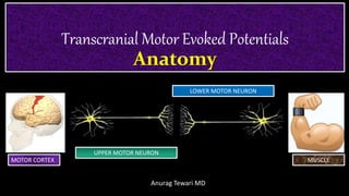
1. Transcranial Motor Evoked Potentials
Anatomy
Anurag Tewari MD
UPPER MOTOR NEURON
LOWER MOTOR NEURON
MOTOR CORTEX MUSCLE
2. Anatomy and Physiology of Motor Systems
• GENERAL ORGANIZATION OF THE SPINAL MOTOR SYSTEMS
• DESCENDING SPINAL PATHWAYS
• LOWER SPINAL MOTONEURON
• MEDIAL SYSTEM
• Miscellaneous
Anurag Tewari MD
3. GENERAL ORGANIZATION
OF THE SPINAL MOTOR SYSTEMS
• The spinal motor system can be divided into ‘UPPER’ and ‘LOWER’ parts:
The UPPER part consists of the
CEREBRAL CORTEX
BASAL GANGLIA
CEREBELLUM
The LOWER part consists of
The SPINAL CORD, including the Alpha Motoneurons (“common final pathway”)
Anurag Tewari MD
5. GENERAL ORGANIZATION
CEREBRAL MOTOR CORTEX
THALAMUS
CEREBELLUM BASAL GANGLIA
BRAIN STEM
SPINAL CORD
MOTOR NEURON
Motor Control
CorticalMotorPathway
Anurag Tewari MD
6. GENERAL ORGANIZATION
OF THE SPINAL MOTOR SYSTEMS
The DESCENDING PATHWAYS start from the motor cortex and the motor nuclei in
the brainstem and cerebellum, and terminate on the neurons in the spinal cord
Anurag Tewari MD
7. GENERAL ORGANIZATION
OF THE SPINAL MOTOR SYSTEMS
• Traditionally, the descending motor pathways have been divided into
Now known as
Anurag Tewari MD
8. GENERAL ORGANIZATION
OF THE SPINAL MOTOR SYSTEMS
LATERAL SYSTEM MEDIAL SYSTEM
The Lateral systems controls muscles in the
distal limbs
The Medial system controls proximal
limb & trunk muscles
Anurag Tewari MD
9. Motor Cortex
• The primary cortex receives input from higher order cortical motor regions such as
• PRIMARY MOTOR CORTICAL
• SUPPLEMENTARY MOTOR AREA
• PRE MOTOR CORTEX
• The motor cortex also receives input from the somatosensory cortical areas
Anurag Tewari MD
10. • The motor cortex generates motor commands
• The primary motor cortex is somatotopically organized in a way similar to the
somatosensory cortex
• The HANDS and the FACE
• comprise the largest parts of the motor cortex
• located on lateral and dorsal surfaces of the brain
• The TRUNK occupies a small part of the
motor cortex
• The DISTAL LEGS are represented by a region
that is hidden between the two hemispheres
Upper Motoneuron
Anurag Tewari MD
11. The Motor Cortex
• RECEIVES INFORMATION via the THALAMUS from
• Brainstem
• Cerebellum
• Basal ganglia
Upper Motoneuron
Anurag Tewari MD
12. Upper Motoneuron
The Motor Cortex
• SENDS INFORMATION to the basal ganglia and brainstem
Anurag Tewari MD
13. Upper Motoneuron
• Main descending pathways from the motor cortex terminate in INTERNEURONS
• INTERNEURONS are found in in different segments of the spinal cord and in
nuclei of cranial motor nerves
Interneurons create neural circuits, enabling communication between SENSORY or MOTOR neurons and the CNS
Anurag Tewari MD
14. Basal Ganglia
• Traditionally, the term “basal ganglia” is used to collectively describe
• THE CAUDATE NUCLEUS
• THE PUTAMEN
• THE GLOBUS PALLIDUS
However, various investigators have included substantia nigra,
the subthalamic nucleus (STN), and the claustrum because they
are related functionally to the other parts of the basal ganglia
Anurag Tewari MD
15. Basal Ganglia
• CAUDATE NUCLEUS and PUTAMEN are referred to as STRIATUM or NEOSTRIATUM
• The PUTAMEN and the GLOBUS PALLIDUS are known as the LENTIFORM NUCLEUS
Anurag Tewari MD
16. Basal Ganglia
• The GLOBUS PALLIDUS consists of
• an external segment (GLOBUS PALLIDUS EXTERNAL part [GPe]), and
• an internal segment (GLOBUS PALLIDUS INTERNAL part [GPi])
• Parts of the SUBSTANTIA NIGRA are
• PARS RETICULATA (SNr)
• SUBSTANTIA NIGRA PARS COMPACTA (SNc)
• These nuclei are of special interest in connection with movement disorders.
Anurag Tewari MD
17. Basal Ganglia
• The basal ganglia processes information from all parts of the cerebral cortex,
including the motor cortex
• And relay information to other subcortical structures and the thalamus
Anurag Tewari MD
18. Basal Ganglia
• The input to the basal ganglia from the primary motor cortex converges on the
• CAUDATE NUCLEUS
• PUTAMEN
• CENTROMEDIAN NUCLEUS (CM) of the thalamus
• SUBSTANTIA NIGRA
Anurag Tewari MD
19. Basal Ganglia
• Thus the basal ganglia receive input from the motor cortex and deliver its
output back to the motor cortex
• Therefore the descending pathways from the motor cortex (corticospinal tract)
contain information from the basal ganglia
Basal ganglia primarily helps in action selection –to decide which of several possible behaviors to execute at any given time
Anurag Tewari MD
20. Basal Ganglia
• All connections between the components of the basal ganglia are INHIBITORY
EXCEPTION: the connections between the STN and the Gpi/SNr (excitatory)
Anurag Tewari MD
21. Basal Ganglia
• The output of GPi and SNr provides tonic inhibition on thalamocortical
neurons
• The direct dopaminergic nigrostriatal pathway from SNc might also modulate
the activity in the two striato-pallidal pathways in two different ways:
• one of which facilitates transmission in the “DIRECT” pathway
• whereas the other is inhibiting transmission in the “INDIRECT” pathway
Anurag Tewari MD
22. Basal Ganglia
• The basal ganglia are associated with movement disorders such as
• Parkinson’s disease (PD)
• Huntington’s disease (HD)
• Gilles de la Tourette syndrome
Anurag Tewari MD
23. Thalamus
(via Latin from Greek thalamos denoting the part of the brain at which a nerve originates)
• The motor portion of the thalamus is involved in processing of movement
information and it links the basal ganglia to the motor cortex
• Lesions made in specific nuclei of the thalamus have been shown effective in
treating movement disorders, as they have been in the treatment of sensory
disorders and pain
• Surgical lesions have now largely been replaced by implantation of electrodes
for chronic electrical stimulation of specific nuclei (DBS).
Anurag Tewari MD
24. Blood Supply to the Basal Ganglia & Thalamus
Anurag Tewari MD
27. DESCENDING SPINAL PATHWAYS
The descending pathways of the spinal cord are organized anatomically together
with ascending sensory pathways
Anurag Tewari MD
28. DESCENDING SPINAL PATHWAYS
The descending pathways are of two main groups:
the MEDIAL systems the LATERAL systems
Cerebrum
Brainstem
Spinal Cord
Anurag Tewari MD
29. LATERAL PATHWAYS
• The (dorso) lateral pathways include the
• CORTICOSPINAL TRACTS
• RUBROSPINAL TRACTS
Anurag Tewari MD
30. DESCENDING SPINAL PATHWAYS
Lateral Pathways
• The corticospinal system is most developed in primates
• Which makes studies of the motor system in other mammals less
representative for humans
• Many aspects of the corticospinal system in humans is incompletely known because of the limited
number of studies of primates and the differences between humans and other primates.
Anurag Tewari MD
31. MEDIAL SYSTEM
• The medial system include the
• VESTIBULOSPINAL TRACTS
• RETICULOSPINAL TRACTS
• TECTOSPINAL TRACTS
Anurag Tewari MD
32. DESCENDING SPINAL PATHWAYS
The fibers of the descending motor pathways terminate on cells in the
VENTRAL HORN of the spinal cord
Anurag Tewari MD
33. DESCENDING SPINAL PATHWAYS
• The pathways of the lateral system provide
• VOLUNTARY
• sophisticated motor control for FINE MOVEMENTS
• Mainly controlling muscles of distal limbs
• It is almost exclusively the LATERAL TRACTS that are monitored in operations
where the spinal cord is at risk of being injured
Anurag Tewari MD
34. DESCENDING SPINAL PATHWAYS
• The pathways of the medial system have general
functions such as
• Control of posture and
• Control of basic function like walking
• The medial group of pathways controls mainly
• Trunk and
• Proximal Limb Muscles
• The medial system activates extensors more than
flexors
Anurag Tewari MD
36. DESCENDING SPINAL PATHWAYS
Lateral Pathways
• The corticospinal tract alone passes through the
PYRAMIDS
• Whereas other descending pathways pass through
other parts of the medulla
This is why the corticospinal tract have been known as the pyramidal
tract and the other tracts were known as the extrapyramidal tracts:
a distinction that is no longer valid
Anurag Tewari MD
37. DESCENDING SPINAL PATHWAYS
Lateral Pathways
• Main parts of lateral pathways cross the midline at level of LOWER MEDULLA
• 10% of the corticospinal fibers in humans do not cross the midline; there are
large individual variations
Anurag Tewari MD
38. DESCENDING SPINAL PATHWAYS
Lateral Pathways
• The corticospinal tract is asymmetric in about 75% of the population,
• RIGHT SIDE often being larger than the left side
• Some of the corticospinal fibers originate in the somatosensory cortex
• which explains why motor responses can be obtained by stimulating the somatosensory cortex
Lateral Corticospinal Tract
Anurag Tewari MD
39. DESCENDING SPINAL PATHWAYS
Lateral Pathways
• Rubrospinal Tract
• The rubrospinal tract originates in the nucleus ruber, which receives indirect
input from the motor cortex
Anurag Tewari MD
40. DESCENDING SPINAL PATHWAYS
Lateral Pathways
• Rubrospinal Tract
• This pathway has very few fibers in humans
• Estimated to be 1% of those of the corticospinal tract in monkey and
man
• The functional importance of the rubrospinal tract in humans has been
questioned
• The tract is responsible for large muscle movement as well as fine motor
control, and it terminates primarily in the cervical spinal cord, suggesting that
it functions in upper limb but not in lower limb control
• It primarily facilitates flexion in the upper extremities
• It is probably of little importance for monitoring purposes
Anurag Tewari MD
41. DESCENDING SPINAL PATHWAYS
Medial Pathways
• The pathways of the medial
system consist of the
• RETICULOSPINAL
• TECTOSPINAL
• VESTIBULAR
Anurag Tewari MD
42. DESCENDING SPINAL PATHWAYS
Medial Pathways
• The tracts of the medial system are, phylogenetically, the oldest motor
pathways
• The tracts of the medial system are less direct motor pathways than those of
the lateral system, and the medial system comprises pathways with different
origins
• The motor tracts that belong to the medial system have both crossed and
uncrossed tracts and their fibers terminate on neurons in the ventromedial
zone of the spinal gray matter
Anurag Tewari MD
43. DESCENDING SPINAL PATHWAYS
Medial Pathways
• These pathways mostly control propriospinal interneurons
• The axons terminate on the motoneurons that control
muscles on the
• Trunk
• Girdle
• Proximal Limb muscles
• The medial motor system mostly controls extensor
muscles and muscles that are involved in posture
(“antigravity” muscles).
Anurag Tewari MD
44. DESCENDING SPINAL PATHWAYS
Medial Pathways
Reticulospinal, Tectospinal, and Vestibular Spinal Pathways
The tectospinal and vestibulospinal fibers are mainly crossed but have small,
uncrossed parts
Anurag Tewari MD
45. DESCENDING SPINAL PATHWAYS
Medial Pathways
Reticulospinal, Tectospinal, and Vestibular Spinal Pathways
• The reticulospinal tract contributes to maintaining posture and can orient the
body in crude stereotyped movements
• The importance in intraoperative monitoring of the reticulospinal tract is
probably mostly related to its role of facilitating the alpha motoneuron
• The reticulospinal tracts are suppressed by many forms of anesthesia
Anurag Tewari MD
46. DESCENDING SPINAL PATHWAYS
Nonspecific Descending Systems
• The NORADRENALIN (NA)–SEROTONIN PATHWAYS belong to a nonspecific
system originating in the raphe nuclei and they project to the spinal cord
• One important function of these descending pathways is
• ADJUSTING MUSCLE TONE, such as suppressing skeletal muscle activity, which occurs, for
example, during rapid eye movement sleep
• These faciliatory systems are sensitive to anesthetic agents
• the reduction in the activity of these systems, caused by anesthetics, is likely to
contribute to the decreased excitability of motor systems that is observed during surgical
operations
Anurag Tewari MD
48. LOWER SPINAL MOTONEURON
• Lower motor neurons consist of
• INTERNEURONS in the spinal cord
• ALPHA MOTONEURON
• The motor nerves emerge and through which
all spinal motor activity must pass
(the “common final pathway”).
Anurag Tewari MD
49. LOWER SPINAL MOTONEURON
Alpha Motoneurons
Alpha motoneurons are located in layer IX of the ventral horn of spinal cord
Anurag Tewari MD
50. LOWER SPINAL MOTONEURON
Alpha Motoneurons
• Their axons form the ventral spinal roots and the motor portions of spinal
nerves that innervate skeletal (extrafusal) muscles
• The motor portion of peripheral nerves also contains the axons of gamma
neurons that innervate the (intrafusal) muscles of muscle spindles
Anurag Tewari MD
51. Extrafusal vs Intrafusal Muscle fibers
• Extrafusal muscle fibers are the skeletal standard
muscle fibers
• Innervated by alpha motor neurons
• Generate tension by contracting
• Thereby allowing for skeletal movement
• Intrafusal muscle fibers are skeletal muscle fibers
• Serve as specialized sensory organs (proprioceptors)
• Detect the amount and rate of change in length of a
muscle
Anurag Tewari MD
52. Blood Supply to the Spinal Cord
The motor (ventral) portion of the spinal cord has a different blood supply than
the dorsal portion of the spinal cord, where the sensory portion of the spinal
cord is located
Anurag Tewari MD
54. Blood Supply to the Spinal Cord
• Compromises of the blood supply to the ventral portion of the spinal cord
might therefore occur without the dorsal part of the spinal cord being affected
and thus go unnoticed if only the SSEP is monitored
• Monitoring of the function of the ventral portion of the spinal cord is important
during operations in which there is risk of ischemia of the spinal cord
Anurag Tewari MD