Short case...tectal plate glioma
•
1 gostou•587 visualizações

Short case...tectal plate glioma http://yassermetwally.com http://yassermetwally.net

Recomendados
Mais conteúdo relacionado
Destaque
Destaque (20)
Semelhante a Short case...tectal plate glioma
Semelhante a Short case...tectal plate glioma (20)
Mais de Professor Yasser Metwally
Mais de Professor Yasser Metwally (20)
Último
Último (20)
Short case...tectal plate glioma
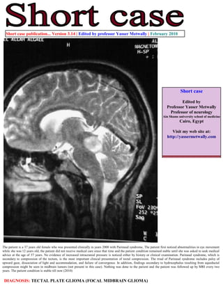
- 1. Short case publication... Version 3.14 | Edited by professor Yasser Metwally | February 2010 Short case Edited by Professor Yasser Metwally Professor of neurology Ain Shams university school of medicine Cairo, Egypt Visit my web site at: http://yassermetwally.com The patient is a 37 years old female who was presented clinically in years 2000 with Parinaud syndrome, The patient first noticed abnormalities in eye movement while she was 12 years old, the patient did not receive medical care since that time and the patient condition remained stable until she was asked to seek medical advice at the age of 37 years. No evidence of increased intracranial pressure is noticed either by history or clinical examination. Parinaud syndrome, which is secondary to compression of the tectum, is the most important clinical presentation of tectal compression. The triad of Parinaud syndrome includes palsy of upward gaze, dissociation of light and accommodation, and failure of convergence. In addition, findings secondary to hydrocephalus resulting from aqueductal compression might be seen in midbrain tumors (not present in this case). Nothing was done to the patient and the patient was followed up by MRI every two years. The patient condition is stable till now (2010) DIAGNOSIS: TECTAL PLATE GLIOMA (FOCAL MIDBRAIN GLIOMA)
- 2. Figure 1. Precontrast MRI T1 images showing an isointense tectal plate glioma. The tumor is showing posterior exophytosis to the left side of the quadrigeminal cistern. The posterior parts of the tumor are showing some hyperintense zones, probably representing hemorrhagic spots. Figure 2. Postcontrast MRI T1 images showing dense ring enhancement within the tectal tumor mass. The mass is seen protruding to the left side of the quadrigeminal cistern.
- 3. Figure 3. The tectal plate glioma is hyperintense in the MRI T2 images, with central hypointensities Figure 4. The tectal plate glioma on postcontrast MRI T1 image (A) and MRI T2 image (B) References 1. Metwally, MYM: Textbook of neurimaging, A CD-ROM publication, (Metwally, MYM editor) WEB-CD agency for electronic publishing, version 11.1a December 2010
- 4. Addendum A new version of short case is uploaded in my web site every week (every Saturday and remains available till Friday.) To download the current version follow the link "http://pdf.yassermetwally.com/short.pdf". You can download the long case version of this short case during the same week from: http://pdf.yassermetwally.com/case.pdf or visit web site: http://pdf.yassermetwally.com To download the software version of the publication (crow.exe) follow the link: http://neurology.yassermetwally.com/crow.zip At the end of each year, all the publications are compiled on a single CD-ROM, please contact the author to know more details. Also to view a list of the previously published case records follow the following link (http://wordpress.com/tag/case- record/) or click on it if it appears as a link in your PDF reader To inspect the patient's full radiological study, click on the attachment icon (the paper clip icon in the left pane) of the acrobat reader then double click on the attached file Click here to download the long case version of this short case in PDF format