Short case...Intramedullary cavernoma
•
3 gostaram•311 visualizações

Short case...Intramedullary cavernoma

Recomendados
Mais conteúdo relacionado
Destaque
Destaque (20)
Semelhante a Short case...Intramedullary cavernoma
Semelhante a Short case...Intramedullary cavernoma (20)
Mais de Professor Yasser Metwally
Mais de Professor Yasser Metwally (20)
Último
Último (20)
Short case...Intramedullary cavernoma
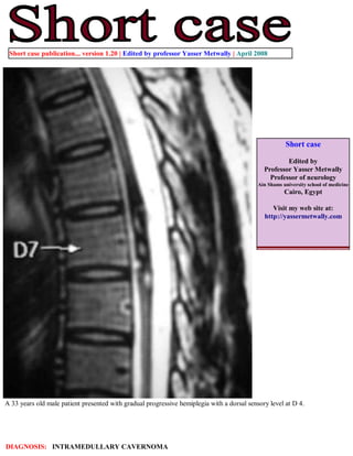
- 1. Short case publication... version 1.20 | Edited by professor Yasser Metwally | April 2008 Short case Edited by Professor Yasser Metwally Professor of neurology Ain Shams university school of medicine Cairo, Egypt Visit my web site at: http://yassermetwally.com A 33 years old male patient presented with gradual progressive hemiplegia with a dorsal sensory level at D 4. DIAGNOSIS: INTRAMEDULLARY CAVERNOMA
- 2. Figure 1. Intramedullary cavernoma. On precontrast T1 images {A) mild dilatation of D5 spinal segment (opposite to D7 vertebral segment) is seen with a markedly hypointense pencil shaped oblong longitudinal center that extends through the whole of the dilated spinal segment with two slightly hyperintense foci seen inside the hypointense longitudinal zone. On postcontrast MRI T1 images {B,C) slight contrast enhancement is seen at the periphery of the hypointense zone and two enhanced spots are seen inside the hypointense zone. Figure 2. Figure 2. Intramedullary cavernoma. On MRI T2 images the intramedullary longitudinal lesion is seen of mixed intensity with mild dilatation of the affected spinal segment. The hypointense longitudinal zone that is appreciated on the MRI T1 images appears hyperintense on the MRI T2 images with irregular contour, This MRI T2 hyperintensity is surrounded by MRI T2 hypointensity that extends around the hyperintense core of the lesion for a variable distance. The MRI T2 mixed signal intensity is much appreciated on cross sectional images. Also notice the asymptomatic vertebral hemangioma at D 8.
- 3. Figure 3. Intramedullary cavernoma. On MRI T2 images (A,B) the intramedullary longitudinal lesion is seen of mixed intensity with mild dilatation of the affected spinal segment. The hypointense longitudinal zone that is appreciated on the MRI T1 (C,D) images appears hyperintense on the MRI T2 images with irregular contour, This MRI T2 hyperintensity is surrounded by MRI T2 hypointensity that extends around the hyperintense core of the lesion for a variable distance. The MRI T2 mixed signal intensity is much appreciated on cross sectional images. Figure 4. Intramedullary cavernoma. On MRI T2 images (B) the intramedullary longitudinal lesion is seen of mixed intensity with mild dilatation of the affected spinal segment. The hypointense longitudinal zone that is appreciated on the MRI T1 images (A) appears hyperintense on the MRI T2 images with irregular contour, This MRI T2 hyperintensity is surrounded by MRI T2 hypointensity that extends around the hyperintense core of the lesion for a variable distance. The MRI T2 mixed signal intensity is much appreciated on cross sectional images. Also notice the asymptomatic vertebral hemangioma at D 8.
- 4. Figure 5. Intramedullary cavernoma. MRI T2 images (A,B) and precontrast MRI T1 images showing a mixed signal intensity intramedullary lesion, The MRI mixed signal intensity of the precontrast MRI T1 images and the MRI T2 images is due to the presence of altered blood of different ages. Conclusion MRI picture of intramedullary cavernoma. The MRI mixed signal intensity of the precontrast MRI T1 images and the MRI T2 images is due to the presence of altered blood of different ages. The MRI T1 precontrast hyperintensity is due to methemoglobin The MRI T2 hypointensity is due to hemosiderin pigments. Addendum A new version of short case is uploaded in my web site every week (every Saturday and remains available till Friday.) To download the current version follow the link quot;http://pdf.yassermetwally.com/short.pdfquot;. You can download the long case version of this short case during the same week from: http://pdf.yassermetwally.com/case.pdf or visit web site: http://pdf.yassermetwally.com To download the software version of the publication (crow.exe) follow the link: http://neurology.yassermetwally.com/crow.zip At the end of each year, all the publications are compiled on a single CD-ROM, please contact the author to know more details. Screen resolution is better set at 1024*768 pixel screen area for optimum display For an archive of the previously reported cases go to www.yassermetwally.net, then under pages in the right panel, scroll down and click on the text entry quot;downloadable short cases in PDF formatquot; References 1. Metwally, MYM: Textbook of neurimaging, A CD-ROM publication, (Metwally, MYM editor) WEB-CD agency for electronic publishing, version 9.1a January 2008