Stages of Normal Labor- easy explanation
•
123 gostaram•54,150 visualizações

Stages of normal labor- easy explanation for Nursing Students(B.Sc & GNM)... Introduction, definition of normal labor, definition of normal labor by WHO, Mechanism of labor, stages of labor, Intrapartum management of Labor, pain control.

Recomendados
Mais conteúdo relacionado
Mais procurados
Mais procurados (20)
Semelhante a Stages of Normal Labor- easy explanation
Semelhante a Stages of Normal Labor- easy explanation (20)
Mais de Swatilekha Das
Mais de Swatilekha Das (20)
Último
Último (20)
Stages of Normal Labor- easy explanation
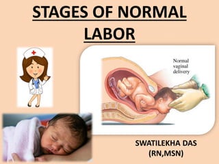
- 1. STAGES OF NORMAL LABOR SWATILEKHA DAS (RN,MSN)
- 2. INTRODUCTION Labor is a physiologic process during which the products of conception (i.e, the fetus, membranes, umbilical cord, and placenta) are expelled outside of the uterus. Labor is achieved with changes in the biochemical connective tissue and with gradual effacement and dilatation of the uterine cervix as a result of rhythmic uterine contractions of sufficient frequency, intensity, and duration.
- 3. DEFINITION OF NORMAL LABOR The onset of labor is defined as regular, painful uterine contractions resulting in progressive cervical effacement and dilatation. Cervical dilatation in the absence of uterine contraction suggests cervical insufficiency, whereas uterine contraction without cervical change does not meet the definition of labor.
- 4. DEFINITION OF LABOR BY WHO The World Health Organization (WHO) defines normal birth as "spontaneous in onset, low-risk at the start of labor and remaining so throughout labor and delivery. The infant is born spontaneously in the vertex position between 37 and 42 completed weeks of pregnancy.
- 5. MECHANISM OF LABOR • The mechanisms of labor, also known as the cardinal movements, involve changes in the position of the fetus’s head during its passage in labor. These are described in relation to a vertex presentation. Although labor and delivery occurs in a continuous fashion, the cardinal movements are described as the following 7 discrete sequences : • Engagement • Descent • Flexion • Internal rotation • Extension • Restitution and external rotation • Expulsion
- 8. 1. FIRST STAGE OF LABOR • Begins with regular uterine contractions and ends with complete cervical dilatation at 10 cm • Divided into a latent phase and an active phase • The latent phase begins with mild, irregular uterine contractions that soften and shorten the cervix • Contractions become progressively more rhythmic and stronger • The active phase usually begins at about 3-4 cm of cervical dilation and is characterized by rapid cervical dilation and descent of the presenting fetal part
- 10. 2. SECOND STAGE OF LABOR • Begins with complete cervical dilatation and ends with the delivery of the fetus • In nulliparous women, the second stage should be considered prolonged if it exceeds 3 hours if regional anesthesia is administered or 2 hours in the absence of regional anesthesia • In multiparous women, the second stage should be considered prolonged if it exceeds 2 hours with regional anesthesia or 1 hour without it
- 12. 3. THIRD STAGE OF LABOR • The period between the delivery of the fetus and the delivery of the placenta and fetal membranes • Delivery of the placenta often takes less than 10 minutes, but the third stage may last as long as 30 minutes • Expectant management involves spontaneous delivery of the placenta • The third stage of labor is considered prolonged after 30 minutes, and active intervention is commonly considered Active management often involves prophylactic administration of oxytocin or other uterotonics (prostaglandins or ergot alkaloids), cord clamping/cutting, and controlled traction of the umbilical cord
- 14. HISTORY TAKING The initial assessment of labor should include : • Frequency and time of onset of contractions • Status of the amniotic membranes (whether spontaneous rupture of the membranes has occurred, and if so, whether the amniotic fluid is clear or meconium stained) • Fetal movements • Presence or absence of vaginal bleeding.
- 15. POINTS TO REMEMBER WHILE HISTORY TAKING Braxton-Hicks contractions must be differentiated from true contractions. Typical features of Braxton-Hicks contractions are as follows: • Usually occur no more often than once or twice per hour, and often just a few times per day • Irregular and do not increase in frequency with increasing intensity • Resolve with ambulation or a change in activity Contractions that lead to labor have the following characteristics: • May start as infrequently as every 10-15 minutes, but usually accelerate over time, increasing to contractions that occur every 2-3 minutes • Tend to last longer and are more intense than Braxton-Hicks contractions • Lead to cervical change
- 16. PHYSICAL EXAMINATION The physical examination should include : • Maternal vital signs • Fetal presentation • Assessment of fetal well-being • Frequency, duration, and intensity of uterine contractions • Abdominal examination with Leopold maneuvers • Pelvic examination with sterile gloves
- 17. Digital examination Digital examination allows the clinician to determine the following aspects of the cervix: • Degree of dilatation, which ranges from 0 cm (closed or fingertip) to 10 cm (complete or fully dilated) • Effacement (assessment of the cervical length, which can be reported as a percentage of the normal 3- to 4-cm–long cervix or described as the actual cervical length) • Position (ie, anterior or posterior) • Consistency (ie, soft or firm) • Palpation of the presenting part of the fetus allows the examiner to establish its station, by quantifying the distance of the body (-5 to +5 cm) that is presenting relative to the maternal ischial spines, where 0 station is in line with the plane of the maternal ischial spines.
- 19. 1. FIRST STAGE OF LABOR On admission to the Labor and Delivery suite, a woman having normal labor should be encouraged to assume the position that she finds most comfortable. Possibilities including the following: • Walking • Lying supine • Sitting • Resting in a left lateral decubitus position
- 20. 1. FIRST STAGE OF LABOR Management includes the following: • Periodic assessment of the frequency and strength of uterine contractions and changes in cervix and in the fetus' station and position • Monitoring the fetal heart rate at least every 15 minutes, particularly during and immediately after uterine contractions; in most obstetric units, the fetal heart rate is assessed continuously
- 21. 2. SECOND STAGE OF LABOR • With complete cervical dilatation, the fetal heart rate should be monitored or auscultated at least every 5 minutes and after each contraction. • Prolonged duration of the second stage alone does not mandate operative delivery if progress is being made, but management options for second-stage arrest include the following:
- 22. 2. SECOND STAGE OF LABOR Continuing observation/expectant management • Operative vaginal delivery by forceps or vacuum-assisted vaginal delivery, or cesarean delivery. Vacuum assisted delivery
- 23. DELIVERY OF THE FETUS • Positioning of the mother for delivery can be any of the following : • Supine with her knees bent (i.e., dorsal lithotomy position; the usual choice) • Lateral (Sims) position • Partial sitting or squatting position • On her hands and knees • Episiotomy used to be routinely performed at this time, but current recommendations restrict its use to maternal or fetal indications
- 24. DELIVERY MANEUVERS ARE AS FOLLOWS: • The head is held in mid position until it is delivered, followed by suctioning of the oropharynx and nares • Check the fetus's neck for a wrapped umbilical cord, and promptly reduce it if possible • If the cord is wrapped too tightly to be removed, the cord can be double clamped and cut • The fetus's anterior shoulder is delivered with gentle downward traction on its head and chin • Subsequent upward pressure in the opposite direction facilitates delivery of the posterior shoulder • The rest of the fetus should now be easily delivered with gentle traction away from the mother • If not done previously, the cord is clamped and cut • The baby is vigorously stimulated and dried and then transferred to the care of the waiting attendants or placed on the mother's abdomen
- 25. 3. THIRD STAGE OF LABOR The following 3 classic signs indicate that the placenta has separated from the uterus : • The uterus contracts and rises • The umbilical cord suddenly lengthens • A gush of blood occurs Delivery of the placenta usually happens within 5- 10 minutes after delivery of the fetus, but it is considered normal up to 30 minutes after delivery of the fetus.
- 26. 3. THIRD STAGE OF LABOR Excessive blood loss during this period or immediately thereafter can be prevented by giving oxytocin, which is a drug that makes the womb contract to close the blood vessels in the placenta and helps it separate from the wall of the uterus. Control Of Postpartum Uterine Bleeding • Intravenous infusion (drip method). If the patient has an intravenous infusion running, 10 to 40 units of oxytocin may be added to the bottle, depending on the amount of electrolyte or dextrose solution remaining (maximum 40 units to 1000 mL) • Intramuscular administration
- 27. 4. PAIN CONTROL Agents given in intermittent doses for systemic pain control include the following : • Meperidine, 25-50 mg IV every 1-2 hours or 50- 100 mg IM every 2-4 hours • Fentanyl, 50-100 mcg IV every hour • Nalbuphine, 10 mg IV or IM every 3 hours • Butorphanol, 1-2 mg IV or IM every 4 hours
- 28. 4. PAIN CONTROL • Morphine, 2-5 mg IV or 10 mg IM every 4 hours • As an alternative, regional anesthesia may be given. Anesthesia options include the following: • Epidural • Spinal • Combined spinal-epidural