SlideShare uma empresa Scribd logo
1 de 39
THREE GOALS:
  •produce a magnified image of the specimen,
  •separate the details in the image,
  •render the details visible to the human eye or
  camera.
The History
 • Hans and Zacharias Janssen of Holland
   in the 1590’s created the “first”
   compound microscope
 • Anthony van Leeuwenhoek and Robert
   Hooke made improvements by working
   on the lenses


Anthony van Leeuwenhoek   Hooke Microscope   Robert Hooke
       1632-1723                             1635-1703
The History


 Zacharias Jansen   The “First” Microscope
    1588-1631
Types of microscopes
 Light microscopes
     Bright field microscope
     Dark field microscope
     Phase contrast microscope
     Fluorescent microscope

 Electron microscopes
   Transmission electron microscope
   Scanning electron microscope
Magnification
• To determine your magnification…you
  just multiply the ocular lens by the
  objective lens
• Ocular 10x Objective 40x:10 x 40 =
  400              So the object is 400 times “larger”

                               Objective Lens have
                               their magnification
                               written on them.


                        Ocular lenses usually magnifies by 10x
RESOLVING POWER/
        RESOLUTION
• ability of a lens to separate or distinguish
  small objects that are close together
• wavelength of light used is major factor in
  resolution
   shorter wavelength ⇒ greater resolution


   Actual                     What We Might See
WAVE LENGTH
• Wave length of light – used for
  illumination
  – Visible range – 400 – 750nm
  (Shorter wave length – high resolution)
  E.g. Blue light has a shorter wave length
    than red light
NUMERIAL APERTURE
• NA – of the objectives (property of lens
  that decides the quantity of light enter
  into it)
  – Two factors 1) Refractive Index 2)Angle
REFRACTION
• The bending of light as it passes from one
  medium to another of different density

• The bending of the light ray gives rise to
  an angle of refraction, the degree of
  bending

• Index of refraction: A measure of the
  speed at which light passes through the
  material
ILLUMINATION
• To collect and reflect the light – two
  devices – 1) Mirror 2) artificial Light
• 1) Mirror – natural source or artificial
  source
  – Two side – reflective- plain – artificial light
  – Concave – natural source
Properties of Light:Light & Objects
  • Reflection: If the light strikes an
    object and bounces back (giving the
    object color)
  • Transmission: The passage of light
    through an object
  • Absorption: The light rays neither
    pass through nor bounce off an
    object but are taken up by the object
OIL IMMERSION
Ocular Lens

Body Tube


Nose Piece
                                 Arm
Objective
Lenses
                                 Stage
Stage
Clips
                                 Coarse Adj.

Diaphragm                       Fine Adjustment

Light Source
                                 Base


            The Parts of a Microscope
Body Tube
   • The body tube holds the objective
     lenses and the ocular lens at the proper
     distance


Diagram
Nose Piece
   • The Nose Piece holds the objective
     lenses and can be turned to increase
     the magnification


Diagram
Objective Lenses
   • The Objective Lenses increase
     magnification (usually from 10x to 40x)


Diagram
Lenses
    • focus light rays at a
      specific place called the
      focal point
    • distance between center
      of lens and focal point is
      the focal length
    • strength of lens related
      to focal length
         – short focal length ⇒more
           magnification
Copyright © The            18
McGraw-Hill
Companies, Inc.
Stage Clips
   • These 2 clips hold the slide/specimen in
     place on the stage.


Diagram
Diaphragm
   • The Diaphragm controls the amount of
     light on the slide/specimen


                           Turn to let more light in or to
                           make dimmer.


Diagram
Light Source
   • Projects light upwards through the
     diaphragm, the specimen and the
     lenses
   • Some have lights, others have mirrors
     where you must move the mirror to
     reflect light


Diagram
Ocular Lens/Eyepiece
   • Magnifies the specimen image


Diagram
Arm
   • Used to support the microscope when
     carried. Holds the body tube, nose
     piece and objective lenses


Diagram
Stage
   • Supports the slide/specimen


Diagram
Coarse Adjustment Knob
   • Moves the stage up and down (quickly)
     for focusing your image


Diagram
Fine Adjustment Knob
   • This knob moves the stage SLIGHTLY
     to sharpen the image


Diagram
Base
   • Supports the microscope


Diagram
How a Microscope Works
Convex Lenses are
curved glass used to
make microscopes
(and glasses etc.)


                       Convex Lenses bend
                       light and focus it in
                       one spot.
How a Microscope Works
Ocular Lens                           Objective Lens
(Magnifies Image)                     (Gathers Light,
                                       Magnifies
                                       And Focuses Image
 Body Tube                             Inside Body Tube)
 (Image Focuses)


•Bending Light: The objective (bottom) convex lens
magnifies and focuses (bends) the image inside the
body tube and the ocular convex (top) lens of a
microscope magnifies it (again).
Caring for a Microscope
• Clean only with a soft cloth/tissue

• Make sure it’s on a flat surface

• Don’t bang it

• Carry it with 2 HANDS…one on the arm
  and the other on the base
Carry a Microscope
Correctly
Using a Microscope
• Start on the lowest magnification
• Don’t use the coarse adjustment knob
  on high magnification…you’ll break the
  slide!!!
• Place slide on stage and lock clips
• Adjust light source (if it’s a mirror…don’t
  stand in front of it!)
• Use fine adjustment to focus
I. Dark field contrast microscopy takes advantage of
objects that “scatter” light - this requires a special
condenser that can “angle” the incident light

II. Phase contrast microscopy takes advantage of
objects that alter the phase of incident light - This
requires “phase rings” in the condenser and in the
objective lens

III. Fluorescence microscopy take advantage of
inherently fluorescent Material of biological
objected that can be fluorescenlty labeled.
DARK FIELD MICROSCOPE
        Contrast
                Bright Field


           Phase Contrast      Dark-Field
Dark field contrast microscopy
takes advantage of objects that
“scatter” light - this requires a
special condenser that can
“angle” the incident light
FLUORESCENT MICROSCOPE
Optical System of a      Fluorescein
Fluorescence          -excited by blue
Microscope            light (450-490)
                      -emits green
                      light (520-560)
[INSERT FIGURE 4.9]


Fluorescence microscopy take advantage of inherently
fluorescent Material of biological objected that can be
fluorescenlty labeled.
Microscopy


      [INSERT FIGURE 4.10]
Applications of different
microscopes

   Bright field – Stained slides
   Dark field – Live unstained cells
   Fluorescent – Microbiological material

Mais conteúdo relacionado

Mais procurados

Microscopes and microscopy
Microscopes and microscopyMicroscopes and microscopy
Microscopes and microscopy
Dakota Boswell
 

Mais procurados (20)

Simple microscope
Simple microscopeSimple microscope
Simple microscope
 
Microscopy
MicroscopyMicroscopy
Microscopy
 
Microscopy and Types of Microscopes
Microscopy and  Types of MicroscopesMicroscopy and  Types of Microscopes
Microscopy and Types of Microscopes
 
basics of microscope - part 1
basics of microscope - part 1basics of microscope - part 1
basics of microscope - part 1
 
Simple & compound microscope PPT
Simple & compound microscope PPTSimple & compound microscope PPT
Simple & compound microscope PPT
 
Introduction to microscopy
Introduction to microscopyIntroduction to microscopy
Introduction to microscopy
 
compound microscope (basic)
compound microscope (basic)compound microscope (basic)
compound microscope (basic)
 
Compound microscope
Compound microscopeCompound microscope
Compound microscope
 
Microscopes and microscopy
Microscopes and microscopyMicroscopes and microscopy
Microscopes and microscopy
 
Microscope
MicroscopeMicroscope
Microscope
 
Fluorescence microscope
Fluorescence microscopeFluorescence microscope
Fluorescence microscope
 
Microscopy
MicroscopyMicroscopy
Microscopy
 
Compound microscope
Compound microscopeCompound microscope
Compound microscope
 
Microscope
MicroscopeMicroscope
Microscope
 
Types of microscope
Types of microscopeTypes of microscope
Types of microscope
 
Light microscopy
Light microscopyLight microscopy
Light microscopy
 
Types and functions of Microscope
Types and functions of MicroscopeTypes and functions of Microscope
Types and functions of Microscope
 
Microscope
MicroscopeMicroscope
Microscope
 
Types of Microscopes with their applications
Types of Microscopes with their applicationsTypes of Microscopes with their applications
Types of Microscopes with their applications
 
Types of microscopy
Types of microscopyTypes of microscopy
Types of microscopy
 

Destaque (7)

B sc biotech i bpi unit 4 microscopy
B sc biotech i bpi unit 4 microscopyB sc biotech i bpi unit 4 microscopy
B sc biotech i bpi unit 4 microscopy
 
Resolving filter grating etc
Resolving filter grating etcResolving filter grating etc
Resolving filter grating etc
 
Compound microscopy
Compound microscopyCompound microscopy
Compound microscopy
 
Electron microscope
Electron microscopeElectron microscope
Electron microscope
 
Electron microscope
Electron microscopeElectron microscope
Electron microscope
 
Scanning Electron Microscopy (SEM) lecture
Scanning Electron Microscopy (SEM) lectureScanning Electron Microscopy (SEM) lecture
Scanning Electron Microscopy (SEM) lecture
 
Process of sugar production from sugarcane - sugar factory
Process of sugar production from sugarcane - sugar factoryProcess of sugar production from sugarcane - sugar factory
Process of sugar production from sugarcane - sugar factory
 

Semelhante a Review of the microscope

Microscopy.ppt Microscopy.pptvMicroscopy.pptMicroscopy.pptMicroscopy.pptMicro...
Microscopy.ppt Microscopy.pptvMicroscopy.pptMicroscopy.pptMicroscopy.pptMicro...Microscopy.ppt Microscopy.pptvMicroscopy.pptMicroscopy.pptMicroscopy.pptMicro...
Microscopy.ppt Microscopy.pptvMicroscopy.pptMicroscopy.pptMicroscopy.pptMicro...
KAOUKAAlaedin
 

Semelhante a Review of the microscope (20)

The microscope
The microscopeThe microscope
The microscope
 
MICROSCOPY.pdf
MICROSCOPY.pdfMICROSCOPY.pdf
MICROSCOPY.pdf
 
Instrumentaion - MICROSCOPE
Instrumentaion - MICROSCOPEInstrumentaion - MICROSCOPE
Instrumentaion - MICROSCOPE
 
Microscope .pptx
Microscope  .pptxMicroscope  .pptx
Microscope .pptx
 
Q2 module-1-2 -microscope
Q2 module-1-2 -microscopeQ2 module-1-2 -microscope
Q2 module-1-2 -microscope
 
Q2 module-1-2 -microscope
Q2 module-1-2 -microscopeQ2 module-1-2 -microscope
Q2 module-1-2 -microscope
 
The microscope
The microscopeThe microscope
The microscope
 
Microscope
MicroscopeMicroscope
Microscope
 
Lab 0-B Microscope Training
Lab 0-B Microscope TrainingLab 0-B Microscope Training
Lab 0-B Microscope Training
 
Prabhakar singh ii sem-unit i-microscopy and centrifugation
Prabhakar singh  ii sem-unit i-microscopy and centrifugationPrabhakar singh  ii sem-unit i-microscopy and centrifugation
Prabhakar singh ii sem-unit i-microscopy and centrifugation
 
B.Sc. Biochemistry II Cellular Biochemistry Unit 4 Basic Techniques in Microb...
B.Sc. Biochemistry II Cellular Biochemistry Unit 4 Basic Techniques in Microb...B.Sc. Biochemistry II Cellular Biochemistry Unit 4 Basic Techniques in Microb...
B.Sc. Biochemistry II Cellular Biochemistry Unit 4 Basic Techniques in Microb...
 
Microscope
MicroscopeMicroscope
Microscope
 
Parts of the microscope and their functions
Parts of the microscope and their functionsParts of the microscope and their functions
Parts of the microscope and their functions
 
Microscopy.ppt Microscopy.pptvMicroscopy.pptMicroscopy.pptMicroscopy.pptMicro...
Microscopy.ppt Microscopy.pptvMicroscopy.pptMicroscopy.pptMicroscopy.pptMicro...Microscopy.ppt Microscopy.pptvMicroscopy.pptMicroscopy.pptMicroscopy.pptMicro...
Microscopy.ppt Microscopy.pptvMicroscopy.pptMicroscopy.pptMicroscopy.pptMicro...
 
Microscope
MicroscopeMicroscope
Microscope
 
microscope.pptx
microscope.pptxmicroscope.pptx
microscope.pptx
 
B sc micro i btm u 1 microscopy and staining
B sc micro i btm u 1 microscopy and staining B sc micro i btm u 1 microscopy and staining
B sc micro i btm u 1 microscopy and staining
 
Chapter 4
Chapter 4Chapter 4
Chapter 4
 
MICROSCOPY.pptx
MICROSCOPY.pptxMICROSCOPY.pptx
MICROSCOPY.pptx
 
The compound light microscope
The compound light microscopeThe compound light microscope
The compound light microscope
 

Último

Salient Features of India constitution especially power and functions
Salient Features of India constitution especially power and functionsSalient Features of India constitution especially power and functions
Salient Features of India constitution especially power and functions
KarakKing
 
Activity 01 - Artificial Culture (1).pdf
Activity 01 - Artificial Culture (1).pdfActivity 01 - Artificial Culture (1).pdf
Activity 01 - Artificial Culture (1).pdf
ciinovamais
 
1029 - Danh muc Sach Giao Khoa 10 . pdf
1029 -  Danh muc Sach Giao Khoa 10 . pdf1029 -  Danh muc Sach Giao Khoa 10 . pdf
1029 - Danh muc Sach Giao Khoa 10 . pdf
QucHHunhnh
 
The basics of sentences session 3pptx.pptx
The basics of sentences session 3pptx.pptxThe basics of sentences session 3pptx.pptx
The basics of sentences session 3pptx.pptx
heathfieldcps1
 

Último (20)

Understanding Accommodations and Modifications
Understanding  Accommodations and ModificationsUnderstanding  Accommodations and Modifications
Understanding Accommodations and Modifications
 
Salient Features of India constitution especially power and functions
Salient Features of India constitution especially power and functionsSalient Features of India constitution especially power and functions
Salient Features of India constitution especially power and functions
 
Application orientated numerical on hev.ppt
Application orientated numerical on hev.pptApplication orientated numerical on hev.ppt
Application orientated numerical on hev.ppt
 
On National Teacher Day, meet the 2024-25 Kenan Fellows
On National Teacher Day, meet the 2024-25 Kenan FellowsOn National Teacher Day, meet the 2024-25 Kenan Fellows
On National Teacher Day, meet the 2024-25 Kenan Fellows
 
Explore beautiful and ugly buildings. Mathematics helps us create beautiful d...
Explore beautiful and ugly buildings. Mathematics helps us create beautiful d...Explore beautiful and ugly buildings. Mathematics helps us create beautiful d...
Explore beautiful and ugly buildings. Mathematics helps us create beautiful d...
 
Holdier Curriculum Vitae (April 2024).pdf
Holdier Curriculum Vitae (April 2024).pdfHoldier Curriculum Vitae (April 2024).pdf
Holdier Curriculum Vitae (April 2024).pdf
 
Kodo Millet PPT made by Ghanshyam bairwa college of Agriculture kumher bhara...
Kodo Millet  PPT made by Ghanshyam bairwa college of Agriculture kumher bhara...Kodo Millet  PPT made by Ghanshyam bairwa college of Agriculture kumher bhara...
Kodo Millet PPT made by Ghanshyam bairwa college of Agriculture kumher bhara...
 
Key note speaker Neum_Admir Softic_ENG.pdf
Key note speaker Neum_Admir Softic_ENG.pdfKey note speaker Neum_Admir Softic_ENG.pdf
Key note speaker Neum_Admir Softic_ENG.pdf
 
Activity 01 - Artificial Culture (1).pdf
Activity 01 - Artificial Culture (1).pdfActivity 01 - Artificial Culture (1).pdf
Activity 01 - Artificial Culture (1).pdf
 
Introduction to Nonprofit Accounting: The Basics
Introduction to Nonprofit Accounting: The BasicsIntroduction to Nonprofit Accounting: The Basics
Introduction to Nonprofit Accounting: The Basics
 
1029 - Danh muc Sach Giao Khoa 10 . pdf
1029 -  Danh muc Sach Giao Khoa 10 . pdf1029 -  Danh muc Sach Giao Khoa 10 . pdf
1029 - Danh muc Sach Giao Khoa 10 . pdf
 
The basics of sentences session 3pptx.pptx
The basics of sentences session 3pptx.pptxThe basics of sentences session 3pptx.pptx
The basics of sentences session 3pptx.pptx
 
FSB Advising Checklist - Orientation 2024
FSB Advising Checklist - Orientation 2024FSB Advising Checklist - Orientation 2024
FSB Advising Checklist - Orientation 2024
 
Unit-IV; Professional Sales Representative (PSR).pptx
Unit-IV; Professional Sales Representative (PSR).pptxUnit-IV; Professional Sales Representative (PSR).pptx
Unit-IV; Professional Sales Representative (PSR).pptx
 
How to Manage Global Discount in Odoo 17 POS
How to Manage Global Discount in Odoo 17 POSHow to Manage Global Discount in Odoo 17 POS
How to Manage Global Discount in Odoo 17 POS
 
Google Gemini An AI Revolution in Education.pptx
Google Gemini An AI Revolution in Education.pptxGoogle Gemini An AI Revolution in Education.pptx
Google Gemini An AI Revolution in Education.pptx
 
Mehran University Newsletter Vol-X, Issue-I, 2024
Mehran University Newsletter Vol-X, Issue-I, 2024Mehran University Newsletter Vol-X, Issue-I, 2024
Mehran University Newsletter Vol-X, Issue-I, 2024
 
Python Notes for mca i year students osmania university.docx
Python Notes for mca i year students osmania university.docxPython Notes for mca i year students osmania university.docx
Python Notes for mca i year students osmania university.docx
 
TỔNG ÔN TẬP THI VÀO LỚP 10 MÔN TIẾNG ANH NĂM HỌC 2023 - 2024 CÓ ĐÁP ÁN (NGỮ Â...
TỔNG ÔN TẬP THI VÀO LỚP 10 MÔN TIẾNG ANH NĂM HỌC 2023 - 2024 CÓ ĐÁP ÁN (NGỮ Â...TỔNG ÔN TẬP THI VÀO LỚP 10 MÔN TIẾNG ANH NĂM HỌC 2023 - 2024 CÓ ĐÁP ÁN (NGỮ Â...
TỔNG ÔN TẬP THI VÀO LỚP 10 MÔN TIẾNG ANH NĂM HỌC 2023 - 2024 CÓ ĐÁP ÁN (NGỮ Â...
 
SOC 101 Demonstration of Learning Presentation
SOC 101 Demonstration of Learning PresentationSOC 101 Demonstration of Learning Presentation
SOC 101 Demonstration of Learning Presentation
 

Review of the microscope

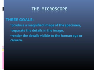
  • 1. THREE GOALS: •produce a magnified image of the specimen, •separate the details in the image, •render the details visible to the human eye or camera.
  • 2. The History • Hans and Zacharias Janssen of Holland in the 1590’s created the “first” compound microscope • Anthony van Leeuwenhoek and Robert Hooke made improvements by working on the lenses Anthony van Leeuwenhoek Hooke Microscope Robert Hooke 1632-1723 1635-1703
  • 3. The History Zacharias Jansen The “First” Microscope 1588-1631
  • 4. Types of microscopes  Light microscopes  Bright field microscope  Dark field microscope  Phase contrast microscope  Fluorescent microscope  Electron microscopes  Transmission electron microscope  Scanning electron microscope
  • 5. Magnification • To determine your magnification…you just multiply the ocular lens by the objective lens • Ocular 10x Objective 40x:10 x 40 = 400 So the object is 400 times “larger” Objective Lens have their magnification written on them. Ocular lenses usually magnifies by 10x
  • 6. RESOLVING POWER/ RESOLUTION • ability of a lens to separate or distinguish small objects that are close together • wavelength of light used is major factor in resolution shorter wavelength ⇒ greater resolution Actual What We Might See
  • 7. WAVE LENGTH • Wave length of light – used for illumination – Visible range – 400 – 750nm (Shorter wave length – high resolution) E.g. Blue light has a shorter wave length than red light
  • 8. NUMERIAL APERTURE • NA – of the objectives (property of lens that decides the quantity of light enter into it) – Two factors 1) Refractive Index 2)Angle
  • 9. REFRACTION • The bending of light as it passes from one medium to another of different density • The bending of the light ray gives rise to an angle of refraction, the degree of bending • Index of refraction: A measure of the speed at which light passes through the material
  • 10. ILLUMINATION • To collect and reflect the light – two devices – 1) Mirror 2) artificial Light • 1) Mirror – natural source or artificial source – Two side – reflective- plain – artificial light – Concave – natural source
  • 11. Properties of Light:Light & Objects • Reflection: If the light strikes an object and bounces back (giving the object color) • Transmission: The passage of light through an object • Absorption: The light rays neither pass through nor bounce off an object but are taken up by the object
  • 12.
  • 14. Ocular Lens Body Tube Nose Piece Arm Objective Lenses Stage Stage Clips Coarse Adj. Diaphragm Fine Adjustment Light Source Base The Parts of a Microscope
  • 15. Body Tube • The body tube holds the objective lenses and the ocular lens at the proper distance Diagram
  • 16. Nose Piece • The Nose Piece holds the objective lenses and can be turned to increase the magnification Diagram
  • 17. Objective Lenses • The Objective Lenses increase magnification (usually from 10x to 40x) Diagram
  • 18. Lenses • focus light rays at a specific place called the focal point • distance between center of lens and focal point is the focal length • strength of lens related to focal length – short focal length ⇒more magnification Copyright © The 18 McGraw-Hill Companies, Inc.
  • 19. Stage Clips • These 2 clips hold the slide/specimen in place on the stage. Diagram
  • 20. Diaphragm • The Diaphragm controls the amount of light on the slide/specimen Turn to let more light in or to make dimmer. Diagram
  • 21. Light Source • Projects light upwards through the diaphragm, the specimen and the lenses • Some have lights, others have mirrors where you must move the mirror to reflect light Diagram
  • 22. Ocular Lens/Eyepiece • Magnifies the specimen image Diagram
  • 23. Arm • Used to support the microscope when carried. Holds the body tube, nose piece and objective lenses Diagram
  • 24. Stage • Supports the slide/specimen Diagram
  • 25. Coarse Adjustment Knob • Moves the stage up and down (quickly) for focusing your image Diagram
  • 26. Fine Adjustment Knob • This knob moves the stage SLIGHTLY to sharpen the image Diagram
  • 27. Base • Supports the microscope Diagram
  • 28. How a Microscope Works Convex Lenses are curved glass used to make microscopes (and glasses etc.) Convex Lenses bend light and focus it in one spot.
  • 29. How a Microscope Works Ocular Lens Objective Lens (Magnifies Image) (Gathers Light, Magnifies And Focuses Image Body Tube Inside Body Tube) (Image Focuses) •Bending Light: The objective (bottom) convex lens magnifies and focuses (bends) the image inside the body tube and the ocular convex (top) lens of a microscope magnifies it (again).
  • 30. Caring for a Microscope • Clean only with a soft cloth/tissue • Make sure it’s on a flat surface • Don’t bang it • Carry it with 2 HANDS…one on the arm and the other on the base
  • 32. Using a Microscope • Start on the lowest magnification • Don’t use the coarse adjustment knob on high magnification…you’ll break the slide!!! • Place slide on stage and lock clips • Adjust light source (if it’s a mirror…don’t stand in front of it!) • Use fine adjustment to focus
  • 33. I. Dark field contrast microscopy takes advantage of objects that “scatter” light - this requires a special condenser that can “angle” the incident light II. Phase contrast microscopy takes advantage of objects that alter the phase of incident light - This requires “phase rings” in the condenser and in the objective lens III. Fluorescence microscopy take advantage of inherently fluorescent Material of biological objected that can be fluorescenlty labeled.
  • 34. DARK FIELD MICROSCOPE Contrast Bright Field Phase Contrast Dark-Field
  • 35. Dark field contrast microscopy takes advantage of objects that “scatter” light - this requires a special condenser that can “angle” the incident light
  • 36. FLUORESCENT MICROSCOPE Optical System of a Fluorescein Fluorescence -excited by blue Microscope light (450-490) -emits green light (520-560)
  • 37. [INSERT FIGURE 4.9] Fluorescence microscopy take advantage of inherently fluorescent Material of biological objected that can be fluorescenlty labeled.
  • 38. Microscopy [INSERT FIGURE 4.10]
  • 39. Applications of different microscopes  Bright field – Stained slides  Dark field – Live unstained cells  Fluorescent – Microbiological material