BLS/BASIC LIFE SUPPORT Module ............
•
1 gostou•792 visualizações

Basic Life Support, or BLS, generally refers to the type of care that first-responders, healthcare providers and public safety professionals provide to anyone who is experiencing cardiac arrest, respiratory distress or an obstructed airway.

Recomendados
Mais conteúdo relacionado
Mais procurados
Mais procurados (20)
Semelhante a BLS/BASIC LIFE SUPPORT Module ............
Semelhante a BLS/BASIC LIFE SUPPORT Module ............ (20)
Mais de DR .PALLAVI PATHANIA
Mais de DR .PALLAVI PATHANIA (20)
Último
Último (20)
BLS/BASIC LIFE SUPPORT Module ............
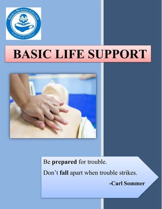
- 1. 0 BASIC LIFE SUPPORT Be prepared for trouble. Don’t fall apart when trouble strikes. -Carl Sommer
- 2. GENERAL CONCEPT This Module booklet explains about Basic Life Support. BLS is the foundation for saving lives after cardiac arrest. You will learn the skills of high-quality cardiopulmonary resuscitation (CPR) for victims of adult both as a single rescuer and as a member of a multirescuer team. The knowledge you learn in this booklet will enable you to recognize cardiac arrest, activate the emergency response system early and respond quickly and confidently. Why do we do what we do? Life is why. Life is a celebration of life. A simple yet powerful answer to the question of why we should all be healthy in heart and mind. I encourage you to discover your Why and share it with others. Ask yourself; what are the moments, people and experiences I live for? What makes me joy, wonder, and happiness? Why am I saves lives? Why is cardiovascular care important to me? The answer to these questions is your Why. Through informational Booklet you will find information that correlates with the importance of cardiovascular care. Module Booklet content: Basic Life Support Introduction of heart……….. Location and surface projection, Layers of heart wall, Chambers of heart, Valves of heart, Normal heart sound, Coronary circulation, Cardiac conduction system Cardiac Arrest…………. Definition, Cause, Symptom, Diagnosis, Management Basic life support……….. Definition, American Heart Association 2020 guideline, Basic Steps Assessment , High quality CPR components for BLS providers, BLS Algorithm, Choking relief for adult Nurses Responsibility…………
- 3. BASIC LIFE SUPPORT INTRODUCTION OF HEART Heart is the vital organ in the circulatory system, primarily responsible for delivering the circulation of blood and transportation of oxygen and nutrients, as well as assisting in the removal of metabolic wastes in all parts of the body. The heart is usually beating about 60 to 100 times per minute. LOCATION AND SURFACE PROJECTION • The heart is the hollow, cone shaped about the size of closed fist • The heart is specifically located in thoracic cavity. It lies in the mediastinum between the lungs and rests upon the diaphragm. • The heart lies within a fluid filled cavity called pericardial cavity. • The heart weighs about 250- 350 gms. LAYERS OF HEART WALLS • Layers of heart: Pericardium/ Epicardium, myocardium, endocardium. 1. PERICARDUIM/ EPICARDUIM: It is the layer immediately outside of the heart muscle proper (the myocardium). The epicardium is largely made of connective tissue and functions as a protective layer. Pericardium is the membrane (sac) that surrounds and protects the heart by the help of two layers.
- 4. BASIC LIFE SUPPORT a. Fibrous pericardium- superficial layer, tough, inelastic, prevents overstretching, provide protection and anchors the heart in place. b. Serous pericardium- 1. Parietal layer- fused to the fibrous pericardium 2. Visceral layer or epicardium- adheres to the heart itself c. Pericardial cavity – Present between two layers is filled with pericardial fluids which reduce friction. 2. MYOCARDUIM The myocardium of the left ventricle is the thickest, as this ventricle is responsible for generating the power needed to pump oxygenated blood from the heart to the rest of the body. 3. ENDOCARDUIM Endocardium (endo-cardium) is the thin inner layer of the heart wall. This layer lines the inner heart chambers, covers heart valves, and is continuous with the endothelium of large blood vessels. The endocardium of heart atria consists of smooth muscle, as well as elastic fibers. CHAMBERS OF HEART There are four chambers of heart: • Right Atrium • Right Ventricle • Left atrium • Left ventricle VALVE OF HEART: • The four valves are the mitral valve, tricuspid valve, pulmonary valve and aortic valve.
- 5. BASIC LIFE SUPPORT NORMAL HEART SOUNDS: • Normal heart sounds are Lubb and Dubb. CORONARY CIRCULATION • It is the circulation of blood in the blood vessels of the heart muscle. The vessels that deliver oxygen- rich (oxygenated) blood to the myocardium are known as coronary arteries. The vessels that remove the deoxygenated blood from the heart muscle are known as cardiac veins. CARDIC CONDUCTION SYSTEM • The cardiac conduction system involves the spread of electrical activity from the sinoatrial node, to the atrioventricular node, down the bundle of His and along the Purkinje fibres. As the electrical activity spreads along the heart’s conduction system it initiates myocardial contraction in the surrounding myocardial tissue.
- 6. BASIC LIFE SUPPORT • The sinoatrial (SA) node or sinus node is the heart's natural pacemaker. The sinoatrial (SA) node or sinus node is the heart's natural pacemaker. The AV node serves as an electrical relay station, slowing the electrical current sent by the sinoatrial (SA) node before the signal is permitted to pass down through to the ventricles. The bundle of His is an important part of the electrical conduction system of the heart, as it transmits impulses from the atrioventricular node, located at the anterior- inferior end of the interatrial septum, to the ventricles of the heart. The left bundle branch further divides into the left anterior and the left posterior fascicles. These bundles and fascicles give rise to thin filaments known as Purkinje fibers. CARDIAC ARREST Cardiac arrest is a sudden loss of blood flow resulting from the failure of the heart to pump effectively. Causes: Abnormal heart rhythm Ventricular fibrillation Arrhythmias develop in people with conditions such as: Heart attack- often due to coronary artery disease Cardiomyopathy Congenital heart disease Heart rhythm abnormalities Coronary heart disease Symptoms: Cardiac arrest does not show warning symptoms in nearly half the cases. The main symptoms are: Loss of consciousness
- 7. BASIC LIFE SUPPORT Collapse/fainting Absence of breathing No pulse Sometimes, chest pain, light-headedness, palpitations or vomiting may be seen before the arrest. DIAGNOSIS: Clinical diagnosis is made when there is an absence of a pulse. The gold standard for diagnosis is a lack of carotid pulse. Additional tests may include: Common tests & procedures Blood test: Check levels of potassium and magnesium and hormones. Hormone test: To check the levels of hormones that may affect the ability of the heart to function. Electrocardiogram (ECG or EKG): Detects abnormal electrical activity and reveals disturbances in heart rhythm. Echocardiogram: Produces images of the heart. Done to identify areas of the heart that has been damaged by the heart attack. Nuclear stress test: To identify problems related to blood flow to the heart. MANAGEMEMT: Cardiac arrest requires immediate action i.e. Basic Life Support.
- 8. BASIC LIFE SUPPORT Medication Beta blockers: Drugs used to manage abnormal heart rhythm. Atenolol, Metoprolol, Thrombolytic agents, Calcium, sodium, bicarbonates. ACE inhibitors: Drugs used to treat hypertension and congestive heart failure. Enalapril, Fosinopril, Epinephrine, Vasopressor, Antiarrhythmic: Amiodarone, Magnesium, Atropine, Defibrillation, Tracheal Intubation, Targeted Temperature management Surgical Procedure: Coronary angioplasty: Opens blocked coronary arteries. Coronary artery bypass graft (CABG): Vein or artery from other parts of the body (usually taken from thigh, arm or mammary vein) is used to bypass a blocked or narrowed artery. Radiofrequency catheter ablation: Used to block a single abnormal electrical pathway.
- 9. BASIC LIFE SUPPORT Dietary modification: To be eaten To be avoided Variety of fruits and vegetables Low fat or fat-free dairy products Poultry Fish Whole grains such as oatmeal and whole- wheat pasta Saturated and trans fat Excess salt and sugar Self care practices: Quit smoking and alcohol. Monitor blood sugar and cholesterol levels. Monitor your blood pressure regularly. Manage stress. Maintain the recommended weight.
- 10. BASIC LIFE SUPPORT BASIC LIFE SUPPORT Basic Life Support, or BLS, generally refers to the type of care that first-responders, healthcare providers and public safety professionals provide to anyone who is experiencing cardiac arrest, respiratory distress or an obstructed airway. It requires knowledge and skills in cardiopulmonary resuscitation (CPR), using automated external defibrillators (AED) and relieving airway obstructions in patients of every age. BLS includes: Airway, Breathing, Circulation, Automated External Defibrillator, and Management of chocking and foreign body obstruction. According to American Heart Association 2020 guideline: Chain of survival: The 5 links in the adult in-the-hospital Chain of Survival are: Surveillance, prevention, and treatment of pre arrest conditions. Immediate Recognition of cardiac arrest and activation of the emergency response system Early cardiopul- monary resuscitati-on with an emphasis on chest compressions Rapid defibrillati- on. Multidisciplin- ary post-cardiac arrest care.
- 11. BASIC LIFE SUPPORT The 5 links in the adult out-the-hospital Chain of Survival are: Recognition of cardiac arrest and activation of the emergency response system Early cardiopul- monary resuscitation with an emphasis on chest compressions Rapid defibrillation . Basic and advanced emergency medical services. Advanced life support and post- cardiac arrest care. BASIC STEPS OF ASSESSMENT: CIRCULA- TION: Check the patient for a carotid pulse for 5-10 seconds. Do not check for more than 10 seconds AIRWAY: Tilt the victim's head back, and lift the chin to open the airway. BREATH-ING: Give mouth to mouth rescue breathing. DEFIBRIL- LATION: They are used to prevent or correct an arrhythmia.
- 12. BASIC LIFE SUPPORT HIGH QUALITY CPR COMPONENTS FOR BLS PROVIDERS COMPONENT DESCRIPTION Scene safety Make sure the environment is safe for the rescuers and victim Recognition of cardiac arrest Check for responsiveness. Tap the victim’s shoulder and shout, “Are you OK?" No breathing or only gasping (i.e.) no normal breathing) Scan the victim’s chest for rise and fall. Check carotid pulse in between the trachea and the muscles at the side of the neck. Feel the pulse for at least 5sec but not more than 10 second. Activation of emergency response system If alone with no mobile phone, leave the victim to activate the emergency response sytem and get the AED before beginning CPR. Otherwise send someone and begin CPR immediately. Compression- ventilation ratio without advanced airway 1 or 2 rescuers 30:2 Compression- ventilation ratio with advanced airway Continuous compressions at a rate of 100-120/min. Give 1 breath every 6 second (10 breaths/ min) Compression rate 100-120/min Compression depth At least 5 cm Continue CPR Do not interrupt resuscitation until: A health professional tells you to stop You become exhausted The victim is definitely waking up, moving, opening eyes and breathing normally It is rare for CPR alone to restart the heart. Unless you are certain the person has recovered, continue CPR Hand placement 2 hands on the lower half of the breastbone (Lower portion of sternum) Chest recoil Allow full recoil of chest after each compression: do not lean on the chest after each compression. Minimizing interruptions Limit interruptions in chest compressions to less than 10 seconds
- 13. BASIC LIFE SUPPORT BLS ALGORITHM
- 14. BASIC LIFE SUPPORT CHOKING RELIEF FOR ADULT Early recognition of foreign-body airway obstruction is the key to successful outcome. It is important to distinguish this emergency from fainting, stroke, heart attack, seizure, drug overdose, or other conditions that may cause sudden respiratory distress but require different treatment. Foreign bodies may cause a range of signs from mild to severe airway obstruction. Signs Rescuer Actions Mild airway obstruction Good air exchange Can cough forcefully May wheeze between coughs As long as good air exchange continues, encourage the victim to continue coughing. Do not interfere with the victim’s own attempts to relieve the obstruction, but stay with victim and monitor the condition. If mild airway obstruction continues or progresses to signs of severe airway obstruction, activate the emergency response system. Severe airway obstruction Clutching the throat with the thumb and fingers, making the universal choking sign. Unable to speak or cry Poor or no air exchange Weak, ineffective cough or no cough at all. Increased respiratory difficulty Possible cyanosis (turning blue) If the victim is an adult, asks him if he victim nods “yes” and cannot talk, severe airway obstruction is present. Take steps immediately to relieve the obstruction. If severe airway obstruction continues and the victim becomes unresponsive, start CPR. If you are not alone, send someone to activate the emergency response system. If you are alone, provide about 2 minutes of CPR before leaving to activate the emergency response system.
- 15. BASIC LIFE SUPPORT Choking relief in a Responsive Adult: With Victim sitting or standing Step Action 1. Stand or kneel behind the victim and wrap your arms around the victim’s waist. 2. Make a fist with one hand. 3. Place the thumb side of your fist against the victim’s abdomen, in the midline, slightly above the navel and well below the breastbone. 4. Grasp your fist with your other hand and press your fist into the victim’s abdomen with a quick, forceful upward thrust. 5. Repeat thrusts until the object is expelled from the airway or the victim becomes unresponsive. 6. Give each new thrust with a separate, distinct movement to relieve the obstruction. Choking relief in an Unresponsive Adult: With Victim sitting or standing Step Action 1. Shout for help. If someone else is available, send that person to activate the emergency response system. 2. Gently lower the victim to the ground if you see that he is becoming unresponsive. 3. Begin CPR, starting with chest compressions. Do not check for a pulse. 4. Each time you open the airway to give breaths, open the victim’s mouth wide. Look for object. If you see an object that can be easily removed, remove it with your fingers. If you see an object, continue CPR. 5. After about 5 cycles or 2 minutes of CPR, activate the emergency response system if someone has not already done so.
- 16. BASIC LIFE SUPPORT Nurses Responsibility Immediate management (Basic Life Support) Administer medications Such as (Anti- anginals) Administer supplemental Oxygen by nasal prongs or face mask as indicated Monitor Vital sign(pulse, blood pressure) Note heart sounds Monitor laboratory Status e.g BUN, Creatinine Measure cardiac output and other functional parameters Monitor closely ECG and Chest x-ray changes and electrolyte and nutrition Administer IV solution as prescribed Encourage rest, semi recumbent in bed or chair, Assist with physical care as indicated. Emotional Support Patient/client and family
- 17. Where to go for more information? If you need more information, please go to www.heart.org www.international.heart.org www.en.wikipedia.org/wiki/IndianHeartAssociation For contacts: +1 (800) 242-8721 +91 44 4202 6644 info@theproject.com