
Recomendados
Mais conteúdo relacionado
Mais procurados
Mais procurados (20)
Destaque
Destaque (20)
Semelhante a Antibiotic-Associated Diarrhea & Clostridium Difficile Infection
Semelhante a Antibiotic-Associated Diarrhea & Clostridium Difficile Infection (20)
Mais de Samir Haffar
Mais de Samir Haffar (20)
Último
Último (20)
Antibiotic-Associated Diarrhea & Clostridium Difficile Infection
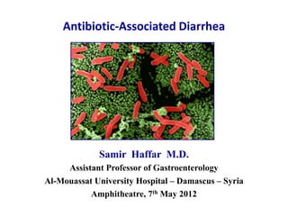
- 1. Antibiotic-Associated Diarrhea Samir Haffar M.D. Assistant Professor of Gastroenterology Al-Mouassat University Hospital – Damascus – Syria Amphitheatre, 7th May 2012
- 2. Antibiotic-Associated Diarrhea (AAD) 5 – 25% depending on causative antibiotic • Motilin receptor agonist Erythromycin • Stimulate bowel motility Clavulanate • Intestinal flora alteration Decreased carbohydrate metab SCFA → Osmotic diarrhea Decreased PBA metabolism PBA → Secretory diarrhea • Overgrowth of pathogenic micro-organisms SCFA: Short Chain Fatty Acid PBA: Primary Biliary Acid Gorkiewicz G. Int J Antimicrobial Agents 2009 ; 33 : S37 – S41.
- 3. Major infectious agent in AAD Causative agent unknown in most cases • Candida overgrowth Controversial • Clostridium Perfringens Food poisoning Necrotizing entero-colitis • Staphylococcus Aureus Food poisoning Diarrhea (majority MRSA*) • Klebsiella Oxytoca Antibiotic-associated hemorrhagic colitis • Clostridium Difficile 30% of AAD in hospitals * MRSA: Methicillin-Resistant Staphylococcus Aureus Gorkiewicz G. Int J Antimicrobial Agents 2009 ; 33 : S37 – S41.
- 4. Candida overgrowth • AAD accompanied by raised candida in stool (> 105 CFU/mL) • Not different from patients taking antibiotics without diarrhea • Not different in diarrhea patients without antibiotic use • Result of antibiotic treatment or diarrhea & not cause of AAD No direct causative role of candida in AAD Gorkiewicz G. Int J Antimicrobial Agents 2009 ; 33 : S37 – S41.
- 5. Enterotoxin-producing C. perfringens • Major cause of food poisoning worldwide • Can cause necrotising enterocolitis in infants & adults • Can cause AAD & sporadic diarrhea 15% of AAD tested positive for C. perfringens or its toxin Mere colonization due to growth after antibiotic treatment • Not generally accepted as causative agent of AAD Testing considered in severe AAD negative for C. difficile * MRSA: Methicillin-Resistant Staphylococcus Aureus Gorkiewicz G. Int J Antimicrobial Agents 2009 ; 33 : S37 – S41.
- 6. Necrotizing colitis Foodborne outbreak of enterotoxigenic C. perfringens type A Residents & staff of care facility for mentally ill in Oklahoma (n=20) 7 cases of enterotoxigenic C. perfringens type A 3 developed acute necrotizing colitis & 2 of them died Bos J et al. Clin Infect Dis 2005 ; 40 : e78 – e83.
- 7. Enterotoxin-producing S. aureus Considered cause of PMC before discovery of CD • Important cause of food poisoning • Can induce diarrhea after antibiotic use 7% of AAD carry significant amounts of S aureus in stool Great proportion positive for S. aureus positive for C. difficile Majority of AAD-associated S. aureus are MRSA strains • Not generally accepted as causative agent of AAD Testing considered in severe AAD negative for C. difficile * MRSA: Methicillin-Resistant Staphylococcus Aureus Gorkiewicz G. Int J Antimicrobial Agents 2009 ; 33 : S37 – S41.
- 8. Klebsiella Oxytoca (AAHC)* First described by Toffler in 1978 • Antibiotic Penicillin-like antibiotic – Intake of NSAID • Presentation Bloody diarrhea – Severe abdominal cramps • Endoscopy Mucosal hemorrhage & edema Segmental: right colon – sparing rectum • Biopsy Colitis induced by toxin-producing bacteria Loss of goblet cells, mucosal hemorrhage Mild to moderate inflammation • Treatment Resolves if antibiotic stopped – Quinolones * AAHC: Antibiotic-Associated Hemorrhagic Colitis Gorkiewicz G. Int J Antimicrobial Agents 2009 ; 33 : S37 – S41.
- 9. Antibiotic-associated hemorrhagic colitis Mucosal hemorrhage – Segmental distribution View of transverse colon Close-up view Mucosal edema & hemorrhage Mucosal hemorrhage Most sensitive test for diagnosis: culture of colonic biopsy Hŏgenauer C et al. N Engl J Med 2006 ; 355 : 2418 – 26.
- 10. Antibiotic-Associated Hemorrhagic Colitis Recto-sigmoid view Sigmoid biopsy (H & E) Hyperemic mucosa Extensive subepithelial hemorrhage Submucosal hemorrhage Consistent with colitis & ischemic injury Chen J et al. Gastrointest Endoscopy 2004 ; 60 : 142 – 145.
- 11. Clostidium Difficile “Difficile”→ Unusual difficulty in isolating & culturing Gram-positive bacterium Obligate anaerobe Spore-forming: Relapse Persistence Toxin-producing: Toxin A & B Rivals MRSA* as most common organism to cause healthcare-associated infections in US * MRSA: Methicillin-Resistant Staphylococcus Aureus
- 12. Clostridium difficile pathogenicity locus Five genes (TcdA – E) TcdA Gene encoding toxin A TcdB Gene encoding toxin B TcdD Gene encoding positive regulator of toxin transcription TcdC Gene encoding negative regulator of toxin production TcdE Gene encoding toxin disrupting bacterial membrane
- 13. Pathogenesis of CD-associated diarrhea & colitis Kyne L et al. Gastroenterol Clin North Am 2001; 30 : 753.
- 14. Clinical practice guidelines for CDI in adults 2010 update Society for Healthcare Epidemiology of America (SHEA) Infectious Diseases Society of America (IDSA) Cohen SH et al. Infect Control Hosp Epidemiol 2010 ; 31(5) : 431 – 455.
- 15. CDI epidemiology Incidence of CDI appears to be increasing in the US 348,950 138,954 Healthcare Cost and Utilization Project (HCUP). http://hcupnet.ahrq.gov.
- 16. Increased incidence especially in patients ≥ 65 years 20.4/1,000 15.2/1,000 8.29/1,000 2.97/1,000 Healthcare Cost and Utilization Project (HCUP). http://hcupnet.ahrq.gov.
- 17. Novel epidemic strain First reported in Canada in 2003 “NAP1 / BI / 027” North American PFGE 1 Restriction endonuclease PCR ribotype (Pulsed-Field Gel Elctrophoresis) analysis pattern BI designation 027 Highly virulent: 16-fold higher levels of toxin A 23-fold higher levels of toxin B Produces binary toxin CD toxin Highly resistant to fluoroquinolones Increased incidence, severity, mortality & relapse rate Cartman ST et al. Int J Med Microbiol 2010 ; 300 : 387 – 395.
- 18. Risk factors for C. difficile infection • Exposure to antimicrobial agents Most important • Advanced age ≥ 65 years • Duration of hospitalization • Cancer chemotherapy Effect of neutropenia • Human immunodeficiency virus • Manipulation of GIT Surgery – Tube feeding • Gastric acid suppression PPIs – H2RA Clinical practice guidelines for Clostridium difficile infection in adults. Infect Control Hosp Epidemiol 2010 ; 31 : 431 – 455.
- 19. Antimicrobial agents predisposing to CDI Classification particularly useful High risk Moderate risk Low risk Cephalosporins Ampicillins Parenteral aminoglycosides Clindamycin Macrolides Vancomycin Tetracyclines Metronidazole Fluoroquinolones Rifampin Co-trimoxazole Rifaximin Any antibiotic, at any dose, for any length of time, potentially allowing C difficile to proliferate & cause disease Shannon-Lowe J et al. BMJ 2010 ; 340 : 641 – 646.
- 20. Risk association of CD with PPI therapy Random effect – 11 studies – 126 999 patients Patients using PPIs who develop diarrhea should be evaluated for CD Leonard J et al. Am J Gastroenterol 2007 ; 102 : 2047 – 2056.
- 21. Range of symptoms possible with CDI Asymptomatic carriage 3 – 5% of adults 25 – 30% of hospitalized adults (acquired in HCF) Mild-to-moderate diarrhea Watery diarrhea, usually not bloody Abdominal cramping may occur No significant abnormalities on colonoscopy CD colitis without PM High-volume diarrhea with trace blood Moderate-to-severe abdominal pain Fever, malaise, & leukocytosis Patchy & moderate colitis on colonoscopy Pseudomembranous colitis Severe diarrhea that may be bloody PMC Abdominal pain & tenderness Fever & marked elevation of WBC (30 – 50 x 109/L) Pseudo-membranes on colonoscopy HCF: Health Care Facilities McFee RB et al. Dis Mon 2009 ; 55 : 439 – 470.
- 22. Clinicians should consider possibility of CDI in hospitalized patients who have unexplained leukocytosis & request stool testing Cohen SH et al. Infect Control Hosp Epidemiol 2010 ; 31(5) : 431 – 455.
- 23. Diagnosis of CDI Tests Sensibility Specificity Time to results Toxin testing Toxin EIA 70 – 85% 95% Hours Toxin A or toxins A & B Least sensitive Cell cytotoxin assay 75 – 85% > 97% 2 – 3 days Gold standard Organism identification Detection of GDH* 75 – 85% 95 – 99% Hours Common antigen detection Good screening test PCR > 90% > 97% Hours Promising test Stool culture > 90% > 95% 3 – 4 days with cytotoxin testing Gold standard * GDH: glutamate dehydrogenase Simor AE. J Am Geriatr Soc 2010 ; 58 : 1556 – 1564.
- 24. 2-step method for diagnosis of CDI Optimal testing method has yet to be established • 1st step EIA for glutamate dehydrogenase Negative assay considered negative Positive assay requires further testing • 2nd step Confirmatory test: CD strain is toxigenic EIA for toxin A & B Cell cytotoxin assay Culture with cytotoxin testing Cohen SH et al. Infect Control Hosp Epidemiol 2010 ; 31(5) : 431 – 455.
- 25. Real time PCR for C. difficile detection in stool Meta-analysis – 10 studies – 7392 samples Parameter Value 95% CI Sensitivity 90% 88 – 91% Specificity 96% 96 – 97% Positive Likelihood Ratio 26.89 20.81 – 34.74 Negative Likelihood Ratio 0.11 .08 – 0.15 Diagnostic Odds Ratio 278.23 213.56 – 362.50 Area under ROC curve 0.98 0.98 – 0.99 Considerable heterogeneity (I2 > 50%) for statistical measures Test accuracy depended on prevalence of C. difficile Deshpande A et al. Clin Infect Dis 2011 ; 53 : e81 – e90.
- 26. Complications of CD associated-disease • Pseudo-membraneous colitis (PMC) • Toxic megacolon • Perforation of colon • Sepsis • Death
- 27. Accordion sign in PMC CT with oral contrast Oral contrast media trapped between swollen haustra Nonspecific: salmonella, shigella, compylobacter, ischemia Macari M et al. Radiology 1999 ; 211 : 743 – 746.
- 28. Accordion sign in PMC CT with only IV contrast Modern CT machine with improved resolution Optimal enhancement of wall with IV contrast Razzaq R & Sukumar SA. Clin Radiol 2006 ; 61 ; 446 – 452.
- 29. Accordion sign in PMC Ultrasound Thickening of bowel wall (edematous mucosal & submucosal layers) Effacement of colonic lumen Prominent haustral pattern Razzaq R & Sukumar SA. Clinical Radiology 2006 ; 61 ; 446 – 452.
- 30. CT in pseudo-menbranous colitis Mucosal enhancement Intraluminal fluid Submucosal oedema (arrowhead) Engorged peri-colonic vessels Razzaq R & Sukumar SA. Clin Radiol 2006 ; 61 ; 446 – 452.
- 31. Endoscopic image of PMC Raised adherent yellow plaques from 2 to 5 mm Erythema of colonic mucosa between pseudomembranes van Nispen tot Pannerden CMF et al. Drugs 2011 ; 71 : 853 – 868.
- 32. Colonic mucosa in pseudomembranous lesions Hematoxylin & eosin stain Volcanic eruption Destruction of mucosa Severe inflammatory response extending deep into lamina propria Expulsion of mucous & cellular debris from crypts into lumen McFee RB et al. Dis Mon 2009 ; 55 : 439 – 470.
- 33. Toxic megacolon in C. difficile infection Plain abdominal X-ray CT scan Dilated loops of large bowel Patient with toxic megacolon without history of IBD should be assumed to have CDI until proven otherwise Gouliouris T et al. Clinical Medicine 2011 ; 11 : 75 – 9.
- 34. Severity of CDI • Mild to moderate WBC ≤ 15,000 cells/μl Cr < 1.5 times pre-morbid level • Severe WBC > 15,000 cells/μl Cr ≥ 1.5 times pre-morbid level • Severe complicated Hypotension Shock Ileus Megacolon Cohen SH et al. Infect Control Hosp Epidemiol 2010 ; 31 : 431 – 455.
- 35. General principles of treatment • Discontinue therapy with antibiotic as soon as possible May influence risk of recurrence • Avoid use of antiperistaltic agents May obscure symptoms & precipitate toxic megacolon • When severe or complicated CDI suspected Empirical treatment as soon as diagnosis suspected Cohen SH et al. Infect Control Hosp Epidemiol 2010 ; 31(5) : 431 – 455.
- 36. CDI management approaches “inside the box” “outside the box” Antimicrobial agents Non-antimicrobial agents Currently available agents Currently available agents Metronidazole Fecal transplants Vancomycin (FDA) IV immunoglobulin Rifaximin Cholesteramine /Colestipol Nitazoxanide Agents under development Tigecycline Bovine whey protein Bacitracin Tolevamer Teicoplanin Nontoxigenic C. difficile Fusidic acid Monoclonal antibodies Agents under development Active vaccine Fidaxomicin (FDA 2011) Ramplanin Gerding DN & Johnson S. Clin Infect Dis 2010 ; 51(11): 1306 – 1313.
- 37. Vancomycin more effective than metronidazole for severe C difficile infection p = 0.4 p = 0.02 90% 98% 76% 97% Zar FA et al. Clin Infect Dis 2007 ; 45 : 302 – 307.
- 38. Equivalent response of low & high dose vancomycin Fekety R et al. Am J Med 1989 ; 86 :15 – 19.
- 39. Treatment of initial CDI Mild-to-moderate CDI Metronidazole is drug of choice 500 mg PO tid for 10 – 14 days Severe CDI Vancomycin is drug of choice 125 mg PO qid for 10 – 14 days Severe complicated CDI Vancomycin (PO, NGT, rectal installation) + metronidazole IV Vancomycin: 500 mg in 100 mL saline qid Metronidazole: 500 mg IV tid Consider subtotal colectomy with preservation of rectum Cohen SH et al. Infect Control Hosp Epidemiol 2010 ; 31(5) : 431 – 455.
- 40. Surgery for C. difficile infection Early surgical consultation for fulminant CDI • Prolonging medical therapy beyond 6 days increases mortality • Indications Megacolon 0.5 – 2.5% Colonic perforation Acute abdomen Septic shock (now also performed) • Predicting factors Leukocytosis >1.5 109/l Elevated serum lactate • Treatment of choice Subtotal colectomy & ileostomy Cohen SH et al. Infect Control Hosp Epidemiol 2010 ; 31(5) : 431 – 455.
- 41. Recurrence of CDI Rate of recurrent CDI 20% after first episode 45% after first recurrence 65% after two or more recurrences Treatment of 1st recurrence (1 month after successful tt) Treat as first episode according to disease severity: Mild-to-moderate – Severe – Severe complicated Treatment of second recurrence Metronidazole should not be used beyond first recurrence Oral vancomycin (prolonged, pulsed, & tapering course) Cohen SH et al. Infect Control Hosp Epidemiol 2010 ; 31(5) : 431 – 455.
- 42. Vancomycin Prolonged, pulse-dosed, & tapering course Week 1 : 125 mg qid Week 2: 125 mg bid Week 3: 125 mg qd Week 4: 125 mg every other day Week 5 – 6: 125 mg every 3 day Management of non-responders or relapsers is challenging Simor AE. J Am Geriatr Soc 2010 ; 58 : 1556 – 1564.
- 43. Treatment of multiple recurrences Clinical challenge • Fecal transplant Most effective treatment Healthy donor (spouses) via enema, colonoscopy or NGT • If patient refuse fecal transplant „Rifaximin chaser‟ approach Vancomycin 125 mg qid po for 2 weeks then Rifaximin 400 mg tid po for 2 weeks IV Immunoglobulin 300 – 500 mg/kg Until resolution (max. 6 doses) Leffler DA & Lamont JT. Gastroenterology 2009 ; 136 : 1899 – 1912. Surawicz CM & Alexander J. Nat Rev Gastroenterol Hepatol 2011 ; 8 : 330 – 339.
- 44. Fecal transplantation for CDI Systematic review • No controlled studies were found • 7 full text case series (124 patients) → weak evidence Recurrent or refractory CDI • Appears to be safe • Symptoms improved immediately after first FT in 83% Some patients stayed diarrhea free for months or years Promising results – RCTs needed Guo B et al. Aliment Pharmacol Ther 2012 ; 35 : 865 – 875.
- 45. Potential future CDI therapies Monoclonal antibodies 200 CDI patients receiving metronidazole or vancomycin Monoclonal antibodies (CDA1 & CDB1) 10 mg/kbw versus placebo 25% P< 0.001 Log rank test 7% Lowy I et al. N Engl J Med 2010 ; 362 : 197 – 205.
- 46. Minimize transmission among healthcare personnel Use of gloves P = 0.015 Four wards randomized Gloves when handling body substances (stool) Gloves placed at bedside Reduction in CDI and colonization on glove wards Johnson S, et al. Am J Med 1990 ; 88 : 137 – 140.
- 47. Hand hygiene methods for removal of C. difficile contamination from hands Oughton M, et al. Infect Control Hosp Epidemiol. 2009 ; 30(10) : 939 – 944.
- 48. If your institution experiences an outbreak Consider using only soap & water for hand hygiene when caring for patients with C. difficile infection
- 49. Guidelines for prevention of C. difficile infection Individual patient Limit antibiotics Institution Hand washing after all patient contact Isolation for patients infected with CD Gloves & gowns when contact patients with CDI Disinfect contaminated objects with: Na hypochlorite, glutaraldehyde, or ethylene oxide Regional Educate medical staff & at-risk population on CDI Leffler DA & Lamont JT. Gastroenterology 2009 ; 136 : 1899 – 1912.
- 50. • antibiotics should be prescribed according to local policies and guidelines for treatment and prophylaxis, • avoiding broad spectrum agents • indication for starting an antibiotic documented in medical record, along with stop or review date; • intravenous antibiotics should be avoided, • shortest treatment course likely to be effective should be prescribed; • prescriptions should be reviewed daily • single antibiotic doses should be used for surgical prophylaxis if possible
- 51. Conclusion • CDI can be effectively reduced by combination of prudent antimicrobial prescribing & infection control measures • Soap & water hand washing more effective than alcohol gel • Wide range of symptoms in CDI • Oral vancomycin is the treatment of choice for severe CDI • Early surgical review is indicated for patients with fulminant colitis & those worsening on medical therapy • Treatment of recurrent CDI remains challenging Gouliouris T et al. Clinical Medicine 2011 ; 11 : 75 – 9.
- 52. Thank You
Notas do Editor
- Residents and staff of a residential care facility for the mentally ill in Oklahoma (n: 20). A total of 7 (3 confirmed and 4 probable) cases of foodborneenterotoxigenicC. perfringens type A were identified (attack rate, 35%) after the consumption of high-risk foods. Three residents developed acute necrotizing colitis; 2 of them died.
- In the majority of patients with AAHC, K. oxytoca is found in stools in significant amounts (>106 CFU/mL).This Gram negative rod is ubiquitous in the environment (e.g. soil, water)but can also be isolated from skin, mucous membranes and theintestines of humans and animals. Similar to K. pneumoniae, it causes human infections of the respiratory and urinarytract (e.g. nosocomial pneumonia), as well as soft-tissue and hepatobiliary infections. It is important to note that K. oxytoca constitutively produces b-lactamases conferring resistance to amino- and carboxy-penicillins.Producing a cytotoxin that caused cell death in HEp-2 cells, Vero cells, CHO-K1 cells, and HeLa cells.Despite unanswered questions regarding several aspects of K. oxytoca’s virulence, it is suggested that, similar to C. difficile-associated disease, AAHC is caused by the cytotoxic effects of K. oxytoca that follow the colonic overgrowth of the bacterium following antibiotic treatment.
- It is characteristically of mild to moderate severity and resolves spontaneously when treatment with the inciting antibiotic is stopped.Treatment with a fluoroquinolone may be needed in select cases.
- This report describes the first case in which K oxytoca colitis was severe and fulminant. Cultures of sigmoid colon biopsy specimens grew K oxytoca (sensitive to ciprofloxacin) and a non-pathogenic Escherichia coli (E coli).Diagnosis: The most sensitive test for the diagnosis of K oxytoca colitis in humans is culture of colonic biopsy specimens. For uncertain reasons, stool cultures do not appear to be as sensitive as culture of a colonic biopsy specimen or aspirated colonic fluid.
- Spore: أبواغ
- Unfortunately, the number of CDI cases appears to be increasing. According to the Nationwide Inpatient Sample of the Healthcare Cost and Utilization Project (HCUP), the total number of patients assigned the ICD-9 code for CDI (008.45) in acute care facilities has increased from 138,954 in 2000 to nearly 350,000 in 2008. Reference:HCUPnet. Healthcare Cost and Utilization Project (HCUP). Rockville (MD): Agency for Healthcare Research and Quality [accessed February 17, 2011]. http://hcupnet.ahrq.gov.
- Elderly patients are at particular risk for recurrence and poorer outcomes from CDI. Rates of CDI in this population have increased exponentially compared to other populations, with the majority of CDI patients being age 60 and up.Reference:HCUPnet. Healthcare Cost and Utilization Project (HCUP). Rockville (MD): Agency for Healthcare Research and Quality [accessed February 17, 2011]. http://hcupnet.ahrq.gov.
- Occurrence of these strains is associated with an excessive use of quinolone antibioticsin North America has been responsible for a 5-fold increase in the historical average of CDAD, more severe disease (complications increased from 7.1% to 18.2%), higher relapse rates (from 20.8% to 47.2%), increased mortality (from 4.7% to 13.8%), and greater resistance to fluoroquinolone antibiotics. International Journal of Medical Microbiology 300 (2010) 387–395.
- Almost all antibiotics and some anti-neoplastic agents have been implicated as factors leading to CDI.CDI occasionally occurs after intestinal obstruction or bowel surgery.
- Different fluoroquinolones differ in propensity to cause C difficile infection, with gatifloxacin having the highest risk.
- Diarrhea can even occur up to 8 weeks after the end of a course of antibiotics.CDI has also been reported as a complication of single dose cephalosporin given preoperatively. Marked elevations of the WBC in the range of 30-50 x 109/L can be observed and may serve as a diagnostic clue.Health Care Facilities (HCF).
- None of current tests were suitable as stand-alone test to diagnose CDI. Toxin EIA:The toxin A/B assay is preferred because 1%–2% of strains in the United States are negative for toxin A.Cytotoxin assayIdentifying C. difficile toxin B in cell culture.Almost all cell lines can be used to detect fecal cytotoxin, but Vero cell lines are considered to be the most sensitive. However, like other tissue culture tests, it is expensive and takes as long as 1-3 days to get the test results.GDH:C. difficile common antigen (GDH).PCR: Polymerase chain reaction (PCR) testing appears to be rapid, sensitive, and specific and may ultimately address testing concerns. More data on utility are necessary before this methodology can be recommended for routine testing.Optimal testing method has yet to be established.
- One potential strategy to overcome this problem is a 2-step method that uses EIA detection of glutamate dehydrogenase (GDH) as initial screening and then uses the cell cytotoxicity assay or toxigenic culture as the confirmatory test for GDH-positive stool specimens only. Results seem to differ based on the GDH kit used; therefore, until more data are available on the sensitivity of GDH testing, this approach remains an interim recommendation (B-II).Polymerase chain reaction (PCR) testing seems to be rapid, sensitive, and specific and may ultimately address testing concerns. More data on utility are necessary before this methodology can be recommended for routine testing.
- There are many in-house & commercial PCR-based amplification methods available for the detection of C. difficile.Only 4 commercial amplification methods have received FDA approval in United States (BD GeneOhm assay, Prodesse assay, Cepheid Gene Xpert assay, Illumigene assay).Two recent systematic reviews evaluated the quality of all currently available diagnostic methodologies including RT-PCR for C. difficile toxin B gene for detecting CDI and concluded that none of current tests were suitable as stand-alone test to diagnose CDI in endemic populations.Infectious Diseases Society of America (IDSA)/Society for Healthcare of America (SHEA) guidelines on diagnostic testing of C. difficile suggest that more data are needed on nucleic acid amplification tests before it can be implemented for wide-scale use.
- 14% of patients with PMC.Haustra coli: قبيبات قولونية
- CT findings in pseudomembranous colitis (Study of 29 patients)CT findings Patients (%)Wall thickening (> 4 mm) 25 (86.2%)Pericolonic stranding 13 (44.8%)Ascites 11 (37.9%)Accordion sign 4 (13.8%)Nodular/polypoid thickening 11 (37.9%)Discontinuous mucosal enhancement 3 (10.3%)Vascular engorgement 5 (17.2%)Normal examination 4 (13.8%)
- On the left: Intact mucosa with normal deep crypts &villiOn the right: destruction of mucosa severe inflammatory response extending deep into lamina propria expulsion of mucous & cellular debris from crypts into lumen of large intestine, giving the appearance of volcanic eruption
- Toxic megacolon is a potential complication of any form of colitis.The differential diagnosis for toxic megacolon includes both infectious and inflammatory causes, including pseudomembranous colitis, salmonella, shigella, amebiasis, campylobacter-related colitis, cytomegalovirus colitis and cryptosporidium in HIV patients, ischemic colitis,and ulcerative colitis (UC) or Crohn’s disease.Cecal perforation imminent when transverse diameter reaches 9 cm (normal diameter of cecum is 5 cm, of transverse colon is 4 cm, of descending colon is 3 cm).This form does not respond to antibiotic management alone and may require surgical intervention. In some cases total colectomymay be lifesaving.
- Monitoring the serum lactate level and the peripheral blood white blood cell count may be helpful in prompting a decision to operate, because a serum lactate level rising to 5 mmol/L and a white blood cell count rising to 50,000 cells per mLhave been associated with greatly increased perioperative mortality.
- Fidaxomicin is the first antimicrobial agent approved by the FDA for treatment of CDI in adults over the last 25 years.Two large, phase 3, randomized, placebo-controlled, double blinded trials comparing fidaxomicin with vancomycin have been completed, including 11000 patients with CDI from North America and Europe. Interim analysis of the phase 3 studies showed that fidaxomicin is not inferior to vancomycin for the primary end point of response to therapy and has a significantly lower recurrence rate.
- Diarrhoea should improve by 48 hours and resolve by 6th-7th day of treatment.Stop antibiotics after 10 days. If no improvement: Call multidisciplinary team for assessment and adviceIf previously on metronidazole change to vancomycin 125 mg orally 6 hourlyIf already on vancomycin, increase dosage to vancomycin 250 mg orally 6 hourly If colectomy not indicated following surgical review, vancomycin dose may be increased to vancomycin 500 mg orally 6 hourlyConsider intracolonic antibiotics, rifampicin, or intravenous immunoglobulin.
- Do not use metronidazole beyond first recurrence of CDI or for long-term chronic therapy because of cumulative neurotoxicity.Usually occurs within one to three weeks but has been described up to two months after the initial episode.
- prolonged, pulse-dosed, and tapering course
- Chaser: يطارد يعقبFecal microbiota transplant was first used for treatment of RCDI in the 1980s.Most effective treatment for patients with multiple recurrences has been replenishment of normal bacterial flora.stool donor should be thoroughly screened for carrier status of pathogens that can be faecally transmitted, such as hepatitis A, B and C virus, HIV (type 1 and 2) and Treponemapallidum. Screening for cytomegalovirus and Epstein-Barr virus is also recommended. Additionally, faeces should be tested for presence of C. difficile & other intestinal pathogens, in particular for Yersinia, Campylobacter, Shigella and Salmonella spp. and parasites.Rifaximin is nonabsorbablerifamycin that has activity against C. difficile in vitro.
- Volume and frequencyVolumes of faecal suspension delivery varied across the studies depending on the method of delivery used. In two studies, 40 a small amount (25–30 mL) faecal suspension was infused through a nasogastric tube, once only.In other studies, larger amount of fecal suspensions (200–600 mL) delivered via gastroscopy, colonoscopy, or rectal enema, once only.The majority of patients received a single FT procedure.Adverse eventsOne study did not report safety outcomes.Four studies observed no adverse events associated with the FT procedure. One study that used nasogastric tubes for feces delivery reported an incident of upper GI hemorrhage which appeared unlikely to relate to the FT procedure.Another study of 18 patients reported a death following the development of peritonitis after FT using a nasogastric tube, and the authors could not exclude the possibility that the use of the nasogastric tube might have contributed to the death.
- The incidence of nosocomialC. difficilediarrhea was monitored by active surveillance for six months before and after an intensive education program regarding glove use on two hospital wards. The interventions included initial and periodic in-services, posters, and placement of boxes of gloves at every patient's bedside. Two comparable wards where no special intervention was instituted served as controls. Results demonstrated a decrease in incidence of C. difficilediarrhea from 7.7 cases/1,000 patient discharges during the six months before intervention to 1.5/1,000 during the six months of intervention on the glove wards was observed (p = 0.015). No significant change in incidence was observed on the two control wards during the same period (5.7/1,000 versus 4.2/1,000). Vinyl glove use was associated with a reduced incidence of C. difficilediarrhea and is indirect evidence for hand carriage as a means of nosocomialC. difficilespread.Reference:Johnson S, Gerding DN, Olson MM, et al. Prospective, controlled study of vinyl glove use to interrupt Clostridium difficilenosocomial transmission. Am J Med. 1990;88:137-140.