Lateral line system

Origin of the Lateral Line System Lateral line is a canal along the side of a fish containing pores that open into tubes supplied with sense organs sensitive to low vibrations. Robert H. Denison explained the origin of the lateral line system. He explained that early vertebrates had a pore-canal system in the dermis which functioned as a primitive sensory system in detecting water movement. Through the evidences from fossils, embryology and comparative anatomy, Denison (1966) established that the inner ear is closely related to the lateral line system. He found a distinct relationship between the pore canal system and the lateral line in Osteotraci. The inner ear and the lateral line are developed from ectodermal thickenings, called dorso-lateral placodes. These have a number of similarities, including receptors with sensory hairs, and are both innervated by fibers in the acoustico-lateral area of the brain. The pore canal system is present and developed in Osteostraci (ostracoderm). It is also present in Heterostraci which is another group of ostracoderms and includes early vertebrates such as lungfishes and crossopterygians. As its presence is extensive, it is reasonable to suggest that the pore canal system was a primitive character in early vertebrates . In transverse sections also , it is very difficult to differentiate the pore canal system from a lateral line canal. Structure of the Lateral Line System Epidermal structures called neuromasts form the peripheral area of the lateral line. Neuromasts consist of two types of cells, hair cells and supporting cells. Hair cells have an epidermal origin and each hair cell has one high kynocyle (5-10 μm) and 30 to 150 short stereocilia (2-3 μm). The number of hair cells in each neuromast depends on its size, and they can range from dozens to thousands. Hair cells can be oriented in two opposite directions with each hair cell surrounded by supporting cells. At the basal part of each hair cell, there are synaptic contacts with afferent and efferent nerve fibers. Afferent fibers, transmit signals to the neural centres of the lateral line and expand at the neuromast base. The regulation of hair cells is achieved by the action of efferent fibers. Stereocilia and kinocilium of hair cells are immersed into a cupula and are located above the surface of the sensory epithelium. The cupula is created by a gel-like media, which is secreted by non-receptor cells of the neuromast.

Recomendados
Mais conteúdo relacionado
Mais procurados
Mais procurados (20)
Semelhante a Lateral line system
Semelhante a Lateral line system (20)
Mais de Govt.college,Nagda, ujjain.M.P
Mais de Govt.college,Nagda, ujjain.M.P (20)
Último
Último (20)
Lateral line system
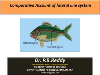
- 1. Comparative Account of lateral line system Dr. P.B.Reddy M.Sc,M.Phil,Ph.D, FIMRF,FICER,FSLSc,FISZS,FISQEM PG DEPARTMENT OF ZOOLOGY GOVERTNAMENT PG COLLEGE, RATLAM.M.P reddysirr@gmail.com
- 3. Origin of the Lateral Line System Lateral line is a canal along the side of a fish containing pores that open into tubes supplied with sense organs sensitive to low vibrations. Robert H. Denison explained the origin of the lateral line system. He explained that early vertebrates had a pore-canal system in the dermis which functioned as a primitive sensory system in detecting water movement. Through the evidences from fossils, embryology and comparative anatomy, Denison (1966) established that the inner ear is closely related to the lateral line system. He found a distinct relationship between the pore canal system and the lateral line in Osteotraci. The inner ear and the lateral line are developed from ectodermal thickenings, called dorso-lateral placodes. These have a number of similarities, including receptors with sensory hairs, and are both innervated by fibers in the acoustico-lateral area of the brain. The pore canal system is present and developed in Osteostraci (ostracoderm). It is also present in Heterostraci which is another group of ostracoderms and includes early vertebrates such as lungfishes and crossopterygians. As its presence is extensive, it is reasonable to suggest that the pore canal system was a primitive character in early vertebrates . In transverse sections also , it is very difficult to differentiate the pore canal system from a lateral line canal.
- 5. The lateral line system consists of 100 or more sensory organs (neuromasts) that are typically arranged in lines on or just under the skin of the head and body. It is absent from all reptiles, birds, and mammals, even those that are aquatic (such as turtles, dolphins, and whales). In the head, the lateral line canal is separated into three canals, one passes forward and above the eye, another forward and below the eye and the other downward and below the jaw. These three canals have numerous pores and together with the lateral line canal, make the lateral line system. Structure of the Lateral Line System
- 6. Structure of the Lateral Line System Epidermal structures called neuromasts form the peripheral area of the lateral line. Neuromasts consist of two types of cells, hair cells and supporting cells. Hair cells have an epidermal origin and each hair cell has one high kynocyle (5-10 μm) and 30 to 150 short stereocilia (2-3 μm). The number of hair cells in each neuromast depends on its size, and they can range from dozens to thousands. Hair cells can be oriented in two opposite directions with each hair cell surrounded by supporting cells. At the basal part of each hair cell, there are synaptic contacts with afferent and efferent nerve fibers. Afferent fibers, transmit signals to the neural centres of the lateral line and expand at the neuromast base. The regulation of hair cells is achieved by the action of efferent fibers. Stereocilia and kinocilium of hair cells are immersed into a cupula and are located above the surface of the sensory epithelium. The cupula is created by a gel-like media, which is secreted by non-receptor cells of the neuromast.
- 7. Figure 2. Lateral line of fish. (a) The figure shows the basic structure of neuromasts and all its components. (b) Hair cell, depicting the innervations of afferent and efferent fibers.
- 8. Superficial and Canal Neuromasts There are two types of neuromasts, superficial or free neuromasts and canal neuromasts. Superficial neuromasts are located at the surface of the head, body and caudal fin and are affected by the environment. They have a cylindrical cupula and a round base with a diameter that can seldom reach 100 μkm. The number of hair cells is small, from several dozens to several hundred. Can be found in lampreys, teleost fishes and in some bony fishes. Superficial neuromasts are categorized into primary or paedomorphic neuromasts and secondary or neomorphic neuromasts. Canal neuromasts are primary neuromasts. These are found inside epidermal or bony canals and are located on the head or body of the fish. In canal neuromasts, the sensory area is situated at the bottom of the canal below the skin. Canal neuromasts have a large range in size, shape and orientation within the canal. Some species have narrow canals and the neuromast can be found in a local constriction with the long axis running parallel to the canal axis. Some other fishes have neuromasts which are found in wide canals and have a different shape. Canal neuromasts allow the efficient detection of pressure differentials, which are created by the current movement across the canal pores.
- 9. The anterior and posterior lateral line cranial nerves collect sensory input from lateral line mechanoreceptors (afferent) on the head and trunk, respectively. Cranial nerve VIII (the octaval or stato-acoustic nerve) is an aggregate of nerve branches ( afferent) that collect input from each of the inner ear sensory end organs. Cranial nerve VIII enters the brain (medial octavo lateralis nucleus (MON) at the level of the medulla oblongata between the entry points of the anterior and posterior lateral line nerves. The lateral line and octaval nerves also contain the axons of central octavolateral efferent neurons that innervate the sensory hair cells. The final pathway for information processing is the relay of information from the midbrain to different diencephalic nuclei. Bending of the cupula caused by the movement of water particles results in bending of the cilia on the hair cell and the sending of signals to the neurons which take signals to the lateral line region of the brain.
- 10. Hair cells utilize a system of transduction that uses rate coding in order to transmit the directionality of a stimulus. Hair cells of the lateral line system produce a constant, tonic rate of firing. As mechanical motion is transmitted through water to the neuromast, the cupula bends and is displaced. Varying in magnitude with the strength of the stimulus, shearing movement and deflection of the hairs is produced, either toward the longest hair or away from it. This results in a shift in the cell's ionic permeability, resulting from changes to open ion channels caused by the deflection of the hairs. Deflection towards the longest hair results in depolarization of the hair cell, increased neurotransmitter release at the excitatory afferent synapse, and a higher rate of signal transduction. Deflection towards the shorter hair has the opposite effect, hyperpolarizing the hair cell and producing a decreased rate of neurotransmitter release. These electrical impulses are then transmitted along afferent lateral neurons to the brain.
- 11. Lateral Line Modifications The lateral line system of elasmobranchs is different to that of teleost fish. Elasmobranchs have superficial neuromasts and two morphological classes of sub-epidermal canals. Elasmobranch canals have skin pores that allow direct contact with the surrounding water. They may also have absent skin pores which prevent the contact of canal fluid with the external environment. In teleost fish, hydrodynamic pressure differences at the skin pores cause fluid motion. This results in pored canal neuromasts being able to detect the acceleration of external water flow and induce behaviours such as hydrodynamic imaging, detection of prey and schooling. Sharks and batoids have non-pored canals which are located on the ventral body surface, rostrum and around the mouth. The absence of skin pores demonstrates that localized weak hydrodynamic flow which causes pressure differences will not produce canal fluid motion directly, as it occurs in the pored canal systems. Ventral non-pored canals are sensitive to small movements of the skin, and primary afferents encode the velocity of fluid induced in the canal by these stimuli.
- 12. Conclusion The lateral line system allows the detection of movement, vibration, and pressure gradients in the water surrounding an animal, providing spatial awareness and the ability to navigate in the environment. The lateral line system which is a sensory system in fish and amphibians has various functions in schooling, navigation, and prey detection. Through paleontology, comparative anatomy and embryology it was demonstrated that there is a phylogenetic connection between the pore canal system in the dermis of early vertebrates and the lateral line. Moreover, through the action of neuromasts and hydrodynamic imaging, the fish is able to detect its surrounding environment.
- 13. The lateral line is a sensory system in fish and amphibians. It is made up of mechanoreceptors called neuromasts which are sensitive to water movement. The lateral line system has an important role in the detection of stationary objects, navigation, prey detection, capture and in swimming in schools. It is used to detect movement, vibration, and pressure gradients in the surrounding water. The sensory ability is achieved via modified epithelial cells, known as hair cells, which respond to displacement caused by motion and transduce these signals into electrical impulses via excitatory synapses. Lateral lines serve an important role in schooling behavior, predation, and orientation. Fish can use their lateral line system to follow the vortices produced by fleeing prey. Lateral lines are usually visible as faint lines of pores running lengthwise down each side, from the vicinity of the gill covers to the base of the tail. In some species, the receptive organs of the lateral line have been modified to function as electroreceptors, which are organs used to detect electrical impulses, and as such, these systems remain closely linked. Most amphibian larvae and some fully aquatic adult amphibians possess mechanosensitive systems comparable to the lateral line.
- 14. Due to many overlapping functions and their great similarity in ultra structure and development, the lateral line system and the inner ear of fish are often grouped together as the octavolateralis system (OLS). Here, the lateral line system detects particle velocities and accelerations with frequencies below 100 Hz. These low frequencies create large wavelengths, which create strong particle accelerations in the near field of swimming fish that do not radiate into the far field as acoustic waves due to an acoustic short circuit. The auditory system detects pressure fluctuations with frequencies above 100 Hz that propagate to the far field as waves. Function It allows the detection of movement, vibration, and pressure gradients in the water. Provides spatial awareness and the ability to navigate the animal in the environment. Plays an essential role in orientation, predatory behavior, defense, and social schooling. Fish are able to detect movement, produced either by prey or a vibrating metal sphere, and orient themselves toward the source before proceeding to make a predatory strike at it. This behavior persists even in blinded fish, but is greatly diminished when lateral line function was inhibited by CoCl2 application. Cobalt chloride treatment results in the release of cobalt ions, disrupting ionic transport and preventing signal transduction in the lateral lines. These behaviors are dependent specifically on mechanoreceptors located within the canals of the lateral line.
- 15. Lateral Line System Function The lateral line system function similar to the senses of touch and hearing. The earliest hypothesis about the function of the lateral line was that it secretes mucus to cover the body. Several years later, it was determined that the lateral line is used to detect water current and stimuli from moving objects. Fish can sense water movements ranging from large-scale currents to small disturbances caused by plankton. This is due to the superficial neuromasts which are able to respond to very weak water currents, with speeds from 0.03 mm/s and higher. Canal neuromasts can respond to current speeds from 0.3 to 20 mm/s. The lateral line has functions in schooling, prey detection, spawning, rheotaxis (which is a form of taxis when fish face an ongoing current), courtship and station holding. It is thought that the lateral line system can create hydrodynamic images of the surrounding area. This can be achieved by detecting moving and stationary objects in active and passive ways.
- 16. Anatomy The receptor organ of the lateral line system is the neuromast. The neuromast is a mechanoreceptive organ which allows the sensing of mechanical changes in water. There are two main varieties of neuromasts located in animals, canal neuromasts and superficial or freestanding neuromasts. Superficial neuromasts are located externally on the surface of the body, while canal neuromasts are located along the lateral lines in sub dermal, fluid filled canals. Each neuromast consists of receptive hair cells whose tips are covered by a flexible and jellylike cupula. Hair cells typically possess both glutamatergic afferent connections and cholinergic efferent connections. The receptive hair cells are modified epithelial cells and typically possess bundles of 40-50 microvilli "hairs" which function as the mechanoreceptors. These bundles are organized in rough "staircases" of hairs of increasing length order. This use of mechanosensitive hairs is homologous to the functioning of hair cells in the auditory and vestibular systems, indicating a close link between these systems.