

Recomendados
Mais conteúdo relacionado
Mais procurados
Mais procurados (20)
Destaque
Destaque (20)
Semelhante a 23204907
Semelhante a 23204907 (20)
Mais de radgirl
Último
Último (20)
23204907
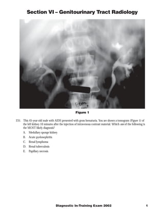
- 1. Section VI – Genitourinary Tract Radiology Figure 1 231. This 45-year-old male with AIDS presented with gross hematuria. You are shown a tomogram (Figure 1) of the left kidney 10 minutes after the injection of intravenous contrast material. Which one of the following is the MOST likely diagnosis? A. Medullary sponge kidney B. Acute pyelonephritis C. Renal lymphoma D. Renal tuberculosis E. Papillary necrosis Diagnostic In-Training Exam 2002 1
- 2. Section VI – Genitourinary Tract Radiology Question #231 Findings: Infundibular strictures with proximal dilatation Rationales: A) Incorrect. Medullary sponge kidney is characterized by dilated renal tubules and medullary nephrocalcinosis, neither of which is present in this case. B) Incorrect. Most cases of acute pyelonephritis are not detectable by excretory urography. When present, the finding of acute pyelonephritis include: Renal enlargement, diminished renal function, and a striated nephrogram. The findings in this case are due to an intrinsic process in the collecting system causing strictures and urothelial edema. C) Incorrect. Renal lymphoma is usually characterized by multiple small, smooth renal masses. The renal collecting system may demonstrate displacement secondary to mass effect, but only very rarely would intrinsic strictures be a prominent feature. Renal lymphoma is more common in immunocompromised individuals, but would not result in hematuria. D) Correct. Renal tuberculosis is a result of a blood borne infection of the kidney by mycobacterium tuberculosis. Concomitant pulmonary infection need not be present. The infection proceeds from renal parenchyma to collecting system to ureter and finally bladder in a step-wise progression. Findings of renal tuberculosis include: irregular calyces secondary to tuberculous papillitis, collecting system and ureteral strictures, parenchymal scarring, and parenchymal calcification. The end stage kidney associated with tuberculosis is termed “putty kidney.” E) Incorrect. While minimal papillary necrosis is seen in the upper pole in this case, the predominant feature is that of urothelial strictures. Papillary necrosis is characterized by collections of contrast material protruding from the calyces on urography, and can be seen in association with tuberculosis. There are many etiologies of papillary necrosis including analgesia abuse, sickle cell disease, and diabetes. 2 American College of Radiology
- 3. Section VI – Genitourinary Tract Radiology Figure 2A Figure 2B 232. A 63-year-old female had flank pain following a motor vehicle accident. A non-contrast CT was performed (Figures 2A and 2B). Which one of the following is the MOST likely diagnosis? A. Adrenal myelolipoma B. Adrenal cortical carcinoma C. Renal laceration D. Adrenal hemorrhage E. Renal angiomyolipoma Diagnostic In-Training Exam 2002 3
- 4. Section VI – Genitourinary Tract Radiology Question #232 Findings: Mixed attenuation adrenal mass, a collapsed inferior vena cava, and strandy changes around the adrenal gland, and blood in the posterior pararenal space. Rationales: A) Incorrect. Adrenal myelolipomas are a benign tumor of the adrenal gland that contain microscopic and soft tissue elements. This case demonstrates a variable high attenuation mass without fat which involves the entire right adrenal gland. B) Incorrect. Most adrenal cortical carcinomas are large (>4.0 cm) at the time of diagnosis, and do not have stranding and blood in the surrounding fat such as in this case. Adrenal carcinomas are active endocrinologically in approximately 50% of cases, but at subclinical levels. C) Incorrect. Renal lacerations are diagnosed when a crack in the renal parenchyma is present. In this case, the slice section is cephalad for the mass to originate from the kidney, and no discrete laceration of renal parenchyma is present. D) Correct. The slice is in the proper anatomic location for the mass to originate from the adrenal gland. The mass is composed of material that is denser than the contralateral kidney and hepatic parenchyma, consistent with hemorrhage. In adults, adrenal hemorrhage may be due to trauma, anticoagulation, sepsis, or adrenal tumor. Adrenal hemorrhage is more common in children than adults, and is often due to birth trauma, sepsis, or asphyxia. Most cases of adrenal hemorrhage do not have accompanying adrenal insufficiency. E) Incorrect. The anatomic location in this case is more consistent with an adrenal mass. In addition, evidence of macroscopic fat is necessary to make the diagnosis of renal angiomyolipoma. 4 American College of Radiology
- 5. Section VI – Genitourinary Tract Radiology Figure 3 233. This 24-year-old male presented with painless gross hematuria. CT showed a clot in the renal pelvis. A selective Renal arteriogram (Figure 3) is shown. Which one of the following is the MOST likely diagnosis? A. Renal angiomyolipoma B. Renal cell carcinoma C. Transitional cell carcinoma D. Congenital renal arterio-venous fistula E. Acquired renal arteries venous fistula Diagnostic In-Training Exam 2002 5
- 6. Section VI – Genitourinary Tract Radiology Question #233 Findings: Saccular vascular lesions with early venous filling Rationales: A) Incorrect. Renal angiomyolipoma is a benign renal tumor composed of fat, vascular, and myeloid elements. On angiography, angiomyolipomas have tortuous, dilated blood vessels without the prominent arteriovenous shunting seen in this case. B) Incorrect. Renal adenocarcinomas are characterized at angiography by tumor neovascularity, vascular encasement, and arteriovenous shunting. In addition, a tumor stain and vascular puddling are common findings. This case demonstrates dilated vascular lakes with rapid and profound arteriovenous shunting without tumor vascularity or staining. C) Incorrect. Transitional cell carcinomas are hypovascular, but neovascularity may be a feature by angiography, especially in large tumors. D) Correct. Congenital arteriovenous malformations are more common in women than men, and are asymptomatic or present with hematuria. The findings at angiography are pathognomonic, and include rapid filling of a circular vascular structure often in a “grape-like” configuration such as in this case. E) Incorrect. Most acquired arteriovenous malformations are due to penetrating renal trauma, such as a renal biopsy. On angiography, they tend to contain straight edges reflecting their etiology, and have rapid arteriovenous shunting. Both congenital and acquired fistulas are able to be diagnosed with color Doppler ultrasound if the degree of shunting is high enough. 6 American College of Radiology
- 7. Section VI – Genitourinary Tract Radiology Figure 4 234. A 75-year-old diabetic male presents with abdominal pain, fever, and normal white blood cell count. A CT scan with intravenous contrast (Figure 4) is obtained. Which one of the following is the MOST likely diagnosis? A. Renal abscess B. Emphysematous pyelonephritis C. Xanthogranulomatous pyelonephritis D. Renal cell carcinoma E. Renal angiomyolipoma Diagnostic In-Training Exam 2002 7
- 8. Section VI – Genitourinary Tract Radiology Question #234 Rationales: A) Correct. The findings in this case include perirenal stranding, and a fluid attenuation mass with a single air bubble within it. In most cases, renal abscess is a complication of acute pyelonephritis with suppuration. As with pyelonephritis, Escherichia coli is the most common etiologic agent. Renal abscesses are treated with antibiotics and catheter drainage if technically feasible. B) Incorrect. Emphysematous pyelonephritis is a severe interstitial renal infection caused by gas-forming organisms. Most cases are seen in diabetics. This case has a well circumscribed abscess cavity with a small amount of gas rather than a gas dissecting throughout renal parenchyma. C) Incorrect. Xanthogranulomatous pyelonephritis is a chronic, low grade infection associated with renal obstruction. It is usually seen in combination with a staghorn calculus. This case has no signs of renal obstruction. D) Incorrect. Renal cell carcinoma usually presents as a solid mass with cystic components. It usually does not present with fever. Air in a renal cell carcinoma is unusual in the absence of a renal interventional procedure, and perinephric stranding is unusual without gross extracapsular invasion. E) Incorrect. Renal angiomyolipomas are a benign renal tumor containing macroscopic fat. 8 American College of Radiology
- 9. Section VI – Genitourinary Tract Radiology Figure 5A Figure 5B 235. A 51-year-old male presents with left flank pain and hematuria. You are shown a scout radiograph (Figure 5A) and post-void view (Figure 5B) of the bladder 30 minutes after the injection of contrast material. Which one of the following is the MOST likely diagnosis? A. Ureteritis cystica B. Pseudoureterocele C. Ectopic ureterocele D. Hutch’s diverticulum E. Tuberculous ureteritis Diagnostic In-Training Exam 2002 9
- 10. Section VI – Genitourinary Tract Radiology Question #235 Findings: Scout view demonstrates a stone in the expected position of the uretero-vesical junction. Film from an excreting urogram shows a typical cobra’s head appearance to a ureterocele with the stone in the lumen. Rationales: A) Incorrect. Ureteritis cystica is a condition defined by multiple small submucosal cysts seen in association with chronic urinary tract infection. This case has a single ureterocele at the ureterovesical junction. B) Correct. Pseudo ureteroceles are acquired lesions at the ureterovesical junction (UVJ) that cause edema of the UVJ, resulting in varying degrees of ureterectasis with a “halo” appearance. Etiologies include: impaction of a stone at the UVJ, transitional cell carcinoma, and radiation therapy. Careful attention should be given to the appearance of the halo. An irregular or thickened appearance should prompt the radiologist to consider malignant etiologies. C) Incorrect. Ectopic ureteroceles are located medial and inferior to the position of orthotopic ureteroceles. Ectopic ureteroceles are associated with obstruction of the renal moiety which drains into it. D) Incorrect. A Hutch’s diverticulum is a congenital bladder diverticulum arising near the ureterovesical junction. E) Incorrect. Tuberculous ureteritis is characterized by ureteral stricture and ureteral wall thickening. Typically, tuberculosis of the urinary tract is a descending process, originating from an infected kidney. 10 American College of Radiology