
Recomendados
Mais conteúdo relacionado
Mais procurados
Mais procurados (20)
Semelhante a Ptosis
Semelhante a Ptosis (20)
Mais de nrvdad
Último
Último (20)
Ptosis
- 2. Functional anatomy • Levator palpebrae superioris (LPS): – It is the primary muscle responsible for lid elevation. – It arises from the back of the orbit and extends forwards over the cone of eye muscles. – It inserts into the eyelid and the tarsal plate, a fibrous semicircular structure which gives the upper eyelid its shape. – The LPS is supplied by the superior division of the oculomotor nerve.
- 4. Muller’s muscles: – The way that the LPS attaches to the tarsal plate is modified by the underlying Müller's muscle. – This involuntary muscle, comprising sympathetically innervated smooth muscle – has the capacity to 'tighten' the attachment and so raise the lid a few millimetres
- 5. • Frontalis & orbicularis oculi: – frontalis muscle and the orbicularis oculi, both supplied by the facial nerve. – Frontalis contraction helps to elevate the lid by acting indirectly on the surrounding soft tissues, – while orbicularis oculi contraction depresses the eyelid
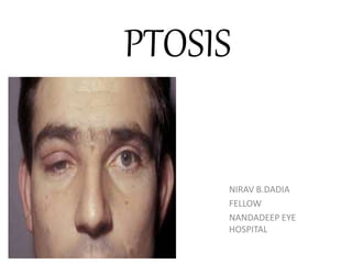
- 7. PTOSIS • Ptosis (from Greek Ptosis -to "fall") is a drooping or falling of the upper or lower eyelid. • Normally upper eyelid covers 1/6th of cornea i.e. 2mm • Therefore in ptosis it covers more than 2mm
- 10. NORMAL POSITION OF EYELIDS • The normal upper eyelid in primary position – crosses the iris between the limbus (junction of the iris and sclera) and the pupil, – usually 1 mm to 2 mm below the limbus; – the lower lid touches or crosses slightly above the limbus. – Normally there is no sclera showing above the iris. • The palpebral fissures: – are normally 9 mm to 12 mm from upper to lower lid margin.
- 12. HISTORY • Age of onset • Duration • Unilateral/bilateral • Weather ptosis worsen through the day • Diplopia • Muscle weakness • trauma/ surgery • lid edema • previous ptosis surgery
- 13. • Presence of any aberrant lid movements • Weather eye movements are impaired • Past medical history • Current medications • Family history • Old photographs
- 14. • Associated history : – Diplopia – Muscle weakness
- 15. EXAMINATION • Head posture • Periocular fullness • Frontalis overaction • Scar mark • Lid skin laxity • Telecanthus, epicanthus inversus
- 16. Ocular Motility • Importance in myogenic ptosis, • To R/O 3rd nerve palsy • presence of strabismus, especially vertical strabismus entails that it be corrected prior to the correction of the ptosis. Pupillary Examination: – TO diagnosis Horner’s syndrome – Involvement in a case of third nerve palsy Visual Acuity
- 17. Refraction- • Cycloplegic test refraction is indicated in all children with ptosis since it is known that a significant number have anisometropia primarily due to astigmatism on the ptotic side. • Any significant refractive error should be corrected • Best-corrected visual acuity should be assessed to record any amblyopia if present, especially in cases of congenital ptosis
- 18. • Complete /incomplete • Total unilateral ptosis – complete third nerve palsy. • Mild to moderate unilateral ptosis – Horner's syndrome, – partial third nerve palsy. • Mild to moderate bilateral ptosis – neuromuscular disorders, such as MG, – muscular dystrophy, – Ocular myopathy.
- 19. Measurements 1. Margin reflex distance 2. Vertical fissure height 3. LPS action 4. Lid crease level 5. Lid level on down gaze
- 20. MEASUREMENTS Margin reflex distance 1(MRD 1)- After shining the torchlight in the patient eye, the distance between the corneal light reflex to the centre of the upper lid margin is measured. Normal value is 4- 4.5mm.
- 22. Margin reflex distance 2 (MRD 2)- • The distance of corneal light reflex to the centre of the lower eyelid margin in primary gaze. • Normal value is 5- 5.5mm
- 24. • Margin reflex distance 3(MRD 3)- the distance between the corneal light reflex and the centre of upper eyelid margin in extreme upgaze.
- 26. • Also it is important to measure the PFH in downgaze. • As reduced ptosis/ lid lag is seen in congenital ptosis as the dystrophic muscle neither contracts nor relaxes.
- 28. Margin crease distance(MCD) • It is an important anatomical landmark, which give clue to levator action • It is measured with patient looking down, distance from the central eyelid margin to the most prominent lid crease. • Normal value in Men 5-7mm, women 8-10mm • Crease is absent in congenital ptosis and higher in aponeurotic ptosis.
- 30. • An absent lid crease is often accompanied by poor levator function. • If a lid crease is present, but higher than normal and if there is a deeper upper lid sulcus on that side these should be noted as signs of levator disinsertion
- 31. MARGIN LIMBAL DISTANCE- • It gives the degree of loss of Levator action. • It is measured as the distance between the centre of upper lid margin to 6o’clock limbus in extreme upgaze • Normally it is 9mm.
- 33. Levator function test- • Excursion of upper eyelid from extreme downgaze to extreme upgaze is a measure of LPS function, negating the action of frontalis muscle (Berke’s method).
- 36. ILLIF’s test • Used in children • Pt upper lid is everted in downgaze. On looking up, the lid should return to normal position if levator action is good
- 37. BELL’S PHENOMENON • The eyes moves generally upwards and outwards on eyelid closure. It is extremely important in assessing post-operative corneal complications. • Poor bells phenomenon invariably warrants under correction.
- 40. • Corneal sensation- always check before planning the surgery. • Schirmers test – to rule out dry eye disease • Pupillary abnormalities-1)miosis in horner’s syndrome 2)mydriasis in 3rd nerve palsy. Look for any associated mass lesion causing mechanical ptosis
- 41. Photographic documentation- • It is the most important aspect of ptosis evaluation. • Review of old photographs gives clue to the duration and nature of ptosis.
- 43. A. Congenital B. Acquired 1) Neurogenic 2)Myogenic 3) Aponeurotic 4)Mechanical 5)Neurotoxic C. Pseudotosis
- 44. CONGENITAL It is associated with congenital weaknes (maldevelopment) of the levator palpebrae superioris (LPS). Simple ptosis Associated with Superior Rectus weakness With blepharophimosis syndrome Synkinetic ptosis – Marcus Gun Jaw Winking ptosis,
- 46. Management • Management of BPES is primarily surgical • Care should be given to treat assoicated amblyopia. • The usual sequence of surgical treatment is -correction of the epicanthic folds at about the age of 3–4 years and - correction of the ptosis about 9–12 months later. Early surgery may be necessary for amblyopia . • Epicanthus fold and telecanthus : • Double Z or Y-Z plasties, transnasal wiring or plating of the medial canthal tendons. • Ptosis: • Usually is corrected with a brow suspension procedure (frontalis sling). • One stage correction of the telecanthus, epicanthic folds and the ptosis has been reported, although often a staged approach is done.
- 47. MARCUS GUNN PHENOMENON – The stimulation of the trigeminal nerve by contraction of the pterygoid muscles of jaw results in the excitation of the branch of the oculomotor nerve that innervates the LPS ipsilaterally, so the patient will have rhythmic upward jerking of their upper eyelid.
- 49. Grading of marcus gunn phenomenon • Mild- elevation of ptotic eyelid to non- ptotic position • Moderate- maximum elevation goes upto superior limbus • Severe- maximum elevation beyond the superior limbus with scleral show
- 50. MARCUS GUNN PHENOMENON • Marcus Gunn Ptosis constitutes about 4-5% of all congenital ptosis. Management of the condition is challenging. • A single stage surgical technique of bilateral levator excision with bilateral fascia lata sling surgery is presented. • The technique eliminates jaw winking, maintains symmetry, provides excellent aesthetics by avoiding contour and lid position abnormalities, as no tarsal fixation of the sling is required.
- 51. ACQUIRED PTOSIS Neurogenic- 3rd nerve palsy, horner syndrome. Myogenic- myasthenia gravis, Myotonic dystrophy. Aponeurotic- involutional, post surgical Mechanical- tumour, swelling.
- 52. Neurogenic ptosis It is caused by innervational defects such • 3rd nerve palsy, • 3rd nerve misdirection • Horner’s syndrome, • Cerebral ptosis • Multiple sclerosis.
- 53. Third nerve palsy • Vascular disorders such as diabetes, heart disease, atherosclerosis and aneurysm, particularly of the posterior communicating artery. • Space occupying lesions or tumours, both malignant and non-malignant. • Inflammation and infection. • Trauma. • Demyelinating disease (multiple sclerosis)
- 54. Management It is recommended to wait at least 6 months for resolution. • The patient needs to be examined periodically by then, and if some improvement is seen, further recovery could be seen up to 1 year post incident. • • With a complete ptosis, diplopia is not an issue but may require occlusion if the ptosis is partial. • In children, amblyopia needs to be addressed with patching and lid elevation with tape or a glasses crutch. • Light sensitivity with a dilated pupil may be controlled with pilocarpine drops. • Botulinum toxin has been used in the lateral rectus with limited usefulness in the short term.
- 55. Management • Patients who do not recover from third cranial nerve palsy after 6-12 months may become candidates for strabismus surgery (eye muscle resection or recession) to treat persistent and stable-angle deviation.
- 57. Horner's syndrome • Horner's syndrome, also known as oculosympathetic paresis, is a combination of symptoms that arises when a group of nerves known as the sympathetic trunk is damaged. • • The signs and symptoms occur on the same side (ipsilateral) • It is characterized by • miosis (a constricted pupil), • partial ptosis (a weak, droopy eyelid), apparent anhydrosis (decreased sweating), • with apparent enophthalmos (inset eyeball).[2]
- 59. Phenyl ephrine test • Patients with minimal ptosis (2 mm or less) should have a phenylephrine test performed in the involved eye • Either 2.5 or 10% phenylephrine is instilled in the affected eye or eyes. Usually two drops are placed and the patient is reexamined 5 minutes later. • The MRD1 is rechecked in the affected and unaffected eyes . • A rise in the MRDl of 1.5 mm or greater is considered a positive test. This indicates that Müller's muscle is viable
- 60. • The treatment of Horner syndrome depends on the location and cause of the lesion or tumor. • In some cases surgical removal of the lesion or growth may be appropriate. • Radiation and chemotherapy may be beneficial to patients with malignant tumors.
- 61. Cerebral ptosis • It is due to supranuclear lesions. • Unilateral cerebral ptosis occurs with lesions, usually ischemic, of the opposite hemisphere, and is more common with right hemisphere lesions. • Bilateral supranuclear ptosis may occur with unilateral or bilateral hemispheric lesions. • Ptosis has been reported in as many as 37.5% of patients with hemispheric strokes.
- 62. MYOGENIC PTOSIS • It is due to acquired disorders of the LPS muscle or of the myoneural junction. 1. Myasthenia gravis 2. Myotonic dystrophy 3. Ocular myopathy 4. following trauma to the LPS muscle.
- 63. Myasthenia Gravis • The ptosis in Myasthenia Gravis is frequently asymmetric and may be unilateral, though it will tend to shift from side to side • It characteristically fluctuates from moment to moment and is worsened by prolonged upgaze (fatiguable ptosis). • Cogan's lid twitch sign, – characteristic of myasthenia, consists of a brief overshoot twitch of lid retraction following sudden return of the eyes to primary position after a period of downgaze.
- 65. • Curtain sign, seesaw ptosis: – • When the ptosis is asymmetric, the driving discharges attempting to keep the more ptotic eyelid open are also transmitted, per Hering's law, to the less ptotic eyelid. • – Manually raising the more ptotic lid causes relaxation and the eye with less ptosis, sometimes even no ptosis, may suddenly crash.
- 67. Myasthenia Gravis 1. Investigations • Edrophonium (Camiston) test • Electromyography to confirm fatigue • Antibodies to acetylcholine receptors • CT or MRI for presence of thymoma
- 70. ICE TEST An ice pack is applied to the affected upper eyelid for 5 minutes. A positive test is the improvement of ptosis by > 2mm or more. This transient improvement in ptosis is due to the cold decreasing the acetylcholinesterase break-down of acetylcholine at the neuromuscular junction. More acetylcholine collects in the junction and therefore increases the muscle contraction
- 72. MYASTHENIA GRAVIS Treatment options • Medical - anticholinesterases, steroids and azathioprine Neostigmine: Initial dose: 0.03 to 0.07 mg/kg IV over a period of at least 1 minute Maximum dose: 0.07 mg/kg IV or up to a total of 5 mg IV, whichever is less • Thymectomy
- 76. APONEUROTIC PTOSIS • It develops due to defects of the levator aponeurosis in the presence of a normal functioning muscle. • It includes – involutional (senile) ptosis, – postoperative ptosis – Post traumatic dehiscence or disinsertion of the aponeurosis.
- 77. Senile ptosis • Senile or involutional ptosis is very common. Asymmetric lids and redundant lid tissue in the elderly. • The levator aponeurosis attaches the levator muscle to the tarsal plate, which forms the eyelid. • Aging may cause levator dehiscence disinsertion (LDD)—with stretching, thinning, or detachment of the aponeurosis. • Normally, with the eyelids gently closed, the upper lid margin lies 5 mm to 7 mm below the upper lid fold (the skin fold at the upper part of the lid). • An increase in this distance suggests LDD.
- 79. MANAGEMENT • Anterior levator aponeurotic-muscle reattachment or resection is effective. • Some surgeons perform a posterior resection of Müller's muscle in patients who demonstrate adequate elevation of the eyelid following instillation of topical phenylephrine.
- 80. Mechanical ptosis • Due to excessive weight on the upper lid – lid tumours – multiple chalazia – lid oedema. • Cicatricial Ptosis – ocular pemphigoid – trachoma.
- 82. Pseudoptosis • Ipsilateral hypotropia • Enopthalmos • Dermatochalasis • Double elevator palsy • Brow ptosis • Blepharospasm • Contralateral lid retraction • Contralateral exopthalmos
- 87. • So we should examine case of ptosis carefully before proceeding for surgical management, to avoid any post operative surprise.