Bone lecture
•Transferir como PPT, PDF•
6 gostaram•2,020 visualizações

lecture

Recomendados
Mais conteúdo relacionado
Mais procurados
Mais procurados (20)
Destaque
Destaque (20)
Semelhante a Bone lecture
Semelhante a Bone lecture (20)
Mais de Minia university, Faculty of Medicine
Mais de Minia university, Faculty of Medicine (14)
Último
Último (20)
Bone lecture
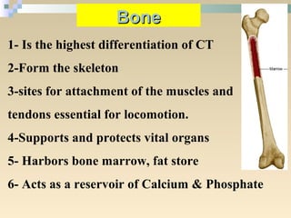
- 1. 1- Is the highest differentiation of CT 2-Form the skeleton 3-sites for attachment of the muscles and tendons essential for locomotion. 4-Supports and protects vital organs 5- Harbors bone marrow, fat store 6- Acts as a reservoir of Calcium & Phosphate BoneBone
- 2. Gross anatomy of bones Compact bone Spongy (trabecular) bone Blood vessels Medullary cavity Membranes Periosteum Endosteum
- 3. Cartilage Bone Water content: ~70% Collagen II: ~40% of organic content. Grows interstitially and by apposition. Avascular Water content: 25% Collagen I: 90% of organic content. Other Ground Substance Osteonectin: anchor collagen to bone mineral. Osteocalcin: Calcium binding protein involved in bone calcification. Osteopontin: Binding of osteoblasts and osteoclasts to bone. Grows only by apposition. Highly vascular
- 4. Types of BoneTypes of Bone (Flat, long, short and irregular bones)(Flat, long, short and irregular bones)
- 6. Flat bones Spongy bone : Compact + spongy Have bone marrow
- 7. Long bones Diaphysis= shaft Epiphyses at the ends: covered with “articular” (=joint) cartilage Epiphyseal line in adults Kids: epiphyseal growth plate (disc of hyaline cartilage that grows to lengthen the bone) Blood vessels Nutrient arteries and veins through nutrient foramen
- 8. Cells Fibers Matrix 1. Osteogenic cells 2. Osteoblasts 3. Osteocytes 4. Osteoclasts Type 1 Collagen (90%) Structure of bone Organic 1.GAGs: Hyaluronan, Chondroitin & Keratan Sulfate 2.Proteoglycans Inorganic Hydroxyapatite crystals [Ca10(PO4)6(OH)2] Calcium phosphate
- 9. 1. Osteogenic or osteoprogenitor cells: - stem cells derived from mesenchyme. - They are spindle-shaped with elongated nuclei. - present in: • inner layer of periosteum • endosteum • lining the vascular canals of compact bone. Function: differentiation into osteoblast cells. Cells of Bone
- 10. 2. Osteoblasts: - young bone cells, located at the surfaces . - known as bone-forming cells. - cuboidal cells with short, slender processes. - has a large nucleus + prominent nucleolus. - The cytoplasm: basophilic, well-developed Golgi apparatus, mitochondria. - can not divide. Functions: synthesis of the organic component and deposition of inorganic components of bone matrix.
- 11. 3. Osteocytes Once the osteoblast cells surrounded by matrix: 1. The cytoplasmic processes become more evident. 2. The cells become flattened. 3. lacunae and canaliculi appear osteoblasts are referred to as Osteocytes. - Processes make contact via gap junctions (?????) - can not divide. - faint basophilic cytoplasm. - Dark nucleus. - Few RER & Golgi. Function: maintenance of the bony matrix.
- 12. 4. Osteoclasts: - Bone-eating cells. - found in depressions known as Howship’s lacunae. - derived from the fusion of blood monocytes. - very large cells (20 – 100 µm). - multinucleated (5 –50). - acidophilic cytoplasm. -The surface facing the bone matrix is irregular. - contain a well-developed Golgi + great number of lysosomes. Function: secrete acid collagenase, and proteolytic enzymes that attack the bone matrix and liberate the calcified ground substance.
- 14. Osteocytes, Lacunae and Canaliculi Source Undetermined
- 15. Types of bone Compact Bone Cancellous bone
- 16. Compact (dense) and Spongy (cancellous) Bone cancellous bone Compact bone • found in diaphysis (shaft) of long bones. • dense, hard, with no cavities. • found in epiphyses (bulbous ends) of long bones, flat (Skull) & irregular (vertebrae) bone • with cavities.
- 17. . 1.Compact bone Compact bone is a solid mass composed of: 1. Haversian system. 2. Circumferential lamellae. 3. Interstitial lamellae (O&I). 4. Periostem 5. Endosteum
- 18. 1. Haversian system (osteon): the structural unit of compact bone, composed of: 1. Haversian canal: it runs parallel to the long axis of bone. It contains loose connective tissue rich in blood vessels and nerves and lined with osteogenic cells. 2. Bone lamellae: they are calcified osteoid tissue arranged in concentric layers (4-20 layers). 3. Osteocytes: they are present in their lacunae between the bone lamellae. 2. Circumferential lamellae: bone lamellae under the periosteum (outer circumferential lamellae) or adjacent to the endosteum (inner circumferential lamellae). 3. Interstitial lamellae: lamellae between Haversian system Volkmann`s canal: transverse or oblique canals that connect haversian canals together.
- 19. Haversian system (osteon), Haversian canal (HC) and Volkmann’s canal (VC) Transverse section Longitudinal section Osteon VC HC VC HC
- 21. •Nutrients diffuse from vessels in central canal •Alternating direction of collagen fibers increases resistance to twisting forces Isolated osteon:
- 22. • Canaliculi •Tiny canals •Radiate from the central canal to lacunae •Form a transport system Figure 5.3
- 23. Dark spots are called ‘lacunae’ and would contain osteocytes in living bone Central canal containing an artery, vein, lymph vessel and nerves.
- 24. Haversian system (osteon) /Harversian canal HC HC
- 25. Periosteum: Connective tissue membrane Covers entire outer surface of bone except at epiphyses Two sublayers 1. Outer fibrous layer 2. Inner (deep) cellular osteogenic layer (Osteoblasts: bone depositing cells, also osteoclasts: bone destroying cells) Secured to bone by perforating fibers (Sharpey’s fibers) Endosteum: Lines the internal surfaces of bones - one cell thick Is also osteogenic • Important roles of the periosteum and endosteum: 1. nutrition 2. histogenesis and repair.
- 26. Periosteum and Endosteum Endosteum Source Undetermined (Both images)
- 27. Cancellous Bone 1- NO Haversian system. 2- Irregularly arranged bone. 3- Irregular marrow spaces. 4- Periostem and endosteum.
- 29. Compact and Cancellous Bone Michigan Medical School Histology Slide Collection