Lumbar disc extrusion –complete absorption
•Transferir como PPTX, PDF•
8 gostaram•5,089 visualizações

lumbar dusc total absorption

Recomendados
Mais conteúdo relacionado
Mais procurados
Mais procurados (20)
Destaque
Semelhante a Lumbar disc extrusion –complete absorption
Semelhante a Lumbar disc extrusion –complete absorption (20)
Mais de vinod naneria
Mais de vinod naneria (20)
Último
Último (20)
Lumbar disc extrusion –complete absorption
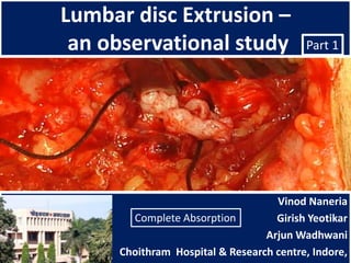
- 1. Lumbar disc Extrusion – an observational study Part 1 Vinod Naneria Complete Absorption Girish Yeotikar Arjun Wadhwani Choithram Hospital & Research centre, Indore,
- 8. Cochrane review • Prolapsed lumbar discs – < 5% of all low-back problems. • Ninety percent of acute attacks of sciatica settle with non-surgical management. • Surgical options are usually considered for more rapid relief in the minority of patients whose recovery is unacceptably slow. The effects of surgical treatments for individuals with 'slipped' lumbar discs Gibson JA, Waddell G
- 12. Saal JA, Saal JS, Herzog RJ: The natural history of lumbar intervertebral disc extrusions treated non-operatively. Spine 15: 683–686, 1990 • Patients with large compressive lesions are also generally believed to be more ideally suited to surgical intervention. These same patients, however, are those most likely to experience spontaneous regression of their lesions and they have a high rate of clinical improvement with noninvasive treatments.
- 13. Asraf Beg – case -summary Case summary 1 • 40 yr, M, short statured, obese • DM + HT • Acute PID L4 – L5 rt<1wk • Nil neurology at admission • MRI – Nov 2010 extruded disc • Tx - Conservative • Develop ankle jerk loss and EHL gr2 in one wk • Follow up MRI – Feb 2011 • Last follow up – Sept 2012 Total absorption
- 16. Case summary - 2 Complete absorption in three months
- 21. Case summary - 3 Complete absorption
- 24. Case summary - 4 Complete absorption
- 30. Case summary - 5 Complete absorption
- 34. Case summary - 6 Complete regression
- 41. Case summary - 7 Complete regression
- 44. Case summary - 8 Absorption of loose piece within one month
- 49. Case summary - 9 Total absorption
- 53. Case summary - 10 Regression of extrusion
- 55. Case summary - 11 Total absorption
- 58. Case summary - 12 Total absorption
- 64. Case summary - -13 Case summary Total absorption
- 67. Case summary - 14 Total regression
- 70. Case summary - 15 Total regression of loose piece
- 72. Total absorption
- 76. Case summary - 16 Total regression
- 82. Case summary - 17
- 85. References – same authors • http://www.slideshare.net/naneria/t1-t2-extruded-disc- case-report-14076043 • http://www.slideshare.net/naneria/lumbar-disk-update- presentation • http://www.slideshare.net/naneria/free-fragments-lumbar- spine-148466 • http://www.slideshare.net/orthonet/lumbar-disc- herniation-naneria-part-1 • http://www.slideshare.net/orthonet/lumbar-disc- herniation-naneria-part-2 • http://www.authorstream.com/Presentation/naneria- 23060-Free-fragments-lumbar-canal-LumbarCanal- fragment-Literature-Migration-Composition-extruded- materia-Education-ppt-powerpoint/
- 86. Disclaimer • All photographs were taken with the consent of the all patients. • Clinical photos were also put with due verbal permission. • This presentation strictly for students of orthopedics with the sole idea of propogating knowledge. • Any objection as for photographs or x-rays, please inform naneria@yahoo.com for prompt deletion. • Material collected from C.H.& R.C., Indore and from private clinics of the authors.
- 87. DISCLAIMER Information contained and transmitted by this presentation is based on personal experience and collection of cases at Choithram Hospital & Research centre, Indore, India, during last 25 years. It is intended for use only by the students of orthopaedic surgery. Views and opinion expressed in this presentation are personal opinion. Depending upon the x- rays and clinical presentations viewers can make their own opinion. For any confusion please contact the sole author for clarification. Every body is allowed to copy or download and use the material best suited to him. I am not responsible for any controversies arise out of this presentation. For any correction or suggestion please contact naneria@yahoo.com
Notas do Editor
- Presence of free fragments of extruded disc material is usually considered as a severe form of PID and it is considered as a strong indication for surgery. How ever when patient is not willing for surgery, a phobia is created into the mind of the patient regarding possible paraplagia (cuada-equina syndrome).
- There are three special situations. One : migration intra-dural. This is always associated with cauda-equina syndrome. Only 60 cases have been reported in the world literature.Second: Migration posterior to the dural sac. It cannot be diagnosed even on MRI and usually it gives a picture of Spinal tumour.A third situation is also commonly seen – Roof disc. It is central extrusion but still contained by the posterior longitudinal ligament. Here symptoms are different and there may not be much root pain but with profound neurological deficit.
- Inui, Yoshihiro et al in Spine. 29(21):2365-2369, November 1, 2004.Fas-Ligand Expression on Nucleus Pulposus Begins in Developing Embryo. Expressed there views as - The present results demonstrated that Fasligand expression is not detected in the notochord, but at the time of intervertebral disc formation, Fasligand expression develops in the nucleus pulposus. These results indicate that the immune privilege of the intervertebral disc may begin in the very early stages of disc formation. Moreover, Fasligand may play an important role in the formation of the intervertebral disc. There are three proposed hypothesis for absorption. The first proposed hypothesis is the retraction back into the intervertebral space. If there is bulging or herniation into the annulus fibrosis, this situation can be encountered theoretically. Second hypothesis is based on the concept of dehydration ( shrinkage with the loss of the water content of the herniated disc material), slowly The recent studies asserted that the spontaneous regression, to be a result of enzymatic degradation and phagocytosis against the extruded disc tissue in the epidural space with inflammatory reaction and neovacularization . There are some pathological and experimental studies supporting this situation. There is also possibility that all 3 of these mechanisms take part in the spontaneous regression and disappearing of the disc material altogether. In journal of Neurological Sciences Volume 23, Number 4, Page(s) 339-343, 2006, Mehmet ŞENOĞLU, KasımZafer YÜKSEL, and Mürvet YÜKSEL reported two cases of spontaneous absorption.It has been made clear in the world literature that the radicular pain from the compressed nerve root is primarily due to sensitization of the nerve root as a result of inflammation induced by antigen – antibody reaction. This is the reason for a quick response to cortisone therapy in acute prolapse. This is the reason why fragments mainly composed of nucleolus pulposus get absorbed early. The annulus is absorbed by granulation formation.Lumbar Disk Herniation:Correlation of Histologic Findings with Marrow Signal Intensity Changes in Vertebral GebhardSchmid, MDEndplates at MR Imaging- GebhardSchmid et al published in “Radiology Vol 231 No.2 - Findings in the current study show that avulsion-type disk herniation with hyaline cartilage material occurs frequently (in nearly 50% of patients). The amount of cartilage may be as much as 50% of the extruded material, and bone fragments were observed in five patients. Our results confirm that there is cartilaginous material in a high proportion of extruded disk herniations. The amount of cartilage in the herniation material is usually less than 10%, but it can be as much as 50%. The association of the amount of cartilaginous material with endplate abnormalities supports the theory that avulsion of the vertebral endplate is one source of disk herniation. The good correlation of marrow signal intensity changes in the middle third of the endplate with cartilaginous material in the disk herniation further supports the histopathologic findings by Tanaka et al (22) that most avulsions occur in the inner or transitional zone of the annulus end plate interface. Carreon LY, Ito T, Yamada M, Uchiyama S, Takahashi HE. Neovascularization induced by annulus and its inhibition by cartilage endplate: its role in disc absorption.Spine 1997; 22:1429–1434.