Extracorporeal shock wave lithotripsy (eswl)
•Transferir como PPTX, PDF•
58 gostaram•20,420 visualizações

Use focusing Shock Waves to breakdown a stone into small pieces. Shock waves are acoustic pulses. Pass through better in water and solid but not in air. Introduce in 1980 by Dornier which is a supersonic aircraft company

Recomendados
Mais conteúdo relacionado
Mais procurados
Mais procurados (20)
Destaque
Semelhante a Extracorporeal shock wave lithotripsy (eswl)
Semelhante a Extracorporeal shock wave lithotripsy (eswl) (20)
Mais de D.A.B.M
Mais de D.A.B.M (20)
Último
Último (20)
Extracorporeal shock wave lithotripsy (eswl)
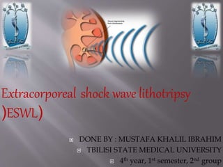
- 1. DONE BY : MUSTAFA KHALIL IBRAHIM TBILISI STATE MEDICAL UNIVERSITY 4th year, 1st semester, 2nd group Extracorporeal shock wave lithotripsy (ESWL(
- 3. Crystallization of minerals inside urine, which act as the nidus for more sedimentation and finally the formation of a stone within the kidney.
- 4. Calcium-containing stone Calcium Oxalate Calcium Phosphate Uric acid stone Cysteine stones
- 5. NO symptom Pain: sudden or severe pain nausea, vomiting Renal colic Frequent and painful urination, hematuria Urinary tract infection: Block the urinary tract
- 7. ESWL Percutaneous nephrolithotomy Ureteroscopy Open surgery
- 8. Use focusing Shock Waves to breakdown a stone into small pieces. Shock waves are acoustic pulses. Pass through better in water and solid but not in air. Introduce in 1980 by Dornier which is a supersonic aircraft
- 9. Contra-indicationIndication Relevant coagulation problems Lung tissue in shock wave path Tumors in shock wave area Aneurysms Polyarthritis (difficult to positioning) Active pyelonephritis Pregnancy • Stones of less than 2 cm in the kidney • Or • less than 1 cm in the ureter.
- 10. 1) A shockwave generator (electromagnetic generator) 2) A focusing system 3) A coupling system 4) An imaging/localization units
- 12. Provide a air-free contact In the propagation and transmission of a wave, energy is lost at interfaces with differing densities. A coupling system is needed to minimize the dissipation of energy of a shockwave as it traverses the skin surface
- 13. Transcranial magnetic stimulation Dornier
- 16. 1) find out the location of stone 2) Fasting 3) Take the blood pressure 4) Check the cardiac physical exam 5) Pre-medication (pain relief) 6) Check LMP for female patients 7) Brief the details of the treatment to the patient
- 17. Lie the patient on the table (Supine oblique or prone(
- 18. 1) Compare with the previous KUB image 2) Using, iliac crest and the spineas landmark 3) Move the patient in the mid level of the removable broad KUB : Kidneys, ureters, and bladder x-ray
- 19. 4)Remove the broad 5)Apply gel to the coupling cushion 6)Move the coupling cushion to treatment position 7) Increase the coupling pressure and touch the patient skin 8) Apply soft pad or sand bag on the opposite side of the patient (immobilize the patient)
- 20. 10)Screening in PA view 11)Move the table to locate the stone in the center 12)Screening in CC view 13)Adjust the height of the table to locate the stone in center 14) Instruct to the patient 15) Call doctor to confirm the position and start the treatment
- 22. Select the suitable parameters 1)Power of shockwave (start from low energy level to high energy level) 2)The frequency of shockwave (ECG gated for patients with cardiac pacemakers or those with arrhythmias who regularly take anti-arrythmic drugs 3) Total energy of shockwave (Renal stone < Ureteric stone( High energy level + high frequency = shorter treatment time Low energy level + low frequency =longer treatment time
- 23. Monitor the patient condition e.g. Blood pressure,heart rate, pain Any abnormality => Stop shock wave! Monitor the position and the progress of stone Move far away from the center => Stop shock wave and make adjustment!
- 26. Patient is being observed for at least an hour in Day ward. Follow up 2 weeks later with X-ray (KUB) Remaining Stone => ESWL again Other treatment
- 27. Hematomas Risk of hemorrhage Hyperventilation tetany Blockage of urinary tract The higher the total energy, the higher risk
- 28. DisadvantagesAdvantages May require repeat procedures Not suitable for all types of stones Cause complications Painful Non-invasive Safe No General anesthesia Short treatment time Convenience
- 29. 1) Presentation powerpoint by Beatrice Pang and Connie Li,2011 2) Dornier Medtech. Operating Manual of Dornier Gemini. 2012 3) JS Rodman et al. No more kidney stones. 2007 4) SWH Chan et al.A report on randomly sampled questionnaire survey about renal stone disease in Hong Kong. HK Med J. 2008 5) B Sturtevant et al. Fracture mechanics model of stone comminutionin ESWL and implications for tissue damage. Phys Med Biol. 2000 6) W Eisenmenger.The mechanisms of stone fragmentation in ESWL. Ultrasound in Med. & Biol. 2001 7) http://zh.wikipedia.org/w/index.php?title=Image:KUB_stone.j pg&variant=zh-tw 8) http://www.medison.ru/uzi/img/p287.jpg 9) http://www.mwstone.com/STONES/equipment.htm 10) http://www.tms-uro.com/eng/physicians/swl/1a_vision_device.htm 11) http://www.dornier.com/EMEA/clinical- solutions/urology/kidney-stones/ 12) http://emedicine.medscape.com/article/444554-overview