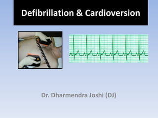
Defibrillation & cardioversion by DJ
•Transferir como PPTX, PDF•
43 gostaram•5,885 visualizações

Defibrillation & cardioversion Presentation by DJ as Medical Officer

Recomendados
Mais conteúdo relacionado
Mais procurados
Mais procurados (20)
Destaque
Semelhante a Defibrillation & cardioversion by DJ
Semelhante a Defibrillation & cardioversion by DJ (20)
Mais de Dharmendra Joshi
Último
Último (20)
Defibrillation & cardioversion by DJ
- 1. Dr. Dharmendra Joshi (DJ) Defibrillation & Cardioversion
- 2. • History • Case & Census • Definition • Types • Principles • Indications • Contraindications • Anaesthesia • Equipments • Positioning • Technique • Safety • Complications • Troubleshooting Topics
- 3. History • 1849: Ludwigg and Hoffa – VF induced by electrical stimuli
- 4. History • 1899: Prevost and Batelli - while a weak stimulus can produce fibrillation, a stimulus of higher strength applied to the heart could arrest ventricular fibrillation and restore normal sinus rhythm.
- 5. History • 1947: First defibrillation on humans.
- 6. History • 1966: Belfast Ambulance transported physicians performed first pre-hospital defibrillation. • 1969: First pre-hospital defibrillation by non physicians. • 1970’s: Diack, Wellborn and Rullman developed first AED’s.
- 7. Chain of Survival • Early Recognition and Assessment • Early Access • Early CPR • Early Defibrillation • Early Advanced Cardiac Life Support
- 8. Chain of Survival Sudden cardiac arrest survival rate: • Pre-Hospital: 10% • In-Hopsital: 10%
- 9. • No record found about patient who was treated by defibrillation or cardioversion in KMCTH, Duwakot in last one month!!! Case Scenario & Census
- 10. • Defibrillation is a non-synchronized delivery of energy during any phase of the cardiac cycle. • Cardioversion is the delivery of energy that is synchronized to the large R waves or QRS complex. Definition
- 11. 11 TYPES OF DEFIBRILLATORS Internal External
- 12. DEFIBRILLATOR ELECTRODES Types of Defibrillator electrodes:- a) Spoon shaped electrode • Applied directly to the heart. b) Paddle type electrode • Applied against the chest wall. c) Pad type electrode • Applied directly on chest wall.
- 14. Fig.- Pad electrode DEFIBRILLATOR ELECTRODES
- 15. PRINCIPLE OF DEFIBRILLATION Energy storage capacitor is charged at relatively slow rate from AC line. Energy stored in capacitor is then delivered at a relatively rapid rate to chest of the patient. Simple arrangement involve the discharge of capacitor energy through the patient’s own resistance.
- 17. PRINCIPLE OF DEFIBRILLATION The discharge resistance which the patient represents as purely ohmic resistance of 50 to 100Ω approximately for a typical electrode size of 80 cm2. This particular waveform Fig 13.9(b) is called ‘ Lown’ waveform. The pulse width of this waveform is generally 10 ms.
- 18. power supply energy storage patient ECG monitor timing circuitry gate charge discharge standby switch is under operator control applies shock about 20 ms after QRS complex, avoids T-wave
- 20. • Minimum defibrillation energy occurs for pulse durations of 3 - 10 ms (for most pulse shapes). • Pulse amplitude in tens of amperes (few thousand volts).
- 21. – intrinsic characteristics of patient – patient’s disease – duration of arrhythmia – patient’s age – type of arrhythmia (more energy required for v. fib.) • Operator selects energy delivered: 50-360 joules, depends on:
- 22. 22 THE POWER OF DEFIBRILLATION • Higher voltages are required for external defibrillation than for internal defibrillation. • A corrective shock of 750-800 volts is applied within a tenth of a second. • That is the same voltage as 500-533 no. of AA batteries!
- 24. Occlusion of the coronary artery leads to ischemia Ischemia leads to infarct which causes interruption of normal cardiac conduction Infarct = VF/VT
- 25. Ventricular Fibrillation Ventricular Tachycardia
- 26. • Indications for synchronized electrical cardioversion: Supraventricular tachycardia Atrial fibrillation Atrial flutter Ventricular tachycardia Reentrant tachycardia with narrow or wide QRS complex. Indications
- 27. 1. Pulse less ventricular tachycardia (VT) 2. Ventricular fibrillation (VF) Indications: Defibrillation
- 28. • Dysrhythmias • Multifocal atrial tachycardia Contraindications
- 29. • Cardioversion: – Almost always under induction or sedation. – Exceptions: hemodynamic instability or if cardiovascular collapse is imminent. • Defibrillation – is an emergent maneuver and performed promptly. Anesthesia
- 30. • Defibrillators (automated external defibrillators [AEDs], semi-automated AED, standard defibrillators with monitors) • Paddle or adhesive patch • Conductive gel or paste • ECG monitor with recorder • Oxygen equipment • Intubation kit • Emergency pacing equipment Equipments
- 31. • Antero-lateral • Antero-posterior Positioning Fig. anterior –apex scheme of electrode placement (AED)
- 32. – Emergent application – Elective cardioversion – Repetitive, futile attempts – Advanced cardiac life support (ACLS) Technique
- 33. Waveforms Monophasic Biphasic Charge Only one direction Two direction (opposite) Effectiveness Less effective More effective and at lower energies Technique Contd…
- 34. Monophasic Defibrillation • Delivers ‘shock’ in one phase • Adult: 200J, 300J, 360J, all subsequent shocks at 360J • Child: 2J/Kg, 2J/Kg, 4J/Kg, all subsequent shocks at 4J/Kg
- 35. Biphasic Defibrillation • Two phases to the delivery of the ‘shock’ • Adjusts ‘shock’ according to thoracic impedance • Adult: 150J, 150J, 150J • Child: 1– 2J/Kg
- 36. Energy selection: Cardioversion: • Begins with 25-50 J for atrial flutter. • 50-100 J (or the biphasic equivalent) to treat atrial fibrillation. Technique Contd…
- 37. Defibrillation: • Immediate • Rapid polymorphic ventricular tachycardia (rate >150 bpm) >> 200-360 J (or biphasic equivalent [100-200 J). • Monomorphic ventricular tachycardia >> 100- 200 J (or biphasic equivalent [50-100 J]). • Ventricular fibrillation >> 200-360 J (or biphasic equivalent [100-200 J]). Technique Contd…
- 38. Classes of discharge waveform Fig:- Generation of bi-phasic waveform
- 39. The biphasic waveform is preferred over monophasic waveform to defibrillate, WHY????? • A monophasic type, give a high-energy shock, up to 360 to 400 joules due to which increased cardiac injury and it burns the chest around the shock pad sites. • A biphasic type, give two sequential lower-energy shocks of 120 - 200 joules, with each shock moving in an opposite polarity between the pads. Classes of discharge waveform
- 41. Rhythm Analysis and the Defibrillator Video Demonstration
- 42. Safety General Safety • Yourself, other staff – Dry surface area – Oxygen • Chest wall – GTN patch – Jewellery – Paddles / Pads not touching • Technique – One Person – Two Person – Adhesive Pads
- 43. Safety Operator Safety • Assertive • Announce: – CHARGING, – ALL CLEAR / STAND CLEAR (Visual Check of Area), – SHOCKING • Check rhythm • Discharge Shock • Continue as per algorithm
- 44. PRECAUTIONS IN DEFIBRILLATION PROCESS The paddles used in the procedure should not be placed:- • on a woman's breasts. • over an internal pacemaker patients. Before the paddle is used, a gel must be applied to the patient's skin.
- 45. Common: • Atrial, Ventricular and Junctional premature beats Serious: • Ventricular fibrillation (VF) • Digitalis toxicity • Severe heart disease • Thromboembolization (1-3%) • Myocardial necrosis • Pulmonary edema • Painful skin burns Complications
- 46. • Attach the external and internal paddles if the monitor reads, "No paddles." • Check to ensure that the leads are securely attached if the monitor reads, "No leads." • Connect the unit to AC power if the message reads, "Low battery." • Verify that the Energy Select control settings are correct if the defibrillator does not charge. TROUBLESHOOTING
- 47. • Change the electrodes and make sure that the electrodes adapter cable is properly connected if you receive a message of "PACER FAILURE." Restart the pacer. • Close the recorder door and the paper roll if the monitor message reads, "Check recorder”. TROUBLESHOOTING
- 49. ‘’Success depends on delivery of current to the myocardium’’
- 50. Thank You!!!
Notas do Editor
- 1849: Written history of fibrillation and defibrillation goes back to the pioneering work of Carl Ludwig’s laboratory. In 1849, Ludwig’s student M. Hoffa was the first to witness and, most importantly, to document the onset of ventricular fibrillation, which he induced by electrical stimulus. This picture from their paper shows rapid contractions produced by electrical stimulation, which resulted in cardiac arrest.
- 1899: Further experiments with “faradization” of the heart were conducted by two physiologists from University of Geneva, Switzerland, J.-L. Prevost and F. Batelli. They discovered that, while a weak stimulus can produce fibrillation, a stimulus of higher strength applied to the heart could arrest ventricular fibrillation and restore normal sinus rhythm. This discovery was made in 1899. Unfortunately, unlike discovery of contemporary electrocardiogram, defibrillation did not enjoy similar attention and success. They did this using dogs!
- Work of Carl J. Wiggers in the Department of Physiology of Western Reserve University was well known to the thoracic surgeon Claude S. Beck from the University Hospitals in Cleveland, which are adjacent to the Western Reserve University. In 1947, Dr. Beck successfully applied defibrillation therapy and saved the first human life by this method (C.S. Beck, W.H. Pritchard, H.S. Feil, Ventricular fibrillation of long duration abolished by electric shock. Jour. Amer. Med. Assoc. 135: 985, 1947). His success triggered the immediate acceptance of this method by the clinical community and started a wide front of basic and clinical research of fibrillation and defibrillation.
- Electrical cardioversion has now become a routine procedure and is used electively or emergently to terminate cardiac arrhythmias. The delivered shock in both defibrillation and cardioversion causes electric current to go from the negative to the positive electrode of the defibrillator, passing the heart on its way. It causes all the heart cells to contract simultaneously, thereby interrupting and terminating the abnormal electrical rhythm without damaging the heart, and thus allowing the sinus node to resume normal pacemaker activity.
- Internal defibrillator Electrodes placed directly to the heart Eg.-Pacemaker External defibrillator Electrodes placed directly on the heart Eg.-AED
- Explain the cardiac conduction pathways with the electrical impulse originating in the SA node (right atrium) which travels to the AV node (division of the right and left ventricles), down along the right and left bundle branches to the Purkinje fibers. Intrinsic heart rates at the SA node are 80 bpm, the AV node is 60 bpm, and the ventricular rate (Purkinje fibers) is 40 bpm.
- Any patient with reentrant tachycardia with narrow or wide QRS complex (ventricular rate >150) who is unstable (eg, chest pain, pulmonary edema, hypotension)
- 3. Cardiac arrest due to or resulting in VF Ventricular fibrillation is a serious cardiac emergency resulting from asynchronous contraction of the heart muscles. Due to ventricular fibrillation, there is an irregular or rapid heart rhythm. Ventricular fibrillation can be converted into a more efficient rhythm by applying a high energy shock to the heart. This sudden surge across the heart causes all muscle fibres to contract simultaneously. Possibly , the fibres may then respond to normal physiological pacemaking pulses. The instrument for administering the shock is called a DEFIBRILLATOR.
- Dysrhythmias due to enhanced automaticity such as in digitalis toxicity and catecholamine-induced arrhythmia. For dysrhythmias due to enhanced automaticity such as in digitalis toxicity and catecholamine-induced arrhythmia, a homogeneous depolarization state already exists. Therefore, cardioversion is not only ineffective but is also associated with a higher incidence of postshock ventricular tachycardia/ventricular fibrillation (VT/VF).
- Cardioversion is almost always performed under induction or sedation (short-acting agent such as midazolam). The only exceptions are if the patient is hemodynamically unstable or if cardiovascular collapse is imminent. Defibrillation is an emergent maneuver and when necessary should be promptly performed in conjunction with or prior to administration of induction or sedative agents.
- The use of hand-held paddle electrodes may be more effective than self-adhesive patch electrodes. The success rates are slightly higher for patients assigned to paddled electrodes because these hand-held electrodes improve electrode-to-skin contact and reduce the transthoracic impedance.
- Paddle placement on the chest wall has 2 conventional positions: anterolateral and anteroposterior. In the anterolateral position, a single paddle is placed on the left fourth or fifth intercostal space on the midaxillary line. The second paddle is placed just to the right of the sternal edge on the second or third intercostal space. In the anteroposterior position, a single paddle is placed to the right of the sternum, as above, and the other paddle is placed between the tip of the left scapula and the spine. An anteroposterior electrode position is more effective than the anterolateral position for external cardioversion of persistent atrial fibrillation. The anteroposterior approach is also preferred in patients with implantable devices, to avoid shunting current to the implantable device and damaging its system.
- Emergent application, which may be lifesaving, and elective cardioversion should be used cautiously, with attention to patient selection and proper techniques. Repetitive, futile attempts at direct current cardioversion should be avoided. Advanced cardiac life support (ACLS) measures should be instituted in preparing the patient, such as obtaining intravenous access and preparing airway management equipment, sedative drugs, and a monitoring device.
- Defibrillators can deliver energy in various waveforms that are broadly characterized as monophasic or biphasic. Monophasic defibrillation delivers a charge in only one direction, while biphasic defibrillation delivers a charge in one direction for half of the shock and in the electrically opposite direction for the second half. Biphasic waveforms defibrillate more effectively and at lower energies than monophasic waveforms
- Synchronized electrical cardioversion begins with 25-50 J (or the biphasic equivalent, which is generally one half of that required with monophasic waveforms) to treat atrial flutter and 50-100 J (or the biphasic equivalent) to treat atrial fibrillation for patients in stable condition, as shown in ECG.
- Rapid polymorphic ventricular tachycardia (rate >150 bpm) associated with hemodynamic instability should be treated with immediate, direct-current, nonsynchronized defibrillation with energies of 200-360 J (or biphasic equivalent [100-200 J). Monomorphic ventricular tachycardia should be treated with a synchronized discharge of 100-200 J (or biphasic equivalent [50-100 J]). Ventricular fibrillation should be treated with unsynchronized electrical countershock with at least 200-360 J (or biphasic equivalent [100-200 J]) administered as rapidly as possible, as shown in ECG.
- The most common complications are harmless arrhythmias, such as atrial, ventricular, and junctional premature beats. Serious complications include ventricular fibrillation (VF) resulting from high amounts of electrical energy, digitalis toxicity, severe heart disease, or improper synchronization of the shock with the R wave. Thromboembolization is associated with cardioversion in 1-3% of patients, especially in patients with atrial fibrillation who have not been anticoagulated prior to cardioversion. Myocardial necrosis can result from high-energy shocks. ST segment elevation can be seen immediately and usually lasts for 1-2 minutes. ST segment elevation that lasts longer than 2 minutes usually indicates myocardial injury unrelated to the shock. Pulmonary edema is a rare complication of cardioversion and is probably due to left ventricular dysfunction or transient left atrial standstill. Painful skin burns can occur after cardioversion or defibrillation; they are moderate to severe in 20-25% of patients. They most likely are due to improper technique and electrode placement.