SlideShare uma empresa Scribd logo
1 de 40
GRAM’S STAIN
Mr. Manoj Mahato
HISTORICAL
BACKGROUND
• Hans Christian Joachim Gram, Danish bacteriologist and
physician.
 Developed Gram staining technique in 1883 and published his
findings in 1884 in Friedlander’s Journal.
 Was working with respiratory disease in lung tissue from
cadaver at Municipal Hospital Berlin and his accidental
spillage of lugol’s iodine over lung tissue sections led to the
development of Gram Staining.
 While examining the lung tissue from the patients who had
died of pneumonia, he observed that certain stains were
preferentially taken up and retained by bacterial cell.
•His initial work with this staining process was performed on
Kleibsella pneumoniae and Streptococcus pneumoniae.
•He did not use a counter stain in his procedure. It was a few
years later, that the German pathologist Carl Weigert (1845-
1904) from Frankfurt, added a final step of staining with
safranin.
STAINING
It is the artificial coloration of a substance
to facilitate examination of tissue,
microorganism or other cells under
microscope .
Classification of staining
On the basis of staining property
a. Positive staining: Gram staining, AFB staining, H/E
staining etc.
b. Negative staining: Stains the background material
e.g. India ink to demonstrate the capsulated yeast cells
in CSF
On the basis of number of stains used
a. Simple staining: e.g. Methylene blue ,Wayson’s stain .
b. Differential staining: e.g. Gram staining , AFB
staining .
Introduction
 Very useful stain in diagnostic Microbiology
 Several modifications after discovery
 No technology developed to substitute Gram’s stain till the
date
 Very easy to perform within short time period
 Helps in rapid diagnosis of several microbial diseases
 Guides physician to start appropriate antimicrobials
providing evidence of Gram positive and Gram negative
bacteria from clinical specimen
Solutions used
I. Primary stain- Crystal violet
II. Mordant- Gram’s Iodine
III. Decolorizer- Acetone or alcohol or mixture of
acetone alcohol
IV. Counter stain- Neutral red or Safranine
Importance of Gram’s stain
 Important test for the rapid presumptive diagnosis of
infectious agent
 To classify the bacteria as a Gram positive or Gram negative
 To study the morphology of bacteria
 To study the arrangement of bacteria
 To find out the evidence of capsule
 To find out the evidence of spore
 To find out the evidence of pus cells
 To find out the evidence of epithelial cells
 To find out the evidence of Yeast cells
Difference betweenGrampositive and Gramnegative bacteria
Peptidoglycan
Lipopolysaccharid
e
Techoic acid/
Techuronic acid
PH of protoplasm
Gram positive
Thick layer
Thin layer
Present
2-3
Gram negative
Thin layer
Thick layer
Absent
4-5
Principle of Gram’s stain
Three hypothesis
a. Cell wall related
b. PH related
c. Magnesium ribonuclease related
PRINCIPLE
Cell wall permeability hypothesis:
 Gram positive cell wall has thicker peptidoglycan layer as
compared to Gram negative cell wall .
 Gram negative cell wall contains a thick layer of
lipopolysaccharide in their cell wall which is absent in Gram
positive bacteria .
 During Gram staining a dye iodine complex (CVI complex)
or lake is formed within the cell wall after staining with crystal
violet and on subsequent treatment with iodine .This complex
is insoluble in water but soluble in alcohol.
 Now when we add decolourizer(Acetone alcohol ),the dye
iodine complex is trapped inside the thick peptidoglycan layer
of Gram positive bacteria so they are not decolorized .
Cont …..
 But outer lipopolysacharide layer of Gram negative cell
wall is easily dissolved by the decolourizer due to which
dye iodine complex is easily washed away from Gram
negative cell.
Principle
Presence of magnesium ribonucleate :
 Magnesium ribonucleate present in Gram positive
bacteria has affinity for basic dyes. As a result Gram
positive bacteria take the color of crystal violet .
 It has also been proved that Gram positive bacteria
become Gram negative upon removing this material.
Principle
 pH hypothesis :
Cytoplasmic pH of Gram positive bacteria is more
acidic (2-3) as compared to Gram negative bacteria(4-
5) and since the primary stain used(crystal violet ) is
basic in nature it develops more affinity with the Gram
positive cytoplasm .
 The difference in pH is mostly due to presence of
Teichoic acid in the cytoplasm of gram positive cell,
which is absent in Gram negative cell .
Reagentpreparation
 Crystal violet –Primary stain (0.5 -2%)
Crystal violet – 20gm
Ammonium oxalate -9gm
Absolute ethanol or methanol -95ml
Distilled water –Upto to 1 liter.
• Lugol’s iodine solution –Mordant
Potassium iodide - 20gm
Iodine - 10gm
Distilled water -Upto 1 liter
Note : Lugol's Iodine = 20g Potassium Iodide + 10g Iodine dissolved in
1L H2O
Gram's Iodine = 6.7g potassium iodide + 3.3g Iodine dissolved
in 1L H2O
Cont…..
 Acetone alcohol -Decolourizer
Acetone -500ml
Ethanol or Methanol absolute -475ml
Distilled water - 25ml
 Neutral red – Counter stain (0.1%)
1:10 diluted carbol fuschin or saffranin (0.5%) can also
be used .The use of dilute carbol fuschin is
recommended for staining Yersinia Campylobacter,
Hemophilus and Vibrio species .
 Precaution :Ethanol and acetone being flammable , has
to be used away from flame .
Storage and stability
 Reagents should be stored in brown glass bottles at
room temperature .
 Crystal violet is stable for 1 year .
 Lugol’s iodine : 6 months .
 Acetone alcohol : Indefinitely .
 Neutral red : 1 year .
The GramStain Procedure
•Step 1 - Prepare a Smear
Watch what happens to the “Bacteria” at each step
“Bacteria”
Slide has to be perfectly clean and smear has to be about
1.5 cm in diameter
Allow to air dry. Heat fix by gently warming
above a flame or other heat source.
Note :For detection of Gonococci and Meningococci
:methanol fix for 5 minutes to avoid damaging of pus
cellls .
The Gram Stain Procedure
•Step 2 - Apply the Primary Stain
Flood the Smear with Crystal Violet
Allow to stand 1 min
Rinse with water to remove excess stain
The Gram Stain Procedure
•Step 3 - Apply the Mordant
Flood the Smear with Iodine solution
Allow to stand 1 min
The Gram Stain Procedure
•Step 4 - Rinse
Rinse with water to remove excess Iodine
The Gram Stain Procedure
•Step 5 - Decolorize
Cover the smear with 70 % Acetone Alcohol for 30 sec
The effluent should appear pale or clear
The Gram Stain Procedure
•Step 6 - Rinse
Rinse with water to remove excess alcohol
The Gram Stain Procedure
•Step 7 - Counterstain
Flood the slide with Safranin solution
Let stand 1 minute.
The Gram Stain
•Step 8 - Rinse, Dry and Observe
Gram-Positive Gram-Negative
Rinse with water to remove excess stain
Blot dry
Observe under Oil Immersion
Examples of Gram Stains
Gram Positive Rods
and Cocci
Gram Negative Rods
and Cocci
RESULTS
Gram positive bacteria………..Dark purple
Yeast cells……………………...Dark purple
Gram negative bacteria…………Pale to dark red
Epithelial cells…………………..Pale red
Reporting of Gramsmears
The report should include the following information:
- Number of bacteria present, whether many, moderate, few
or scanty
- -Gram reaction of the bacteria, whether Gram positive or
Gram negative.
- Morphology of the bacteria whether cocci, diplococci,
streptococci, rods or coccobacilli.
- Whether the organisms are intracellular.
- Presence and number of pus cells.
- Presence of yeast cell and epithelial cells
Result and interpretation
a. Evaluate the general nature of the smear under
low-power magnification (10X).
Determine if smear has been properly decolorized.
Determine if thickness of smear is appropriate.
b. Examine under low power for the evidence of
inflammation
Relative amount of WBCs
Relative amount of epithelial cell
c. Location and arrangement of microorganism
Variation in Gram reaction.
Gram positive organism may lose their ability to retain crystal violet and stain Gram
negative for the following reasons:
Cell wall damage due to antibiotic therapy or excessive heat fixation of the
smear.
Over de-colorization of the smear
Use of an iodine solution which is too old
Smear that has been prepared from an old culture
Gram negative organisms may not be fully decolorized and appear as gram positive
when a smear is too thick.
Recording observations
a. Record relative amount of observed cells and
microorganism
 Numerical
1+ (< 1 per oil immersion field, 100x)
2+ (1 per oil immersion field)
3+ (2 to 10 per oil immersion field)
4+ (predominant or >10 per oil immersion field)
 Descriptive
Rare (<1 per oil immersion field)
Few (1 to 5 per oil immersion field)
Moderate (5 to 10 per oil immersion field)
Many (>10 per oil immersion field)
Quality control
 Check appearances of chemicals daily.
 Daily and when a new lot of stain is used, prepare a
smear of E. coli, S. epidermidis or S. aureus, fix and
stain.
 Use a set of reference slide.
 To avoid the errors resulting from dirty slides use the
slides previously dipped in 95% Ethanol and soak prior
to use.
Limitations of Gram stain
 The number of microorganism required is
very high, for visualizing with Gram stain
(>10000 organisms/ml)
 Can not give good results under following
conditions:
a. Cell wall damage due to antibiotic
therapy or over heat fixation of smear
b. Use of old cultures
c. Uneven thickness of smear
d. Use of old iodine solution
e. Over decolorization of smear
Gram stain modifications and uses
Stain and use Original Hucker’s
(1921)
Kopeloff and
Beerman’s
Primary stain Gentian violet Crystal violet Alkaline crystal
violet
Mordant Lugol’s iodine Gram’s iodine Kopeloff’s iodine
Decolourizer Absolute
alcohol
Acetone
alcohol
3:7 acetone alcohol
Counter stain Bismark
brown
Safranin Kopeloff’s safranin
Use General
bacteriology
Anaerobes , For
diagnosis of
bacterial vaginosis .
Gram stain modifications and uses
Stain and use Jensen’s Preston and
Morrell’s(1962
)
Brown and
Brenn
Weigert’s
Primary stain 0.5%methyl
violet
Ammonium
oxalate-crystal
violet
1%Crystal
violet
Carbol gentian
violet
Mordant 1%Lugol’s
iodine
1%Lugol’s
iodine
Lugol’s Iodine Gram’s iodine
Decolourizer Absolute
alcohol
Iodine –
acetone
Acetone Aniline xylene
Counter stain 0.1% Neutral
red
Dilute carbol
fuschin
0.25%Basic
fuschin
Carminic acid
and potassium
alum in water .
Use For Gonococci
and
Meningococci
Bacteroides
spp,Fusobacte
rium species ,
For
Demonstrating
Bacteria in
tissue .
Pneumocyst
,Nocardia .
Applications of Gram’s Staining
 Used to guide initial therapy until definitive
identification of microorganism obtained.
 Morphology of stained bacteria can sometimes be
diagnostic. For example Gram negative intracellular
diplococci in urethral pus provides a presumptive
diagnosis of Gonorrhea .
 Sometimes specimens may show organisms under
microscope but appear sterile in culture media .In
these cases Gram stain is the only clue to the nature
,variety and relative proportion of infecting organism
.
 Aids in interpretation of culture reports .

Mais conteúdo relacionado

Mais procurados

Mais procurados (20)

Laboratory diagnosis of fungal infections
Laboratory diagnosis of fungal infectionsLaboratory diagnosis of fungal infections
Laboratory diagnosis of fungal infections
 
Automated methods in the microbiology lab
Automated methods in the microbiology labAutomated methods in the microbiology lab
Automated methods in the microbiology lab
 
Gram’s staining
Gram’s  stainingGram’s  staining
Gram’s staining
 
Capsule staining
Capsule stainingCapsule staining
Capsule staining
 
Staphylococcus aureus
Staphylococcus aureusStaphylococcus aureus
Staphylococcus aureus
 
Gram staining
Gram stainingGram staining
Gram staining
 
Gram Stain Practical Briefing
Gram Stain Practical BriefingGram Stain Practical Briefing
Gram Stain Practical Briefing
 
Capsule staining
Capsule stainingCapsule staining
Capsule staining
 
Negative staining
Negative stainingNegative staining
Negative staining
 
Staining Techniques in Microbiology
Staining Techniques in MicrobiologyStaining Techniques in Microbiology
Staining Techniques in Microbiology
 
Spore staining
Spore stainingSpore staining
Spore staining
 
Oxidase test
Oxidase testOxidase test
Oxidase test
 
Fungal media
Fungal mediaFungal media
Fungal media
 
Lab diagnosis of fungal infections, Dr Naveen Reddy
Lab diagnosis of fungal infections, Dr Naveen ReddyLab diagnosis of fungal infections, Dr Naveen Reddy
Lab diagnosis of fungal infections, Dr Naveen Reddy
 
Ziehl neelsen staining
Ziehl neelsen stainingZiehl neelsen staining
Ziehl neelsen staining
 
Streptococcus
StreptococcusStreptococcus
Streptococcus
 
2 specimen collection
2 specimen collection2 specimen collection
2 specimen collection
 
Streptococcus
Streptococcus Streptococcus
Streptococcus
 
Flagella Staining.pptx
Flagella Staining.pptxFlagella Staining.pptx
Flagella Staining.pptx
 
Enterobacteriaceae
EnterobacteriaceaeEnterobacteriaceae
Enterobacteriaceae
 

Semelhante a Gram stain by manoj

bacterial different staining technique
bacterial different staining techniquebacterial different staining technique
bacterial different staining technique
Koi Bdk Pahang
 
Gram stain for microbiology student
Gram stain for microbiology studentGram stain for microbiology student
Gram stain for microbiology student
RaNa MB
 

Semelhante a Gram stain by manoj (20)

Gram staining
Gram stainingGram staining
Gram staining
 
Gram’s Staining for medical students.pptx
Gram’s Staining for medical students.pptxGram’s Staining for medical students.pptx
Gram’s Staining for medical students.pptx
 
Presentation on Gram Staining.pptx
Presentation on Gram Staining.pptxPresentation on Gram Staining.pptx
Presentation on Gram Staining.pptx
 
Gram staining technique.pptx
Gram staining technique.pptxGram staining technique.pptx
Gram staining technique.pptx
 
Grams staining
Grams stainingGrams staining
Grams staining
 
Bacteria - FULL INFO-
Bacteria - FULL INFO-Bacteria - FULL INFO-
Bacteria - FULL INFO-
 
Assessing Gram-Stain Error Rates Within The Pharmaceutical Microbiology Labor...
Assessing Gram-Stain Error Rates Within The Pharmaceutical Microbiology Labor...Assessing Gram-Stain Error Rates Within The Pharmaceutical Microbiology Labor...
Assessing Gram-Stain Error Rates Within The Pharmaceutical Microbiology Labor...
 
bacterial different staining technique
bacterial different staining techniquebacterial different staining technique
bacterial different staining technique
 
Microbiology 2 unit (1).pptx
Microbiology 2 unit (1).pptxMicrobiology 2 unit (1).pptx
Microbiology 2 unit (1).pptx
 
Gram staining
Gram stainingGram staining
Gram staining
 
Gram stain for microbiology student
Gram stain for microbiology studentGram stain for microbiology student
Gram stain for microbiology student
 
bacterial staining.pptx
bacterial staining.pptxbacterial staining.pptx
bacterial staining.pptx
 
Gram stain demonstration
Gram stain demonstrationGram stain demonstration
Gram stain demonstration
 
Bacteriology Update 2020
Bacteriology Update 2020Bacteriology Update 2020
Bacteriology Update 2020
 
Gram staining
Gram stainingGram staining
Gram staining
 
Staining techniques
Staining techniquesStaining techniques
Staining techniques
 
Detailed examination of tuberculosis in microbiology laboratory including zn ...
Detailed examination of tuberculosis in microbiology laboratory including zn ...Detailed examination of tuberculosis in microbiology laboratory including zn ...
Detailed examination of tuberculosis in microbiology laboratory including zn ...
 
INTRODUCTION TO MICRO LAB, STAINING TECHNIQUES & MORPHOLOGY OF BACTERIA
INTRODUCTION TO MICRO LAB, STAINING TECHNIQUES & MORPHOLOGY OF BACTERIAINTRODUCTION TO MICRO LAB, STAINING TECHNIQUES & MORPHOLOGY OF BACTERIA
INTRODUCTION TO MICRO LAB, STAINING TECHNIQUES & MORPHOLOGY OF BACTERIA
 
GRAM STAINING- GLM.pptx
GRAM STAINING- GLM.pptxGRAM STAINING- GLM.pptx
GRAM STAINING- GLM.pptx
 
Gram Staining.pptx
Gram Staining.pptxGram Staining.pptx
Gram Staining.pptx
 

Mais de Manoj Mahato

Corona Virus.pptx Msc. clinical Microbiology
Corona Virus.pptx Msc. clinical MicrobiologyCorona Virus.pptx Msc. clinical Microbiology
Corona Virus.pptx Msc. clinical Microbiology
Manoj Mahato
 
Infection.pptx Manoj Mahato Clinical Micro
Infection.pptx Manoj Mahato Clinical MicroInfection.pptx Manoj Mahato Clinical Micro
Infection.pptx Manoj Mahato Clinical Micro
Manoj Mahato
 
leishmania.pptx by Manoj Mahato Clinical Microbiologist
leishmania.pptx by Manoj Mahato Clinical Microbiologistleishmania.pptx by Manoj Mahato Clinical Microbiologist
leishmania.pptx by Manoj Mahato Clinical Microbiologist
Manoj Mahato
 
Antimicrobial agent.pptx Manoj Mahato Micro
Antimicrobial agent.pptx Manoj Mahato MicroAntimicrobial agent.pptx Manoj Mahato Micro
Antimicrobial agent.pptx Manoj Mahato Micro
Manoj Mahato
 
plasmodium.pptx Manoj Mahato Clinical Micro
plasmodium.pptx Manoj Mahato Clinical Microplasmodium.pptx Manoj Mahato Clinical Micro
plasmodium.pptx Manoj Mahato Clinical Micro
Manoj Mahato
 
Equipments.ppt by Manoj Mahato, Clinical
Equipments.ppt by Manoj Mahato, ClinicalEquipments.ppt by Manoj Mahato, Clinical
Equipments.ppt by Manoj Mahato, Clinical
Manoj Mahato
 
Safety precautions and infection control by Manoj.pptx
Safety precautions and infection control by Manoj.pptxSafety precautions and infection control by Manoj.pptx
Safety precautions and infection control by Manoj.pptx
Manoj Mahato
 
Safety precautions and infection control by Manoj.pptx
Safety precautions and infection control by Manoj.pptxSafety precautions and infection control by Manoj.pptx
Safety precautions and infection control by Manoj.pptx
Manoj Mahato
 
Replication of virus.pptx by Manoj mahato
Replication of virus.pptx by Manoj mahatoReplication of virus.pptx by Manoj mahato
Replication of virus.pptx by Manoj mahato
Manoj Mahato
 

Mais de Manoj Mahato (20)

Corona Virus.pptx Msc. clinical Microbiology
Corona Virus.pptx Msc. clinical MicrobiologyCorona Virus.pptx Msc. clinical Microbiology
Corona Virus.pptx Msc. clinical Microbiology
 
Infection.pptx Manoj Mahato Clinical Micro
Infection.pptx Manoj Mahato Clinical MicroInfection.pptx Manoj Mahato Clinical Micro
Infection.pptx Manoj Mahato Clinical Micro
 
leishmania.pptx by Manoj Mahato Clinical Microbiologist
leishmania.pptx by Manoj Mahato Clinical Microbiologistleishmania.pptx by Manoj Mahato Clinical Microbiologist
leishmania.pptx by Manoj Mahato Clinical Microbiologist
 
Antimicrobial agent.pptx Manoj Mahato Micro
Antimicrobial agent.pptx Manoj Mahato MicroAntimicrobial agent.pptx Manoj Mahato Micro
Antimicrobial agent.pptx Manoj Mahato Micro
 
plasmodium.pptx Manoj Mahato Clinical Micro
plasmodium.pptx Manoj Mahato Clinical Microplasmodium.pptx Manoj Mahato Clinical Micro
plasmodium.pptx Manoj Mahato Clinical Micro
 
Hepatitis B Virus.ppt Manoj Mahato Micro
Hepatitis B Virus.ppt Manoj Mahato MicroHepatitis B Virus.ppt Manoj Mahato Micro
Hepatitis B Virus.ppt Manoj Mahato Micro
 
Equipments.ppt by Manoj Mahato, Clinical
Equipments.ppt by Manoj Mahato, ClinicalEquipments.ppt by Manoj Mahato, Clinical
Equipments.ppt by Manoj Mahato, Clinical
 
Safety precautions and infection control by Manoj.pptx
Safety precautions and infection control by Manoj.pptxSafety precautions and infection control by Manoj.pptx
Safety precautions and infection control by Manoj.pptx
 
Safety precautions and infection control by Manoj.pptx
Safety precautions and infection control by Manoj.pptxSafety precautions and infection control by Manoj.pptx
Safety precautions and infection control by Manoj.pptx
 
Equipments.ppt by Manoj Mahato, Clinical
Equipments.ppt by Manoj Mahato, ClinicalEquipments.ppt by Manoj Mahato, Clinical
Equipments.ppt by Manoj Mahato, Clinical
 
Replication of virus.pptx by Manoj mahato
Replication of virus.pptx by Manoj mahatoReplication of virus.pptx by Manoj mahato
Replication of virus.pptx by Manoj mahato
 
Vral disease
Vral disease Vral disease
Vral disease
 
Viral disease
Viral diseaseViral disease
Viral disease
 
Respiratory disease
Respiratory diseaseRespiratory disease
Respiratory disease
 
Coombs test
Coombs testCoombs test
Coombs test
 
Echinococcusgranulosusandmultilocularis
EchinococcusgranulosusandmultilocularisEchinococcusgranulosusandmultilocularis
Echinococcusgranulosusandmultilocularis
 
Entamoeba by-manoj
Entamoeba by-manojEntamoeba by-manoj
Entamoeba by-manoj
 
Intro paraby manoj
Intro paraby manojIntro paraby manoj
Intro paraby manoj
 
Bacterial physiologogy
Bacterial physiologogyBacterial physiologogy
Bacterial physiologogy
 
Bacterial culture media by manoj
Bacterial culture media by manojBacterial culture media by manoj
Bacterial culture media by manoj
 

Último

🌹Attapur⬅️ Vip Call Girls Hyderabad 📱9352852248 Book Well Trand Call Girls In...
🌹Attapur⬅️ Vip Call Girls Hyderabad 📱9352852248 Book Well Trand Call Girls In...🌹Attapur⬅️ Vip Call Girls Hyderabad 📱9352852248 Book Well Trand Call Girls In...
🌹Attapur⬅️ Vip Call Girls Hyderabad 📱9352852248 Book Well Trand Call Girls In...
Call Girls In Delhi Whatsup 9873940964 Enjoy Unlimited Pleasure
 
Call Girl in Indore 8827247818 {LowPrice} ❤️ (ahana) Indore Call Girls * UPA...
Call Girl in Indore 8827247818 {LowPrice} ❤️ (ahana) Indore Call Girls  * UPA...Call Girl in Indore 8827247818 {LowPrice} ❤️ (ahana) Indore Call Girls  * UPA...
Call Girl in Indore 8827247818 {LowPrice} ❤️ (ahana) Indore Call Girls * UPA...
mahaiklolahd
 

Último (20)

Call Girls Varanasi Just Call 8250077686 Top Class Call Girl Service Available
Call Girls Varanasi Just Call 8250077686 Top Class Call Girl Service AvailableCall Girls Varanasi Just Call 8250077686 Top Class Call Girl Service Available
Call Girls Varanasi Just Call 8250077686 Top Class Call Girl Service Available
 
Coimbatore Call Girls in Thudiyalur : 7427069034 High Profile Model Escorts |...
Coimbatore Call Girls in Thudiyalur : 7427069034 High Profile Model Escorts |...Coimbatore Call Girls in Thudiyalur : 7427069034 High Profile Model Escorts |...
Coimbatore Call Girls in Thudiyalur : 7427069034 High Profile Model Escorts |...
 
🌹Attapur⬅️ Vip Call Girls Hyderabad 📱9352852248 Book Well Trand Call Girls In...
🌹Attapur⬅️ Vip Call Girls Hyderabad 📱9352852248 Book Well Trand Call Girls In...🌹Attapur⬅️ Vip Call Girls Hyderabad 📱9352852248 Book Well Trand Call Girls In...
🌹Attapur⬅️ Vip Call Girls Hyderabad 📱9352852248 Book Well Trand Call Girls In...
 
Call Girls Mysore Just Call 8250077686 Top Class Call Girl Service Available
Call Girls Mysore Just Call 8250077686 Top Class Call Girl Service AvailableCall Girls Mysore Just Call 8250077686 Top Class Call Girl Service Available
Call Girls Mysore Just Call 8250077686 Top Class Call Girl Service Available
 
Call Girls Rishikesh Just Call 8250077686 Top Class Call Girl Service Available
Call Girls Rishikesh Just Call 8250077686 Top Class Call Girl Service AvailableCall Girls Rishikesh Just Call 8250077686 Top Class Call Girl Service Available
Call Girls Rishikesh Just Call 8250077686 Top Class Call Girl Service Available
 
Kollam call girls Mallu aunty service 7877702510
Kollam call girls Mallu aunty service 7877702510Kollam call girls Mallu aunty service 7877702510
Kollam call girls Mallu aunty service 7877702510
 
Call Girls in Delhi Triveni Complex Escort Service(🔝))/WhatsApp 97111⇛47426
Call Girls in Delhi Triveni Complex Escort Service(🔝))/WhatsApp 97111⇛47426Call Girls in Delhi Triveni Complex Escort Service(🔝))/WhatsApp 97111⇛47426
Call Girls in Delhi Triveni Complex Escort Service(🔝))/WhatsApp 97111⇛47426
 
Call Girls Rishikesh Just Call 9667172968 Top Class Call Girl Service Available
Call Girls Rishikesh Just Call 9667172968 Top Class Call Girl Service AvailableCall Girls Rishikesh Just Call 9667172968 Top Class Call Girl Service Available
Call Girls Rishikesh Just Call 9667172968 Top Class Call Girl Service Available
 
Russian Call Girls Service Jaipur {8445551418} ❤️PALLAVI VIP Jaipur Call Gir...
Russian Call Girls Service  Jaipur {8445551418} ❤️PALLAVI VIP Jaipur Call Gir...Russian Call Girls Service  Jaipur {8445551418} ❤️PALLAVI VIP Jaipur Call Gir...
Russian Call Girls Service Jaipur {8445551418} ❤️PALLAVI VIP Jaipur Call Gir...
 
Call Girls Jaipur Just Call 9521753030 Top Class Call Girl Service Available
Call Girls Jaipur Just Call 9521753030 Top Class Call Girl Service AvailableCall Girls Jaipur Just Call 9521753030 Top Class Call Girl Service Available
Call Girls Jaipur Just Call 9521753030 Top Class Call Girl Service Available
 
Call Girls Service Jaipur {9521753030} ❤️VVIP RIDDHI Call Girl in Jaipur Raja...
Call Girls Service Jaipur {9521753030} ❤️VVIP RIDDHI Call Girl in Jaipur Raja...Call Girls Service Jaipur {9521753030} ❤️VVIP RIDDHI Call Girl in Jaipur Raja...
Call Girls Service Jaipur {9521753030} ❤️VVIP RIDDHI Call Girl in Jaipur Raja...
 
Call Girls Hyderabad Just Call 8250077686 Top Class Call Girl Service Available
Call Girls Hyderabad Just Call 8250077686 Top Class Call Girl Service AvailableCall Girls Hyderabad Just Call 8250077686 Top Class Call Girl Service Available
Call Girls Hyderabad Just Call 8250077686 Top Class Call Girl Service Available
 
Top Rated Call Girls Kerala ☎ 8250092165👄 Delivery in 20 Mins Near Me
Top Rated Call Girls Kerala ☎ 8250092165👄 Delivery in 20 Mins Near MeTop Rated Call Girls Kerala ☎ 8250092165👄 Delivery in 20 Mins Near Me
Top Rated Call Girls Kerala ☎ 8250092165👄 Delivery in 20 Mins Near Me
 
9630942363 Genuine Call Girls In Ahmedabad Gujarat Call Girls Service
9630942363 Genuine Call Girls In Ahmedabad Gujarat Call Girls Service9630942363 Genuine Call Girls In Ahmedabad Gujarat Call Girls Service
9630942363 Genuine Call Girls In Ahmedabad Gujarat Call Girls Service
 
Independent Call Girls Service Mohali Sector 116 | 6367187148 | Call Girl Ser...
Independent Call Girls Service Mohali Sector 116 | 6367187148 | Call Girl Ser...Independent Call Girls Service Mohali Sector 116 | 6367187148 | Call Girl Ser...
Independent Call Girls Service Mohali Sector 116 | 6367187148 | Call Girl Ser...
 
Call Girl in Indore 8827247818 {LowPrice} ❤️ (ahana) Indore Call Girls * UPA...
Call Girl in Indore 8827247818 {LowPrice} ❤️ (ahana) Indore Call Girls  * UPA...Call Girl in Indore 8827247818 {LowPrice} ❤️ (ahana) Indore Call Girls  * UPA...
Call Girl in Indore 8827247818 {LowPrice} ❤️ (ahana) Indore Call Girls * UPA...
 
Call Girls Mumbai Just Call 8250077686 Top Class Call Girl Service Available
Call Girls Mumbai Just Call 8250077686 Top Class Call Girl Service AvailableCall Girls Mumbai Just Call 8250077686 Top Class Call Girl Service Available
Call Girls Mumbai Just Call 8250077686 Top Class Call Girl Service Available
 
8980367676 Call Girls In Ahmedabad Escort Service Available 24×7 In Ahmedabad
8980367676 Call Girls In Ahmedabad Escort Service Available 24×7 In Ahmedabad8980367676 Call Girls In Ahmedabad Escort Service Available 24×7 In Ahmedabad
8980367676 Call Girls In Ahmedabad Escort Service Available 24×7 In Ahmedabad
 
Premium Call Girls In Jaipur {8445551418} ❤️VVIP SEEMA Call Girl in Jaipur Ra...
Premium Call Girls In Jaipur {8445551418} ❤️VVIP SEEMA Call Girl in Jaipur Ra...Premium Call Girls In Jaipur {8445551418} ❤️VVIP SEEMA Call Girl in Jaipur Ra...
Premium Call Girls In Jaipur {8445551418} ❤️VVIP SEEMA Call Girl in Jaipur Ra...
 
Saket * Call Girls in Delhi - Phone 9711199012 Escorts Service at 6k to 50k a...
Saket * Call Girls in Delhi - Phone 9711199012 Escorts Service at 6k to 50k a...Saket * Call Girls in Delhi - Phone 9711199012 Escorts Service at 6k to 50k a...
Saket * Call Girls in Delhi - Phone 9711199012 Escorts Service at 6k to 50k a...
 

Gram stain by manoj

  • 2. HISTORICAL BACKGROUND • Hans Christian Joachim Gram, Danish bacteriologist and physician.  Developed Gram staining technique in 1883 and published his findings in 1884 in Friedlander’s Journal.  Was working with respiratory disease in lung tissue from cadaver at Municipal Hospital Berlin and his accidental spillage of lugol’s iodine over lung tissue sections led to the development of Gram Staining.  While examining the lung tissue from the patients who had died of pneumonia, he observed that certain stains were preferentially taken up and retained by bacterial cell.
  • 3. •His initial work with this staining process was performed on Kleibsella pneumoniae and Streptococcus pneumoniae. •He did not use a counter stain in his procedure. It was a few years later, that the German pathologist Carl Weigert (1845- 1904) from Frankfurt, added a final step of staining with safranin.
  • 4. STAINING It is the artificial coloration of a substance to facilitate examination of tissue, microorganism or other cells under microscope .
  • 5. Classification of staining On the basis of staining property a. Positive staining: Gram staining, AFB staining, H/E staining etc. b. Negative staining: Stains the background material e.g. India ink to demonstrate the capsulated yeast cells in CSF
  • 6. On the basis of number of stains used a. Simple staining: e.g. Methylene blue ,Wayson’s stain . b. Differential staining: e.g. Gram staining , AFB staining .
  • 7. Introduction  Very useful stain in diagnostic Microbiology  Several modifications after discovery  No technology developed to substitute Gram’s stain till the date  Very easy to perform within short time period  Helps in rapid diagnosis of several microbial diseases  Guides physician to start appropriate antimicrobials providing evidence of Gram positive and Gram negative bacteria from clinical specimen
  • 8. Solutions used I. Primary stain- Crystal violet II. Mordant- Gram’s Iodine III. Decolorizer- Acetone or alcohol or mixture of acetone alcohol IV. Counter stain- Neutral red or Safranine
  • 9. Importance of Gram’s stain  Important test for the rapid presumptive diagnosis of infectious agent  To classify the bacteria as a Gram positive or Gram negative  To study the morphology of bacteria  To study the arrangement of bacteria  To find out the evidence of capsule  To find out the evidence of spore  To find out the evidence of pus cells  To find out the evidence of epithelial cells  To find out the evidence of Yeast cells
  • 10.
  • 11. Difference betweenGrampositive and Gramnegative bacteria Peptidoglycan Lipopolysaccharid e Techoic acid/ Techuronic acid PH of protoplasm Gram positive Thick layer Thin layer Present 2-3 Gram negative Thin layer Thick layer Absent 4-5
  • 12.
  • 13. Principle of Gram’s stain Three hypothesis a. Cell wall related b. PH related c. Magnesium ribonuclease related
  • 14. PRINCIPLE Cell wall permeability hypothesis:  Gram positive cell wall has thicker peptidoglycan layer as compared to Gram negative cell wall .  Gram negative cell wall contains a thick layer of lipopolysaccharide in their cell wall which is absent in Gram positive bacteria .  During Gram staining a dye iodine complex (CVI complex) or lake is formed within the cell wall after staining with crystal violet and on subsequent treatment with iodine .This complex is insoluble in water but soluble in alcohol.  Now when we add decolourizer(Acetone alcohol ),the dye iodine complex is trapped inside the thick peptidoglycan layer of Gram positive bacteria so they are not decolorized .
  • 15. Cont …..  But outer lipopolysacharide layer of Gram negative cell wall is easily dissolved by the decolourizer due to which dye iodine complex is easily washed away from Gram negative cell.
  • 16. Principle Presence of magnesium ribonucleate :  Magnesium ribonucleate present in Gram positive bacteria has affinity for basic dyes. As a result Gram positive bacteria take the color of crystal violet .  It has also been proved that Gram positive bacteria become Gram negative upon removing this material.
  • 17. Principle  pH hypothesis : Cytoplasmic pH of Gram positive bacteria is more acidic (2-3) as compared to Gram negative bacteria(4- 5) and since the primary stain used(crystal violet ) is basic in nature it develops more affinity with the Gram positive cytoplasm .  The difference in pH is mostly due to presence of Teichoic acid in the cytoplasm of gram positive cell, which is absent in Gram negative cell .
  • 18. Reagentpreparation  Crystal violet –Primary stain (0.5 -2%) Crystal violet – 20gm Ammonium oxalate -9gm Absolute ethanol or methanol -95ml Distilled water –Upto to 1 liter. • Lugol’s iodine solution –Mordant Potassium iodide - 20gm Iodine - 10gm Distilled water -Upto 1 liter Note : Lugol's Iodine = 20g Potassium Iodide + 10g Iodine dissolved in 1L H2O Gram's Iodine = 6.7g potassium iodide + 3.3g Iodine dissolved in 1L H2O
  • 19. Cont…..  Acetone alcohol -Decolourizer Acetone -500ml Ethanol or Methanol absolute -475ml Distilled water - 25ml  Neutral red – Counter stain (0.1%) 1:10 diluted carbol fuschin or saffranin (0.5%) can also be used .The use of dilute carbol fuschin is recommended for staining Yersinia Campylobacter, Hemophilus and Vibrio species .  Precaution :Ethanol and acetone being flammable , has to be used away from flame .
  • 20. Storage and stability  Reagents should be stored in brown glass bottles at room temperature .  Crystal violet is stable for 1 year .  Lugol’s iodine : 6 months .  Acetone alcohol : Indefinitely .  Neutral red : 1 year .
  • 21. The GramStain Procedure •Step 1 - Prepare a Smear Watch what happens to the “Bacteria” at each step “Bacteria” Slide has to be perfectly clean and smear has to be about 1.5 cm in diameter Allow to air dry. Heat fix by gently warming above a flame or other heat source. Note :For detection of Gonococci and Meningococci :methanol fix for 5 minutes to avoid damaging of pus cellls .
  • 22. The Gram Stain Procedure •Step 2 - Apply the Primary Stain Flood the Smear with Crystal Violet Allow to stand 1 min Rinse with water to remove excess stain
  • 23. The Gram Stain Procedure •Step 3 - Apply the Mordant Flood the Smear with Iodine solution Allow to stand 1 min
  • 24. The Gram Stain Procedure •Step 4 - Rinse Rinse with water to remove excess Iodine
  • 25. The Gram Stain Procedure •Step 5 - Decolorize Cover the smear with 70 % Acetone Alcohol for 30 sec The effluent should appear pale or clear
  • 26. The Gram Stain Procedure •Step 6 - Rinse Rinse with water to remove excess alcohol
  • 27. The Gram Stain Procedure •Step 7 - Counterstain Flood the slide with Safranin solution Let stand 1 minute.
  • 28. The Gram Stain •Step 8 - Rinse, Dry and Observe Gram-Positive Gram-Negative Rinse with water to remove excess stain Blot dry Observe under Oil Immersion
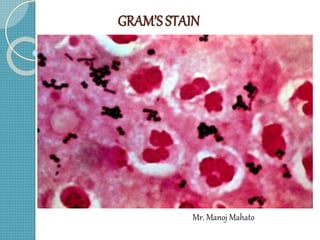
  • 29. Examples of Gram Stains Gram Positive Rods and Cocci Gram Negative Rods and Cocci
  • 30. RESULTS Gram positive bacteria………..Dark purple Yeast cells……………………...Dark purple Gram negative bacteria…………Pale to dark red Epithelial cells…………………..Pale red
  • 31. Reporting of Gramsmears The report should include the following information: - Number of bacteria present, whether many, moderate, few or scanty - -Gram reaction of the bacteria, whether Gram positive or Gram negative. - Morphology of the bacteria whether cocci, diplococci, streptococci, rods or coccobacilli. - Whether the organisms are intracellular. - Presence and number of pus cells. - Presence of yeast cell and epithelial cells
  • 32. Result and interpretation a. Evaluate the general nature of the smear under low-power magnification (10X). Determine if smear has been properly decolorized. Determine if thickness of smear is appropriate. b. Examine under low power for the evidence of inflammation Relative amount of WBCs Relative amount of epithelial cell c. Location and arrangement of microorganism
  • 33. Variation in Gram reaction. Gram positive organism may lose their ability to retain crystal violet and stain Gram negative for the following reasons: Cell wall damage due to antibiotic therapy or excessive heat fixation of the smear. Over de-colorization of the smear Use of an iodine solution which is too old Smear that has been prepared from an old culture Gram negative organisms may not be fully decolorized and appear as gram positive when a smear is too thick.
  • 34. Recording observations a. Record relative amount of observed cells and microorganism  Numerical 1+ (< 1 per oil immersion field, 100x) 2+ (1 per oil immersion field) 3+ (2 to 10 per oil immersion field) 4+ (predominant or >10 per oil immersion field)
  • 35.  Descriptive Rare (<1 per oil immersion field) Few (1 to 5 per oil immersion field) Moderate (5 to 10 per oil immersion field) Many (>10 per oil immersion field)
  • 36. Quality control  Check appearances of chemicals daily.  Daily and when a new lot of stain is used, prepare a smear of E. coli, S. epidermidis or S. aureus, fix and stain.  Use a set of reference slide.  To avoid the errors resulting from dirty slides use the slides previously dipped in 95% Ethanol and soak prior to use.
  • 37. Limitations of Gram stain  The number of microorganism required is very high, for visualizing with Gram stain (>10000 organisms/ml)  Can not give good results under following conditions: a. Cell wall damage due to antibiotic therapy or over heat fixation of smear b. Use of old cultures c. Uneven thickness of smear d. Use of old iodine solution e. Over decolorization of smear
  • 38. Gram stain modifications and uses Stain and use Original Hucker’s (1921) Kopeloff and Beerman’s Primary stain Gentian violet Crystal violet Alkaline crystal violet Mordant Lugol’s iodine Gram’s iodine Kopeloff’s iodine Decolourizer Absolute alcohol Acetone alcohol 3:7 acetone alcohol Counter stain Bismark brown Safranin Kopeloff’s safranin Use General bacteriology Anaerobes , For diagnosis of bacterial vaginosis .
  • 39. Gram stain modifications and uses Stain and use Jensen’s Preston and Morrell’s(1962 ) Brown and Brenn Weigert’s Primary stain 0.5%methyl violet Ammonium oxalate-crystal violet 1%Crystal violet Carbol gentian violet Mordant 1%Lugol’s iodine 1%Lugol’s iodine Lugol’s Iodine Gram’s iodine Decolourizer Absolute alcohol Iodine – acetone Acetone Aniline xylene Counter stain 0.1% Neutral red Dilute carbol fuschin 0.25%Basic fuschin Carminic acid and potassium alum in water . Use For Gonococci and Meningococci Bacteroides spp,Fusobacte rium species , For Demonstrating Bacteria in tissue . Pneumocyst ,Nocardia .
  • 40. Applications of Gram’s Staining  Used to guide initial therapy until definitive identification of microorganism obtained.  Morphology of stained bacteria can sometimes be diagnostic. For example Gram negative intracellular diplococci in urethral pus provides a presumptive diagnosis of Gonorrhea .  Sometimes specimens may show organisms under microscope but appear sterile in culture media .In these cases Gram stain is the only clue to the nature ,variety and relative proportion of infecting organism .  Aids in interpretation of culture reports .