Healt assessment - Eyes
•Transferir como PPTX, PDF•
11 gostaram•4,059 visualizações


Recomendados
Mais conteúdo relacionado
Mais procurados
Mais procurados (20)
Destaque
Destaque (20)
Semelhante a Healt assessment - Eyes
Semelhante a Healt assessment - Eyes (20)
Healt assessment - Eyes
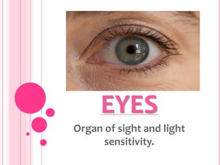
- 1. EYES Organ of sight and light sensitivity.
- 2. PHYSICAL EXAMINATION OF EYES Physical examination of the eyes provides information about the patient’s nutritional, endocrine, cardiovascular, gastrointestinal, and neurological systems.
- 3. EXTERNAL STRUCTURES Eyelids Consist of smooth muscle covered with a very thin layer of skin. They admit light to the eye while protecting and maintaining lubrication of the eye. Palpebral Conjunctiva Pink mucous membrane that covers the interior surface of lid muscle. Bulbar Conjunctiva Folds back over the anterior surface of the eyeball and merges with the cornea. Limbus Junction of the sclera and the cornea. o Conjunctiva Contains blood vessels and pain receptors that respond quickly to outside insult.
- 4. Palpebral Fissure Opening between eyelids. o Meibomian Glands Secrete lubricating substance onto the surface of the eye. o Tarsal Plates Are connective tissues that gives shape to the upper eyelids. o Lacrimal Apparatus Made up of the lacrimal glands and ducts. o Lacrimal Glands Responsible for the production of tears which lubricate the eyes. o Caruncle Which contains sebaceous glands. o Sebaceous Glands Is a round, red structure in the inner canthus.
- 5. EXTRAOCULAR MUSCLES There are six Extraocular muscles. These muscles work in concert to move the eyes with great precision in several directions to provide a single image to the brain. These muscles are the Superior, Inferior, Medial, Lateral Recti, and Superior and Inferior Obliques. INTERNAL STRUCTURES The eye itself is approximately 1 inch in diameter and has three layers: a tough, outer, fibrous tunic (sclera); a middle, vascular tunic; and the innermost layer, which contains the retina.
- 7. OUTER LAYER Sclera An opaque material that appears white and covers the structure inside the eye. It protects the eye and is a surface for the attachment of the Extraocular muscle. Cornea Nonvascular, transparent surface that covers the iris and is continuous with the conjunctiva epithelium. MIDDLE LAYER Choroid Vascular tissue that lines the inner surface of the eye just beneath the retina. It provides nutrition to the retinal pigment epithelium and helps absorb excess light.
- 8. Ciliary Body It siphons serum from the systemic blood flow to produce the aqueous humor needed to nourish the corneal endothelium. Iris Provides a distinctive color of the eye. It is comprised of smooth muscle that regulates the entrance of light. Pupil Regulates the amount of light that enters the eye. o Vitreous Humor Maintain the shape of the eye and position of the internal structures.
- 9. INNER LAYER Retina An extension of the optic nerve, which lines the inside of the globe and receives light impulses to be transmitted to the occipital lobe of the brain. Optic Disk A round or oval area with distinct margins located on the nasal side of the retina. Physiologic Cup A pale, central area in the optic disc occupying one-third to one-fourth of the disc. Cones Necessary for color vision, reading ability, and other tasks requiring fine visual discrimination. Fovea Area of sharpest vision. Rods Provide dark and light discrimination and peripheral vision.
- 10. General Approach to Eye Assessment 1. Greet the patient and explain the assessment techniques that you will be using. 2. Use a quiet room that will be free from interruptions. 3. Ensure that the light in the room provides sufficient brightness to allow adequate observation of the patient. 4. Place the patient in an upright sitting position on the examination table. 5. Visualize the underlying structures during the assessment process to allow adequate description of findings. 6. Always compare right and left eyes. 7. Use a systematic approach that is followed consistently each time the assessment is performed.
- 11. Equipments • Ophthalmoscope • Penlight • Snellen Chart Snellen E Chart
- 12. Rosenbaum near vision pocket screening card • Vision Occluder • Cotton-tipped applicator
- 13. Assessment Of The Eye Assessment of the eyes should be carried out in an orderly fashion, moving from the extraocular structures to the intraocular structures. The eye assessment usually includes testing of associated cranial nerves and can be performed in the following order. 1. Determination of the Visual Acuity 2. Determination of the Visual Fields 3. Assessment of the external eye and lacrimal apparatus 4. Evaluation of extraocular muscle function 5. Assessment of the anterior segment structures 6. Assessment of the posterior segment structures
- 14. Visual Fields The entire area that can be seen when the eye is directed forward, including that which is seen with peripheral vision.
- 15. Visual Acuity (Cranial Nerve II) A simple, noninvasive procedure that is carried out with the use of snellen chart and occluder to cover the patient’s eye. Snellen Chart Contains letters of various sizes with standardized visual acuity numbers at the end of each lines of letters.
- 16. DISTANCE VISION E 1. Ask the patient to stand or sit facing the snellen chart at a distance of 20 feet. 2. If the patient normally wears glasses, ask that they be removed. Contact lenses may be left in the eyes. 3. Instruct the patient to cover the left eye with the occluder and to read as many lines on the chart as possible. 4. Note the number at the end of the last line the patient was able to read. 5. If the patient is unable to read the letters at the top of the chart, move the patient closer to the chart. Note the distance at which the patient is able to read the top line. 6. Repeat the test, occluding the right eye. 7. Repeat the test, using both eyes. 8. If the patient normally wears glasses, the test should be repeated with the patient wearing the glasses, and it should be so noted (corrected or uncorrected)
- 17. N The patient who has a visual acuity of 20/20 is considered to have a normal visual acuity. A The patient is unable to read the chart with an uncorrected visual acuity of 20/30 in one eye, vision in both eyes is different by two lines or more, or acuity is absent. P The patient may have a refractive error related to a difference in the refractive power of the cornea. In myopia, light rays focus in front of retina. It can be changed by corrective lenses. In amblyopia, permanent loss of visual acuity is seen resulting from strabismus that was not corrected in early childhood, or certain medical conditions (alcoholism, uremia, diabetes mellitus). No corrective lenses can improve this vision. P The patient may have corneal opacities that are congenital, from lesions that have scarred the cornea like herpes simplex, from trauma or from degeneration and dystrophies. P Visual Acuity can be decreased because of opacities of the lens caused by senile or traumatic cataracts. P Inflammation of the retina caused by toxoplasmosis or by the presence of blood in the vitreous humor following hemorrhage can be responsible for decreased visual acuity. P Systemic diseases such as hypertension or diabetes mellitus, and trauma may damage the choroid and retina, causing decreased visual acuity.
- 18. Myopia Amblyopia
- 19. Near Vision E 1. Use a pocket snellen chart, Rosenbaum card or any printed material written at an appropriate reading level. 2. Have the patient sit comfortably and hold the card 14 inches from the face without moving it. 3. Ask the patient to read the smallest line. If other printed material is used, you will be able to gain only a general understanding of the patient’s near vision. N Until the patient is in the late 30’s to the late 40’s, reading is generally possible at a distance of 14 inches. A A patient is this age range who cannot read at 14 inches is considered presbyopic. Younger persons may have difficulty seeing up close because they have hyperopia. P The normal aging process causes the lens to harden (nuclear sclerosis), decreasing its ability to change shape and therefore focus on near objects.
- 20. Presbyopic Hyperopia
- 21. Color Vision E Color vision is usually tested in young children. If there is suspicion that the patient has a color vision deficit, have the patient identify the primary colors on snellen chart or colors in the examining room. N The patient should be able to identify colors correctly. A The color vision defect is designated as red/green, blue/yellow, or complete when the patient sees only shade of gray. P Defects in color vision can result from diseases of the optic nerve, macular degeneration, pathology of the fovea centralis, nutritional deficiency, or heredity.
- 22. Visual Fields The confrontation technique is used to test visual test of each eye. The visual field of each eye is divided into quadrants, and a stimulus is presented in each quadrant. E 1. Sit or stand approximately 2 to 3 feet away from and opposite the patient, with your eyes at the same level as the patient. 2. Have the patient cover the right eye with the right hand or an occluder. 3. Cover your left eye in the same manner. 4. Have the patient look at your uncovered eye with his or her uncovered eye. 5. Hold your free hand at arm’s length equidistant from you and the patient and move it or a held object such as pen into your and the patient’s field of vision from nasal, temporal, superior, inferior, and oblique angles. 6. Ask the patient to say “now” when your hand is seen moving into the field of vision. Use your own visual fields as the control for comparison of the patient’s. 7. Repeat the procedure for the other eye.
- 23. N The patient is able to see the stimulus at about 90 temporally, 60 nasally, 50 superiorly, and 70 inferiorly. A If the patient is unable to identify movement that you perceive, a defect in the visual field is presumed. The portion of the visual field loss should be noted. P Defects in the patient’s visual field can be associated with tumors, strokes, or neurological diseases such as glaucoma or retinal detachment.
- 24. Eyelids, Eyebrows, and Eyelashes E 1. Ask the patient to sit facing you. 2. Observe the patient’s eyelids for drooping, infections, tumors, or other abnormalities. 3. Note the distribution and symmetry of the eyelashes and eyebrows. Note any lesions. 4. Instruct the patient to focus on an object or a finger held about 10 to 12 inches away and slightly above eye level. 5. Move the object or fingers slowly downward and observe for white space of sclera between upper lid and limbus. 6. Observe the blinking of the eye. N Ask the patient to elevate eyelids. The eyelid should appear symmetrical with no drooping, infection, or tumors of the lid. When the eyes are focused in a normal frontal gaze, the lids should cover the upper portion of the iris. Slight ptosis, or drooping of the lid can be normal. When the eye is closed. No portion of the cornea should be exposed. Normal lid margins are smooth, with the lashes evenly distributed and sweeping upward from the upper lids and downward the lower lids. Eyebrows present bilaterally and are symmetrical and without lesions or scaling.
- 25. A The patients has unilateral or bilateral, constant or intermittent ptosis of the lid. If part of the pupil is occluded, there may be wrinkling of the forehead above the affected eye in an attempt to compensate by using the frontalis muscle to lift the lid. P Ptosis can be congenital or acquired. In congenital, there is failure of the elevator muscle to develop. This can be associated with pathology of the superior rectus muscle as well. If it is acquired, it is related to one of three factors. 1. Mechanical: heavy lids from lesions, adipose tissue, swelling or edema. 2. Myogenic: muscular diseases such as myasthenia gravis or multiple sclerosis. 3. Neurogenic: paralysis from damage or interruption of the neutral pathways. A An area of white sclera appears between the upper lid and the limbus, widening as the object is moved downward. P This condition is called lid lag and may indicate the presence of thyrotoxicosis or increased circulating levels of free thyroxin or triiodothyronine. A The patient is unable to bring about complete lid closure. P This condition is referred to as lagophthalmos and can be associated with Bell’s palsy, stroke, trauma, or ectropion (everted eyelid) A During inspection of the lids, a disparity of the Palpebral fissure is noted with apparent lid retraction, indicating a protrusion of the globe. This condition may be unilateral or bilateral.
- 26. P This abnormality is exophthalmos (or proptosis) and can be present unilaterally in orbital tumors, thyroid disease, trauma, or inflammation. Bilateral exophthalmos is related to thyroid disease. A There is apparent disparity in the size of the globe, manifested by a narrowing of the Palpebral fissure. P Enophthalmos is a backward displacement of the globe in the orbit, generally caused by orbital blowout fracture due to trauma. When this occurs, the orbital contents herniated through the fracture site. A The turning inward, or inversion, of the lower lid is referred to as entropion and can cause severe discomfort to the patient as the eyelashes abrade the cornea (trichiasis). If left untreated, it can cause inflammation, corneal scarring, and eventual ulceration. P Entropion is caused by spasms or advancing age (senile). In senile entropion, there is a loss of muscle tone, which causes the lid to fold inward. A The turning outward, or eversion, of the lower lid is referred to as ectropion and may be unilateral or bilateral. With ectropion, the lower lids appear to be sagging outward. P The normal aging process can cause the muscles to lose their tone and relax, or may be affected by Bell’s palsy.
- 27. A The patient exhibits excessive blinking that may or may not be accompanied by increased tearing and pain. P The causes of excessive blinking are: 1. Voluntary: irritation to the cornea or the conjunctiva, or stress and anxiety (usually disappears when stimulus is removed) 2. Involuntary: tonic spasm of the orbicularis oculi muscle called blepharopasm; often seen in elderly individuals as well as in patients with CN VII lesions, irritation of the eye, fatigue, and stress. A The lids are black and blue, bluish, yellow, or red< depending on race and skin color. P Color changes from the lids can causes the following: 1. Redness: generalized redness is nonspecific; however, redness in the nasal half of the lid may indicate frontal sinusitis. Redness adjacent to the lower lid can indicate disease of the larcimal sac or nasolacrimal duct, such as dacryocystitis; redness in the temporal portion of the lid can result from dacryoadenitis, an inflammation of the lacrimal gland. 2. Bluish: cyanosis can result from orbital vein thrombosis, orbital tumors, or aneurysms in the orbit. 3. Black and Blue: ecchymosis is caused by bleeding into the surrounding tissues following trauma (black eye) A Swelling or edema is located in the eyelid.
- 28. P Swelling or edema may be noted in nonocular conditions such as inflammation associated with allergies, herpes simplex virus, systemic diseases, medications that contribute to swelling from fluid overload, trichinosis, early myxedema, thyrotoxicosis, or contact dermatitis. A There is an acute localized inflammation, tenderness, and redness, with the patient complaining of pain in the infected area. This is called a hordeolum. P Staphylococcus is generally the infecting organism that causes a hordeolum. There are two types of hordeolum: 1. Internal: affects the Meibomian glands, is usually large, and can point either to the skin or to the conjunctival side of the lid. 2. External: often called a “style”, an infection of a sebaceous gland that is usually points to the skin side of the lid, and extends to the lid margin. Infections of the glands of the eyelid can be caused by improper removal of makeup, dry eyes, or seborrhea. There may be some connection between a hordeolum and increased handling of the lids in activities such as inserting and removing contact lenses. A There is a chronic inflammation of the Meibomian gland in either the upper or lower lid. It generally forms over several weeks and in many cases, points toward the conjunctival side of the lid, usually not on the lid margin. There is no redness or tenderness.
- 29. P This inflammation is referred to as chalazion and its cause is unknown. A The lids are inflamed bilaterally and are red rimmed, with scales clinging to both the upper and lower lids. The patient complaints of itching and burning along the lid margins. There may also be some loss of the eyelashes. P This is blepharitis, which may be either staphylococcal or seborrheic. Often a patient has both eyes simultaneously. If the patient has seborrheic infections elsewhere (scalp or eyebrows), it is more likely that the blephritis is of the seborrheic type. A Raised, yellow, non painful plaques are present in upper and lower lids near the inner canthus. P These lesions are xanthelasma, a form of xanthoma frequently associated with hypercholesterolemia. A The eyebrows have scaling areas. P This is caused by seborrhea.
- 30. Ptosis Exophthalmos Ectropion Enophthalmos Entropion Hordeolum
- 31. Chalazion Blepharitis Xanthelasma
- 32. Lacrimal Apparatus Inspection E 1. Have the patient sit facing you. 2. Identify the area of the lacrimal gland. Note any swelling or enlargement of the gland or elevation of the eyelid. Note any enlargement, swelling, redness, increased tearing or exudate in the area of the lacrimal sac at the inner canthus. 3. Compare to the other eye in order to determine whether there Is unilateral or bilateral involvement. N There should be no enlargement, swelling, or redness; no large amount of exudate; and minimal tearing. A There is inflammation and swelling in the upper lateral aspect of one or both eye, and the patient complains of pain in the affected area. P Acute inflammation of the lacrimal gland is called dacryoadenitis and does not occur commonly. Dacryoadenitis may result from trauma or may be found in association with measles, mumps, and mononucleosis. A There is inflammation and painful swelling beside the nose and near the inner canthus and possibly extending to eyelid. P Dacryocystitis is caused by inflammatory or neoplastic obstruction of the lacrimal duct.
- 33. Dacryoadenitis Dacryocystitis
- 34. Palpation E 1. To assess the lacrimal sac for obstruction, don the gloves. 2. Gently press the index finger near the inner canthus, just inside the rim of the bony orbit of the eye. 3. Note any discharge of the punctum. N There should not be excessive tearing or discharge from the punctum. A Mucopurulent discharge is noted. P Obstruction anywhere along the system of lacrimal sac to the point at which the ducts empty below the inferior nasal turbinate can cause mucopurulent discharge. A There is overflowing of tears from the eye. P This condition is epiphora, which is caused by the obstruction of the lacrimal duct. Epiphora
- 35. Corneal Light Reflex (Hirshberg Test) E 1. Instruct the patient to look straight ahead. 2. Focus a penlight on the corneas from a distance of 12 to 15 inches away at the midline. 3. Observe the location of reflected light on the cornea. N The reflected light (light reflex) should be seen symmetrically in the center of each cornea. A There is a discrepancy in the placement of one of the light reflections. P Asymmetrical corneal light reflexes indicate an extraocular muscle imbalance that may be related to a variety of causes, depending on the patient’s age and medical condition. The condition of one eye constantly being deviated is called strabismus, or tropia: esotropia is an inward turning of the eye: exotropia is an outward turning of the eye. Esotropia Exotropia
- 36. Cover/Uncover Test E 1. Ask the patient to look straight ahead and to focus on an object in the distance. 2. Place an Occluder over the left eye for several seconds and observe for movement in the uncovered right eye. 3. As the Occluder is removed, observe the covered eye for a moment. 4. Repeat the procedure with the same eye, having the patient focus on an object held close to the eye. 5. Repeat on the other side. N If the eyes are in alignment , there will be no movement of either eye. A If the uncovered eye shifts position as the other eye is covered, or if the covered eye shifts position at it is uncovered, a phoria, or latent misalignment of an eye, exists. P This condition is a mild weakness elicited by the cover/uncover test and has two forms: esophoria, nasal inward drift, and exophoria, a temporal outward drift.
- 37. Cardinal Fields of Gaze (Extraocular Muscle Movement) E 1. Place the patient in a sitting position facing you. 2. Place the nondominant hand just under the patient’s chin or on top of the patient’s head as a reminder to hold the head still. 3. Ask the patient to follow an object (finger, pencil, or penlight) with the eyes. 4. Move the object through the fields of gaze in a smooth and steady manner, pausing at each extreme position to detect any nystagmus, or involuntary movement, and returning to the center after each field is tested. 5. Note the patient’s ability to move the eyes in each direction. 6. Move the object forward to about % inches in front of the patient’s nose at the midline. 7. Observe for convergence of gaze.
- 38. N Both eyes should move smoothly and symmetrically in each of the sic fields of gaze and converge on the held object as it moves toward the nose. A few beats of nystagmus with extreme lateral gaze can be normal. A There is a lack of symmetrical eye movement in a particular direction. P Inability to move the eye in a given direction indicates a weakness in the muscle responsible for moving the eye in that direction. A Abnormal eye movements consist of failure of an eye to move outward, inability of eye to move downward when deviated inward, or other defects in movement. P Traumatic ophthalmoplegia may be caused by fracture of the orbit near foramen magnum causing damage to extraocular muscle. Basilar skull fractures that involve the cavernous sinus may also cause extraocular muscle palsy. P Vitamin deficiency, especially thiamine may cause extraocular muscle palsy and nystagmus. CN VI is affected. A Ophthalmoplegia is paralysis of one or more optic muscles. P Increased intracranial pressure may cause strangulation of CN VI. CN VI palsy occurs late after the onset of increased intracranial pressure. A Vertical gaze is a paralysis of upward gaze and its abnormal. P Destruction at the area of the midbrain-diencephalic junction or the medial longitudinal fascilicus or tumors of the pineal gland that press on the brain stem at the superior colliculus can cause a vertical gaze deviation.
- 39. A Paralysis of horizontal gaze is abnormal. P Damage to the motor areas of the cerebral cortex causes the loss of the ability of both eyes to look to the contra lateral side, so the eyes tend to deviate toward the side of the lesion. A With internuclear ophthalmoplegia, the eyes are unable to look medially, but convergence may be maintained because the pathway for convergence is different from that for conjugate gaze. P The medial rectus muscle is involved, so the eyes are unable to look medially. Internuclear ophthalmoplegia may be caused by demyelinization due to multiple sclerosis. A If one eye deviates down and the other eye deviates up, it is called skew deviation. P Cerebellar disease or a lesion in the pons on the same side as the eye that is deviated down may cause skew deviation. A There is rhythmic, beating, involuntary oscillation of the eyes as the objects is held at points away from the midline. Movements is usually lateral, vertical, or rotary. Nystagmus can be jerky, with fast and slow components, or rhythmic, similar to the pendulum of a clock. P Nystagmus may be caused by a lesion in the brain stem, cerebellum, vestibular system, or along the visual pathways in the cerebral hemispheres.
- 40. Anterior Segment Structures Conjunctiva To assess the bulbar conjunctiva: E 1. Separate the lid margins with fingers. 2. Have the patient look up, down, and to the right and left. 3. Inspect the surface of the bulbar conjunctiva for color, redness, swelling, exudate, or foreign bodies. Note whether injection or redness is around the cornea or toward the periphery. 4. With the thumb, gently pull the lower lid toward the cheek and inspect the surface of the bulbar conjunctiva for a color, inflammation, edema, lesions, or foreign bodies. N The bulbar conjunctiva is transparent, with small blood vessels visible in it. It should appear white except for a few small blood vessels, which are normal. No swelling, injection, exudate, foreign bodies, or lesions are noted. A/P Refer to palpebral conjunctiva. • The palpebral conjunctiva is examined only when there is a concern about its condition. To examine the palpebral conjunctiva of the upper lid.
- 41. E 1. Explain the procedure to the patient to alleviate the fear of pain or damage to the eye. 2. Don gloves. Have the patient look down to relax the levator muscle. 3. Gently pull the eyelashes downward and place a sterile, cotton-tipped applicator above 1 cm above the lid margin. 4. Gently exert downward pressure on the applicator while pulling the eyelashes upward to evert the lid. 5. Inspect the palpebral conjunctiva for injection, swelling or chemosis, exudate, and foreign bodies. 6. Return the lid to its normal position by instructing the patient to look up and then pulling the eyelid outward and removing the cotton-tipped applicator. Ask the patient to blink. N The palpebral conjunctiva should appear pink and moist. It is without swelling, lesions, injection, exudate, or foreign bodies. A Bilateral injected conjunctiva with purulent sticky discharge and lid edema are noted. P These findings usually indicate the presence of bacterial conjunctivitis. Bacterial Conjunctivitis
- 42. A A yellow nodule is noted on the nasal side of the bulbar conjunctiva adjacent to the cornea. Can be on temporal side. This is painless unless it becomes inflamed. P This is called pinguecula. A nodular degeneration and is thought to be a result of increased exposure to UV rays. A A unilateral or bilateral triangle shaped encroachment onto conjunctiva is abnormal. This always occurs nasally and remains painless unless it becomes ulcerated. If it covers the cornea, loss of vision may occur. P This is called pterygium and is caused by excessive UV light exposure. A The patient exhibits sudden onset of a painless, bright red appearance on the bulbar conjunctiva. P This is subconjunctival hemorrhage and may result from the pressure exerted during coughing, sneezing, or a valsalva maneuver. It can also be anticoagulant medications or uncontrolled hypertension.
- 43. Sclera E Inspect the sclera for color, exudate, lesions, and foreign bodies. N In light-skinned persons, slera shoud be white with some small, superficial vessels and without exudate, lesion, or foreign bodies. In dark-skinned persons, sclera may have tiny brown patches of melanin or a grayish-blue or “muddy” color. A The color of the sclera is uniformly yellow. P This condition is known as jaundice or scleral icterus. This is an early manifestation of systemic conditions such as hepatitis, sickel cell disease, gallstones, and physiological jaundice of the newborn. A The sclera is blue. P This is a distinctive feature of osteogeneses imperfecta and is due to the thinning of sclera which allows the choroid to show through.
- 44. Cornea E 1. Stand in front of the patient. 2. Shine a penlight directly on the cornea. 3. Move the light laterally and view the cornea from that angle, noting color, discharge and lesions. N The corneal surface should be moist and shiny, with no discharge, cloudiness, opacities, or irregularities. A A grayish, well-circumscribed ulcerated area on the cornea is abnormal. P The most common cause of this is a corneal ulceration resulting from a bacterial invasion. A There is a hazy gray ring about 2 mm in width just inside the limbus. P This is arcus senilis, a bilateral benign degeneration of the peripheral cornea. Can be found in any age but most common in older person. If a young person, it may be associated with hypercholesterolemia.
- 45. A A steamy or cloudy cornea is abnormal. The patient has also ocular pain. P Glaucoma is caused by increased intraocular pressure. A Any irregularities in the appearance of the cornea are abnormal. P Keratoconus is the conical protrusion of the center of the cornea. It is noninflammatory condition in which the cornea thins, leading to the need for corneal transplant surgery. P A corneal scar forms at the site of past injury or inflammation. P Corneal laceration can occur secondary to trauma.
- 46. Anterior Chamber The anterior chamber is that compartment of the eye found between the cornea and iris. To differentiate a normal from narrowed angle: E 1. Face the patient and shine a light obliquely through the anterior chamber from the lateral side toward the nasal side. 2. Observe the distribution of light in the anterior chamber. 3. Repeat the procedure with the other eye N In a normal eye, the entire iris will be illuminated. A The eye has a narrow angle, with the decreased space between the iris and the cornea appearing as a crescent shaped shadow on the far portion of the iris. P The narrow angle is an anatomic variant that can predispose an individual to the development of angle-closure glaucoma. As aging progresses, the lens thickens, which may cause even further narrowing of the angle.
- 47. Iris E With the penlight, inspect the iris for colors, nodules, and vascularity. N Normally, the color is evenly distributed over the iris, although there can be a mosaic variant. It is normally smooth and without apparent vascularity. A There is a heavily pigmented, slightly elevated area visible in the iris. P This lesion can be a benign iris nevus or a malignant melanoma. An iris nevus is much more common than melanoma. A The inferior portion of the iris is obscured by blood. P This is hyphema and is caused by bleeding from vessels in the iris as a result of direct trauma to the globe. It can also occur as a result of eye surgery. A An absent wedge portion of the iris is abnormal P The shape of the iris changes after surgical removal of a cataract; the pupil may also have an irregular shape.
- 48. Pupil E 1. Stand in front of the patient in a darkened room. 2. Note the shape and size of the pupils in millimeters. 3. Move a penlight from the side to the front of one eye without allowing the light to shine on the other eye. 4. Observe the pupillary reaction in that eye. Note the size of the pupil receiving light stimulus and the speed of pupillary response to light. 5. Repeat in other eye. 6. Move the penlight in front of one eye, observe the other eye for pupillary constriction. 7. Repeat the procedure on other eye. 8. Instruct the patient to shift gaze to a distant object for 30 seconds. 9. Instruct the patient to then look at you finger or an object held in your hand about 10 cm from the patient. 10. Note the reaction and size of the pupils. Accomodation occurs when pupils constrict and converge to focus on objects at close range. N The pupils should be deep black, round, and equal diameter, ranging from 2 to 6 mm. A The pupil dilates to more than 6mm in diameter is termed mydriatic. P These abnormalities may also be due to nerve damage or trauma.
- 49. Lens E 1. Stand in front of the patient. 2. Shine a penlight directly on the pupil. The lens in behind the pupil. 3. Note the color. N The lens is transparent in color. A One or more of the pupils are not deep black. P n adult, a pearl gray appearance of one or both pupils may indicate an opacity in the I lens called cataract. P A senile cataract is the most common type. P A unilateral cataract may occur soon after eye injury caused by a foreign body.
- 50. Bilateral Cataract P Bilateral cataracts that are found in infants or young children are congenital cataracts. Posterior Segment Structure The funduscopic assessment requires the use of direct ophthalmoscope to assess the structures in the posterior segment of the eye. The ophthalmoscope consist of two parts; the head and the handle to active light source in the head and depress the rheostat button and it is far as possible. Move the aperture selector to produce the largest beam of light on the palm of the hand. The larger the beam is preferred when assessing an average-sized pupil and the smaller beam makes assessment of a small pupil easier. The lens selector allows you to choose lenses varying power for different parts of the assessment. These lenses are marked with red and black numbers, signifying different focal lengths. In some ophthalmoscope there is no color designation and the lens power is signified by + or – sign in front of numbers. A + sign, or diopter is equivalent to black and focuses closer to the ophthalmoscope. This is used to correct for farsightedness or hyperopia. A – sign or diopter is equivalent to red and focuses farther from the instrument. This is used to correct nearsightedness or myopia.
- 51. Apertures of the Ophthalmoscope Small round light – Used to examine eyes with small, undilated pupils Large round light – Used for routine eye examinations and examination of dilated eyes Grid – Used to access size and location of funduscopic lesions. Slit light – Assesses anterior eye and determines elevation of funduscopic lesions. Green light (red-free filter) – Used to assess retinal hemorrhages (which appear black) and small vessel changes.
- 52. Retinal Structures In a darkened room, ask the patient to remove eyeglasses; contact lenses may be left in place. N The red reflex is present. The optic disc is pinkish-orange in color with a yellow white excavated center known as the physiologic cup. The ratio of the cup diameter to that of the entire disc is 1:3. the border of the disc may range from a sharp round demarcation from the surrounding retima to a more blended border, but should be on the same place as the retina. In general, there are four main vascular vbranches emanating from the sisc, each branch consist of an arteriole and venule. The venules are approximately four time size of the accompnying arterioles and are darker in color. Light often produces a glistening “light reflex” from the arteolar vessel. Normal arterial-to-venos width is a ratio of 2:3 or 4:5 Findings Characteristics Fair-skinned individual •Retina appears a lighter red-orange color •Tessellated appearance of the fundi Dark-skinned individual •Fundi appear darker, grayish-purple to brownish •No tessellated appearance •Choroidal vessels usually obscure Aging individual •Vessels are straighter and narrower •Choroidal vessels are easily visualized •Retinal pigment epithelium atrophies and causes the retina color to become paler
- 53. A The red reflex is absent. P The present of cataracts can prevent the red reflex from being observe due to the opacity of the lens. A The red reflex is absent and the pupil appears white. P Leucoria, or white reflex is found in retinoblastoma, congenital cataracts, and retina detachment. This often referred to as the cat’s eye reflex. A The optic disc is pale. P Pallor is due to optic atrophy caused by increased intracranial pressure or from congenital syphilis; an intracranial space-occupying lesion. A Fibrous white bands that obscure the retinal vessels are abnormal. Neovascularization may also be present. P These finding occur in proliferative diabetic retinopathy.
- 54. P Glaucomatous cupping occurs due to increased intraocular pressure. A Fluffy white gray, slightly irregular areas that appear on the retina and are ovoid in shape are abnormal. P Cotton wool spots represent microscopic infarcts of the nerve fiber layer and are due to diabetic or hypertensive retinopathy. A A cleft defect of the choroid and retina is abnormal. P Coloboma is a congenital, pathological, or surgically induced abnormality.
- 55. Macula N The macula is darker, avascular area with a pinpoint reflective center known as the fovea centralis. The fovea centralis is present. A The retina is pale with the macular region appearing as a cherry red spot. P This finding is central retina artery occlusion, an indication of tay-sac disease. A An enlarge macula is abnormal. P Macular edema is caused by the leakage of fluid from retinal blood vessels. A Sharply defined, small red spots are found in and around the macula. P These microaneurysms are pathognomonic of diabetes mellitus.