1. 20 Animal Development:
From Genes to Organism
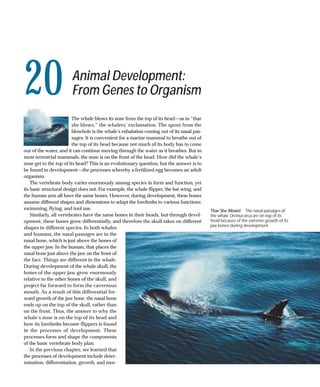
The whale blows its nose from the top of its head—as in “thar
she blows,” the whalers’ exclamation. The spout from the
blowhole is the whale’s exhalation coming out of its nasal pas-
sages. It is convenient for a marine mammal to breathe out of
the top of its head because not much of its body has to come
out of the water, and it can continue moving through the water as it breathes. But in
most terrestrial mammals, the nose is on the front of the head. How did the whale’s
nose get to the top of its head? This is an evolutionary question, but the answer is to
be found in development—the processes whereby a fertilized egg becomes an adult
organism.
The vertebrate body varies enormously among species in form and function, yet
its basic structural design does not. For example, the whale flipper, the bat wing, and
the human arm all have the same bones. However, during development, these bones
assume different shapes and dimensions to adapt the forelimbs to various functions:
swimming, flying, and tool use. Thar She Blows! The nasal passages of
Similarly, all vertebrates have the same bones in their heads, but through devel- the whale Orcinus orca are on top of its
opment, these bones grow differentially, and therefore the skull takes on different head because of the extreme growth of its
jaw bones during development.
shapes in different species. In both whales
and humans, the nasal passages are in the
nasal bone, which is just above the bones of
the upper jaw. In the human, that places the
nasal bone just above the jaw on the front of
the face. Things are different in the whale.
During development of the whale skull, the
bones of the upper jaw grow enormously
relative to the other bones of the skull, and
project far forward to form the cavernous
mouth. As a result of this differential for-
ward growth of the jaw bone, the nasal bone
ends up on the top of the skull, rather than
on the front. Thus, the answer to why the
whale’s nose is on the top of its head and
how its forelimbs become flippers is found
in the processes of development. These
processes form and shape the components
of the basic vertebrate body plan.
In the previous chapter, we learned that
the processes of development include deter-
mination, differentiation, growth, and mor-
2. ANIMAL DEVELOPMENT 409
phogenesis. In this chapter we will see how these processes
are carried out in the early stages of development.
Development begins with the joining of sperm and egg.
The fertilized egg goes through an initial rapid series of cell
divisions without growth that subdivides the egg cytoplasm
into a mass of smaller undifferentiated cells. Although this
mass of cells shows no hints of the eventual body plan, the
uneven distribution of molecules in the cytoplasm of the fer-
tilized egg provides positional information that will result in
the determination of cells and set up the body plan. The body
plan then unfolds through orderly movements of cells that
create multiple cell layers and set up new cell-to-cell contacts
that trigger signal transduction cascades and further steps of
determination. These inductive interactions influence the
temporal and spatial expression of the genes that control the
growth and differentiation of cells, leading to the emergence
of the organs of the new individual.
To appreciate both the diversity and the similarity in the 20.1 Sperm and Egg Differ Greatly in Size This artificially colored
development of different animals, we will discuss these early micrograph of human fertilization illustrates the size difference
developmental steps in a few model organisms that have between the two types of gametes in mammals. The large egg (blue)
been studied extensively by developmental biologists: sea contributes more cytoplasm to the zygote than the much smaller
sperm (yellow).
urchins (invertebrates), and frogs, chickens, and humans (all
vertebrates).
comes the centrosome of the zygote, which produces the mi-
totic spindles for subsequent cell divisions.
Development Begins with Fertilization It had long been assumed that the one thing that sperm
Fertilization is the union of a haploid sperm and a haploid and egg contributed equally to the zygote was their haploid
egg to produce a diploid zygote. Fertilization does more, nuclei. However, we now know that even though they are
however, than just restore a full complement of maternal equivalent in terms of genetic material, mammalian sperm
and paternal genes. The entry of a sperm into an egg acti- and eggs are not equivalent in terms of their roles in devel-
vates the egg metabolically and initiates the rapid series of opment. In the laboratory, it is possible to construct zygotes
cell divisions that produce a multicellular embryo. Also, in in which both haploid nuclei come from the mother or both
many species, the point of entry of the sperm creates an come from the father. In neither case does development
asymmetry in the radially symmetrical egg. This asymme- progress normally. Apparently, in mammals at least, certain
try is the initiating event that enables a bilateral body plan genes involved in development are active only if they come
to emerge from the radial symmetry of the egg. We will de- from a sperm, and others are active only if they come from
scribe the mechanisms of fertilization in Chapter 43. Here an egg. This phenomenon, called genomic imprinting, was de-
we take a closer look at the cellular and molecular interac- scribed in Chapter 17.
tions of sperm and egg that result in the first steps of devel-
opment.
Fertilization causes rearrangements of egg cytoplasm
The entry of the sperm into the egg stimulates changes in and
The sperm and the egg make different contributions rearrangements of the egg cytoplasm that establish the po-
to the zygote larity of the embryo. The nutrients and molecules in the cy-
Nearly all of the cytoplasm of the zygote comes from the egg toplasm of the zygote are not homogeneously distributed,
(Figure 20.1). Egg cytoplasm is well stocked with nutrients, and therefore, they are not divided equally among all daugh-
ribosomes, and a variety of molecules, including mRNAs. Be- ter cells when cell divisions begin. This unequal distribution
cause the sperm’s mitochondria degenerate, all of the mito- of cytoplasmic factors sets the stage for the signal transduc-
chondria (and therefore all of the mitochondrial DNA) in the tion cascades that orchestrate the sequential steps of devel-
zygote come from the mother. In addition to its haploid nu- opment: determination, differentiation, and morphogenesis.
cleus, the sperm makes one other important contribution to Let’s examine these earliest developmental events in the frog,
the zygote in some species: a centriole. This centriole be- an organism in which they have been well studied.
3. 410 CHAPTER T WENT Y
The rearrangements of egg cytoplasm in some frog species sperm centriole rearranges the microtubules in the vegetal
are easily observed because of pigments in the egg cyto- hemisphere cytoplasm into a parallel array that presumably
plasm. The nutrient molecules in an unfertilized frog egg are guides the movement of the cortical cytoplasm. Organelles
dense, and they are therefore concentrated by gravity in the and certain proteins from the vegetal hemisphere move to
lower half of the egg, which is called the vegetal hemisphere. the gray crescent region even faster than the cortical cyto-
The haploid nucleus of the egg is located at the opposite end plasm rotates.
of the egg, in the animal hemisphere. The outermost (cortical) As a result of these movements of cytoplasm, proteins, and
cytoplasm of the animal hemisphere is heavily pigmented, organelles, changes in the distribution of critical developmen-
and the underlying cytoplasm has more diffuse pigmenta- tal signals occur. A key transcription factor in early develop-
tion. The vegetal hemisphere is not pigmented. ment is β-catenin, which is produced from maternal mRNA
The surface of the frog egg has specific sperm-binding and is found throughout the cytoplasm of the egg. Also pres-
sites located only in the animal hemisphere, so sperm always ent throughout the egg cytoplasm is a protein kinase called
enter the egg in that hemisphere. When a sperm enters, the GSK-3, which phosphorylates and thereby targets β-catenin
cortical cytoplasm rotates toward the site of sperm entry. This for degradation. However, an inhibitor of GSK-3 is segregated
rotation reveals a band of diffusely pigmented cytoplasm on in the vegetal cortex of the egg. After sperm entry, this inhibitor
the side of the egg opposite the site of sperm entry. This band, is moved along microtubules to the gray crescent, where it pre-
called the gray crescent, will be the site of important devel- vents the degradation of β-catenin. As a result, the concentra-
opmental events (Figure 20.2). tion of β-catenin is higher on the dorsal side than on the ven-
The cytoplasmic rearrangements that create the gray cres- tral side of the developing embryo (Figure 20.3).
cent bring different regions of cytoplasm into contact on op- Evidence supports the hypothesis that β-catenin is a key
posite sides of the egg. Therefore, bilateral symmetry is im- player in the cell–cell signaling cascade that begins the
posed on what was a radially symmetrical egg. In addition process of cell determination and the formation of the em-
to the up–down difference of the animal and vegetal hemi- bryo in the region of the gray crescent. But before there can
spheres, the movement of the cytoplasm sets the stage for be cell–cell signaling, there must be multiple cells, so let’s
the creation of the anterior–posterior and left–right axes. In turn first to the early series of cell divisions that transforms
the frog, the site of sperm entry will become the ventral the zygote into a multicellular embryo.
(belly) region of the embryo, and the gray crescent will be-
come the dorsal (back) region. Since the gray crescent also
marks the posterior end of the embryo, these relationships
Cleavage: Repackaging the Cytoplasm
specify the anterior–posterior and left–right axes as well. The transformation of the diploid zygote into a mass of cells
occurs through a rapid series of cell divisions, called cleav-
age. Because the cytoplasm of the zygote is not homoge-
Rearrangements of egg cytoplasm set the stage neous, these first cell divisions result in the differential dis-
for determination tribution of nutrients and cytoplasmic determinants among
The molecular mechanisms underlying the first steps in frog the cells of the early embryo. In most animals, cleavage pro-
embryo formation are beginning to be understood. The ceeds with rapid DNA replication and mitosis, but no cell
growth and little gene expression. The em-
bryo becomes a solid ball of smaller and
The cortical cytoplasm smaller cells, called a morula (from the Latin
Animal
cortical rotates relative to the word for “mulberry”). Eventually, this ball
Animal inner cytoplasm.
cytoplasm forms a central fluid-filled cavity called a
(pigmented) pole
A blastocoel, at which point the embryo is
called a blastula. Its individual cells are
Inner called blastomeres.
cytoplasm The pattern of cleavage, and therefore
Sperm
entry the form of the blastula, is influenced by
point
Vegetal
two major factors. First, the amount of nu-
cortical V trient material, or yolk, stored in the egg
Vegetal cytoplasm The gray crescent is
pole differs among species. Yolk influences the
(unpigmented) created by the rotation.
pattern of cell divisions by impeding the
20.2 The Gray Crescent Rearrangements of the cytoplasm of frog eggs after fertilization pinching in of the plasma membrane to
create the gray crescent. form a cleavage furrow between the daugh-
4. ANIMAL DEVELOPMENT 411
ter cells. Second, cytoplasmic determinants stored in the egg
(a) Fertilization by the mother guide the formation of mitotic spindles and
Egg
Animal pole the timing of cell divisions.
β-Catenin (orange)
is distributed throughout
cytoplasm.
Sperm
The amount of yolk influences cleavage
GSK-3 (blue), which targets
β-catenin for degradation, In embryos with little or no yolk, there is little interference
is also found throughout with cleavage furrow formation, and all the daughter cells are
cytoplasm.
of similar size; the sea urchin egg provides an example (Fig-
Vegetal pole ure 20.4a). More yolk means more resistance to cleavage fur-
A protein that inhibits row formation; therefore, cell divisions progress more rapidly
GSK-3 is contained in
vegetal pole vesicles. in the animal hemisphere than in the vegetal hemisphere,
(b) Cortical rotation where the yolk is concentrated. As a result, the cells derived
from the vegetal hemisphere are fewer and larger; the frog
egg provides an example of this pattern (Figure 20.4b).
Ventral Dorsal
In spite of this difference between sea urchin and frog
(V) (D) eggs, the cleavage furrows completely divide the egg mass
Vesicles in vegetal pole
move on microtubule in both cases; thus these animals are said to have complete
tracks to side opposite cleavage. In contrast, in eggs that contain a lot of yolk, such as
sperm entry.
the chicken egg, the cleavage furrows do not penetrate the
yolk. As a result, cleavage is incomplete, and the embryo
(c) Dorsal enrichment
forms as a disc of cells, called a blastodisc, on top of the yolk
inhibitor mass (Figure 20.4c). This type of incomplete cleavage, called
The vesicles release
GSK-inhibiting protein…
discoidal cleavage, is common in fishes, reptiles, and birds.
Another type of incomplete cleavage, called superficial
V D cleavage, occurs in insects such as the fruit fly (Drosophila). In
the insect egg, the mass of yolk is centrally located (Figure
20.4d). Early in development, cycles of mitosis occur without
cytokinesis. Eventually the resulting nuclei migrate to the pe-
riphery of the egg, and after several more mitotic cycles, the
plasma membrane of the egg grows inward, partitioning the
(d) Dorsal inhibition nuclei into individual cells.
of GSK-3 …so GSK-3 cannot
degrade β-catenin
on the dorsal side…
The orientation of mitotic spindles influences
V D
…but does degrade it
the pattern of cleavage
on the ventral side. The positions of the mitotic spindles during cleavage are not
random; rather, they are defined by cytoplasmic determi-
nants that were produced from the maternal genome and
stored in the egg. The orientation of the mitotic spindles de-
(e) Dorsal enrichment termines the planes of cleavage and, therefore, the arrange-
of b-catenin
Thus there is a higher ment of the daughter cells.
β-catenin concentration If the mitotic spindles of successive cell divisions form
in the dorsal cells of the parallel or perpendicular to the animal–vegetal axis of the
V D early embryo.
zygote, the cleavage pattern is radial, as in the sea urchin and
the frog. In these organisms, the first two cell divisions are
parallel to the animal–vegetal axis and the third is perpen-
dicular to it (Figure 20.4a,b). Another cleavage pattern, spi-
20.3 Cytoplasmic Factors Set Up Signaling Cascades ral cleavage, results when the mitotic spindles are at oblique
Cytoplasmic movement changes the distributions of critical develop- angles to the animal–vegetal axis. Mollusks have spiral
mental signals. In the frog zygote, the interaction of the protein
kinase GSK-3, its inhibitor, and the protein β-catenin are crucial in cleavage, and a visible expression of this is the coiling of
specifying the dorsal–ventral (back–belly) axis of the embryo. snail shells.
5. 412 CHAPTER T WENT Y
FERTILIZED 2-CELL 4-CELL 8-CELL
EGG STAGE STAGE STAGE
(a) Sea urchin Animal Blastomeres
(lateral view) pole
Yolk platelets are
Early cleavage results
evenly distributed.
in blastomeres of
similar size.
Complete
cleavage 0.15 mm Vegetal
pole
(b) Frog Animal pole Cleavage
(lateral view) furrow
Blastomeres at the animal
pole are smaller, and those at
the vegetal pole are larger.
Gray
Yolk is concentrated crescent
at the vegetal pole.
Vegetal pole
0.5–1 mm
(c) Chick Blastomeres
The embryo develops
(view from top)
on top of the yolk as a
disc of cells, called a
Incomplete blastodisc.
cleavage
Cleavage is
incomplete.
~25 mm
Single
(d) Drosophila cell layer Yolk core
(lateral section)
Superficial
cleavage
Nucleus Yolk Multiple The nuclei migrate to the periphery, and
0.5 mm nuclei plasma membranes form between them.
20.4 Patterns of Cleavage in Four Model Organisms Differences in patterns of early
embryonic development reflect differences in the way the egg cytoplasm is organized.
Cleavage in mammals is unique in cleavage. In species such as sea urchins and frogs, gene ex-
Several features of mammalian cleavage are very different pression does not occur in the blastomeres, and cleavage is
from those seen in other animal groups. First, the pattern of directed exclusively by molecules that were present in the
cleavage in mammals is rotational: the first cell division is par- egg prior to fertilization.
allel to the animal–vegetal axis, yielding two blastomeres. As in other animals that have complete cleavage, the early
The second cell division occurs at right angles: one blas- cell divisions in a mammalian zygote produce a loosely as-
tomere divides parallel to the animal–vegetal axis, while the sociated ball of cells. However, at about the 8-cell stage, the
other divides perpendicular to it (Figure 20.5a). behavior of the mammalian blastomeres changes. They
Cleavage in mammals is very slow; cell divisions are 12–24 change shape to maximize their surface contact with one an-
hours apart, compared with tens of minutes to a few hours in other, form tight junctions, and become a very compact mass
non-mammalian species. Also, the cell divisions of mam- of cells (Figure 20.5b).
malian blastomeres are not in synchrony with each other. Be- At the transition from the 16-cell to the 32-cell stage, the
cause the blastomeres do not undergo mitosis at the same cells separate into two groups. The inner cell mass will be-
time, the number of cells in the embryo does not progress in come the embryo, while the surrounding cells become an
the regular (2, 4, 8, 16, 32, etc.) progression typical of other encompassing sac called the trophoblast, which will be-
species. come part of the placenta. Trophoblast cells secrete fluid,
Another unique feature of the slow mammalian cleavage creating a cavity (blastocoel) with the inner cell mass at one
is that the products of genes expressed at this time play roles end (see Figure 20.5b). At this stage, the mammalian embryo
6. (a) ANIMAL DEVELOPMENT 413
Parallel Plane of first
plane cell division
A 20.5 The Mammalian Zygote Becomes a Blastocyst
(a) Mammals have rotational cleavage, in which the plane of
Perpendicular the first cleavage is parallel to the animal–vegetal (A, V) axis,
plane but the planes of the second cell division (shown in beige) are
at right angles to each other. (b) Starting late in the 8-cell stage,
the mammalian embryo undergoes compaction of its cells,
resulting in a blastocyst—a dense inner cell mass on top of a
hollow blastocoel, completely surrounded by trophoblast cells.
V
(b)
Later 8-cell stage Blastocyst
Early 8-cell stage (compaction) 16-cell stage (32-cell stage)
Blastocoel
Tight junctions have The inner cell mass will
Zona pellucida Trophoblast
formed between the cells. form the embryo.
is called a blastocyst to distinguish it from the blastulas of The blastocoel prevents cells from different regions of the
other animals. blastula from interacting, but that will soon change. During
Fertilization in mammals occurs in the upper reaches of the the next stage of development, the cells of the blastula will
mother’s oviduct, and cleavage occurs as the zygote travels move around and come into new associations with one an-
down the oviduct to the uterus. When the blastocyst arrives in other, communicate instructions to one another, and begin to
the uterus, the trophoblast adheres to the endometrium (the differentiate. In many animals, these movements of the blas-
uterine wall). This event begins the process of implantation that tomeres are so regular and well orchestrated that it is possible
embeds the embryo in the wall of the uterus (see Figure 20.14). to label a specific blastomere with a dye and identify the tis-
In humans, implantation begins on about the sixth day after sues and organs that form from its progeny. Such labeling ex-
fertilization. As the blastocyst moves down the oviduct to the periments produce fate maps of the blastula (Figure 20.6).
uterus, it must not embed itself in the oviduct wall, or the re-
sult will be an ectopic or tubal pregnancy—a very dangerous
condition. Early implantation is normally prevented by an ex-
ternal proteinaceous layer called the zona pellucida, which sur- Animal pole
rounds the egg and remains around the cleaving ball of cells. Ectoderm will
form epidermal
At about the time the blastocyst reaches the uterus, it hatches layer of skin. The neural ectoderm will
from the zona pellucida, and implantation can occur. form the nervous system.
The gray crescent is
Specific blastomeres generate specific the site where major
cell movement will
tissues and organs begin.
In all animal species, cleavage results in a repackaging of the
egg cytoplasm into a large number of small cells surround-
ing a central cavity. Little cell differentiation occurs during Endoderm will form Vegetal pole Mesoderm will form muscle,
cleavage, and in most nonmammalian species, none of the the lining of the gut, bone, kidneys, blood, gonads,
the liver, and the lungs. and connective tissues.
genome of the embryo is expressed. Nevertheless, cells in dif-
ferent regions of the resulting blastula possess different com-
20.6 Fate Map of a Frog Blastula The colors indicate the portions
plements of the nutrients and cytoplasmic determinants that of the blastula that will form the three germ layers, and subsequently
were present in the egg. the frog’s tissues and organs.
7. 414 CHAPTER T WENT Y
20.7 Twinning in Humans
Division of blastomeres during …produces monozygotic
Because humans have regulative early blastula formation… twins with separate placentas. Two chorions
development, remaining cells can
compensate when cells are lost in Inner cell mass
Uterus
early cleavages. Monozygotic (identi-
cal) twins can result when cells in the
early blastula become physically sep-
arated and each group of cells goes
on to produce a separate embryo. Embryos
2-cell embryo Two amnions
Trophoblasts
Blastomeres become determined—committed to specific
the digestive tract, respiratory tract, and circulatory sys-
fates—at different times in different species. In some species,
tem and make up other internal tissues such as the pan-
such as roundworms and clams, blastomeres are determined
creas and liver.
by the 8-cell stage. If one of these blastomeres is experimen-
The cells remaining on the outside of the embryo become
tally removed, a particular portion of the embryo will not
the outer germ layer, the ectoderm. The ectoderm will
form. This type of development has been called mosaic de-
give rise to the nervous system, the skin, hair, and nails,
velopment because each blastomere seems to contribute a
sweat glands, oil glands, and milk secretory ducts.
specific set of “tiles” to the final “mosaic” that is the adult an-
Other cells migrate between the endoderm and the ecto-
imal. In contrast, other species, such as sea urchins and ver-
derm to become the middle germ layer, or mesoderm.
tebrates, have regulative development: The loss of some cells
The mesoderm will contribute tissues to many organs,
during cleavage does not affect the developing embryo be-
including blood vessels, muscle, bones, liver, and heart.
cause the remaining cells compensate for the loss.
If some blastomeres can change their fate to compensate Some of the most challenging and interesting questions in
for the loss of other cells during cleavage and blastula for- animal development have concerned what directs the cell
mation, are those cells capable of forming an entire embryo? movements of gastrulation and what is responsible for the
To a certain extent, they are. During cleavage or early blas- resulting patterns of cell differentiation and organ formation.
tula formation in mammals, for example, if the blastomeres In the past 25 years, scientists have answered many of these
are physically separated into two groups, both groups can questions at the molecular level. In the discussion that fol-
produce complete embryos (Figure 20.7). Since the two em- lows, we’ll consider the similarities and differences among
bryos come from the same zygote, they will be monozygotic gastrulation in sea urchins, frogs, reptiles, birds, and mam-
twins—genetically identical. Non-identical twins occur when mals. We’ll also review some of the exciting discoveries about
two separate eggs are fertilized by two separate sperm. Thus, the mechanisms underlying these phenomena.
while identical twins are always of the same sex, non-identi-
cal twins have a 50 percent chance of being the same sex.
Invagination of the vegetal pole characterizes
gastrulation in the sea urchin
Gastrulation: Producing the Body Plan The sea urchin blastula is a simple, hollow ball of cells that is
The blastula is typically a fluid-filled ball of cells. How does this only one cell thick. The end of the blastula stage is marked by
simple ball of cells become an embryo, made up of multiple tis- a dramatic slowing of the rate of mitosis, and the beginning of
sue layers, with head and tail ends and dorsal and ventral gastrulation is marked by a flattening of the vegetal hemisphere
sides? Gastrulation is the process whereby the blastula is trans- (Figure 20.8). Some cells at the vegetal pole bulge into the blas-
formed by massive movements of cells into an embryo with tocoel, break free, and migrate into the cavity. These cells be-
multiple tissue layers and visible body axes. The resulting spa- come primary mesenchyme cells—cells of the middle germ layer,
tial relationships between tissues make possible the inductive the mesoderm. (Mesenchyme cells are unconnected to one an-
interactions that trigger differentiation and organ formation. other and act as independent units, in contrast to epithelial cells,
During gastrulation, the animal body forms three germ which are tightly packed into sheets or tubes.)
layers (also called cell layers or tissue layers): The flattening at the vegetal pole results from changes in
the shape of the individual blastomeres. These cells shift
Some blastomeres move together as a sheet to the inside from being rather cuboidal to become wedge-shaped, with
of the embryo, creating an inner germ layer called the constricted outer edges and expanded inner edges. As a re-
endoderm. The endoderm will give rise to the lining of sult of these shape changes, the vegetal pole bulges inward,
8. ANIMAL DEVELOPMENT 415
1 The vegetal 2 Some cells change 3 Other cells break 4 More cells break free, 5 The archenteron 6 The mouth will form
pole of the shape and move free, becoming forming secondary elongates by where the archenteron
blastula flattens. inward to form primary mesenchyme. Thin rearrangement meets ectoderm.
the archenteron. mesenchyme. extensions of these of cells.
cells attach to the
Animal overlying ectoderm. Secondary
hemisphere mesenchyme
Ectoderm
Endoderm
Archenteron
Vegetal Primary
hemisphere Blastopore mesenchyme
7 The blastopore will
20.8 Gastrulation in Sea Urchins During gastrulation, cells move to new positions form the anus of
and form the three germ layers from which differentiated tissues develop. the mature animal.
or invaginates, as if someone were poking a finger into a hol- velopment of a complete larva. It has been proposed that the
low ball. The cells that invaginate become the endoderm and reason for these differences is an uneven distribution of var-
form the primitive gut, the archenteron. At the tip of the ious transcriptional regulatory proteins in the egg cytoplasm.
archenteron more cells break free, entering the blastocoel to As cleavage progresses, these proteins end up in different
form more mesoderm, the secondary mesenchyme. combinations in different groups of cells. Therefore, specific
The early invagination of the archenteron is due to the sets of genes are activated in different cells, determining their
changes in cell shapes, but eventually it is pulled by the sec- different developmental capacities. Let’s turn now to gastru-
ondary mesenchyme cells. These cells, attached to the tip of lation in the frog, in which a number of key signaling mole-
the archenteron, send out extensions that adhere to the over- cules have been identified.
lying ectoderm and contract. Where the archenteron eventu-
ally makes contact with the ectoderm, the mouth of the ani-
mal will form. The opening created by the invagination of the Gastrulation in the frog begins at the gray crescent
vegetal pole is called the blastopore; it will become the anus Amphibian blastulas have considerable yolk and are more
of the animal. than one cell thick; therefore, gastrulation is more complex
What mechanisms control the various cell movements of in amphibians than in sea urchins. Furthermore, there is con-
sea urchin gastrulation? The immediate answer is that spe- siderable variation among different species of amphibians.
cific properties of particular blastomeres change. For exam- In this brief account, we will mix results from studies done
ple, some vegetal cells migrate into the blastocoel to form the on different species to produce a generalized picture of am-
primary mesenchyme because they lose their attachments to phibian development.
neighboring cells. Once they bulge into the blastocoel, they Amphibian gastrulation begins when certain cells in the
move by extending long processes called filopodia along an gray crescent change their shape and their cell adhesion
extracellular matrix of proteins that is laid down by the ec- properties. The main bodies of these cells bulge inward to-
todermal cells lining the blastocoel. ward the blastocoel while they remain attached to the outer
A deeper understanding of gastrulation requires that we surface of the blastula by slender necks. Because of their
discover the molecular mechanisms whereby certain blas- shape, these cells are called bottle cells.
tomeres develop properties different from those of others. The bottle cells mark the spot where the dorsal lip of the
Cleavage divides up the cytoplasm of the egg in a very sys- blastopore will form (Figure 20.9). As the bottle cells move
tematic way. The sea urchin blastula at the 64-cell stage is ra- inward, they create this lip, over which successive sheets of
dially symmetrical, but it has polarity. It consists of tiers of cells will move into the blastocoel in a process called involu-
cells. As in the frog blastula, the top is the animal pole and tion. The first involuting cells are those of the prospective en-
the bottom the vegetal pole. doderm, and they form the primitive gut, or archenteron.
If different tiers of blastula cells are separated, they show Closely following are the cells that will form the mesoderm.
different developmental potentials (see Figure 19.7). Only As gastrulation proceeds, cells from the animal hemisphere
cells from the vegetal pole are capable of initiating the de- move toward the site of involution in a process called epiboly.
9. 416 CHAPTER T WENT Y
Animal pole
Blastocoel 20.9 Gastrulation in the Frog Embryo The
colors in this diagram are matched to those in the
frog fate map (Figure 20.6).
1 Gastrulation begins when
cells just below the center of
the gray crescent move Bottle cells
inward to form the dorsal lip
of the future blastopore.
The blastopore lip widens and eventually forms a
Dorsal lip of
blastopore complete circle surrounding a “plug” of yolk-rich
cells. As cells continue to move inward through the
Vegetal pole
blastopore, the archenteron grows, gradually dis-
placing the blastocoel.
As gastrulation comes to an end, the amphibian
Blastocoel embryo consists of three germ layers: ectoderm on
the outside, endoderm on the inside, and meso-
derm in the middle. The embryo also has a dor-
sal–ventral and anterior–posterior organization.
Most importantly, however, the fates of specific re-
Dorsal lip of gions of the endoderm, mesoderm, and ectoderm
blastopore
have been determined. The discovery of the events
whereby determination takes place in the amphib-
ian embryo is one of the most exciting stories in an-
Blastocoel Archenteron imal development.
displaced
2 Cells of the animal pole Mesoderm
spread out, pushing surface
cells below them toward and The dorsal lip of the blastopore organizes
across the dorsal lip. These embryo formation
cells involute into the interior Dorsal lip of
of the embryo, where they blastopore In the 1920s, the German biologist Hans Spemann
form the endoderm and was studying the development of salamander
mesoderm. eggs. He was interested in finding out whether the
Endoderm
nuclei of blastomeres remain totipotent—capable of
directing the development of a complete embryo.
Archenteron Ectoderm With great patience and dexterity, he formed loops
from a single human baby hair to constrict fertil-
Mesoderm ized eggs, effectively dividing them in half.
3 Involution creates the
(notochord)
archenteron and destroys the When Spemann’s loops bisected the gray cres-
blastocoel. The blastopore lip
forms a circle, with cells Dorsal lip of
cent, both halves of the zygote gastrulated and de-
moving to the interior all blastopore veloped into complete embryos (Experiment 1 in
around the blastopore; the Figure 20.10). But when the gray crescent was on
yolk plug is visible through Yolk plug
the blastopore. only one side of the constriction, only that half of the
Ventral lip of
zygote developed into a complete embryo. The half
blastopore lacking gray crescent material became a clump of
undifferentiated cells that Spemann called the “belly
piece” (Experiment 2 in Figure 20.10). Spemann thus
Neural plate
of brain Neurula Notochord hypothesized that cytoplasmic determinants in the
region of the gray crescent are necessary for gas-
Endoderm trulation and thus for the development of a normal
Neural plate
Mesoderm organism.
Ectoderm To test his hypothesis, Spemann and his student
Hilde Mangold conducted a series of delicate tissue
4 Gastrulation is followed by transplantation experiments. They transplanted
neurulation, which is marked by
the development of the nervous
pieces of early gastrulas to various locations on
system from ectoderm. Blastopore other gastrulas. Guided by fate maps (see Figure
20.6), they were able to take a piece of ectoderm
10. ANIMAL DEVELOPMENT 417
EXPERIMENT they knew would develop into skin and transplant it to a re-
Question: Are cytoplasmic factors necessary for development gion that normally becomes part of the nervous system, and
segregated within the fertilized egg? vice versa.
Experiment 1 Experiment 2 When they performed these transplants in early gastrulas,
the transplanted pieces always developed into tissues that
were appropriate for the location where they were placed.
Using a baby’s hair, the Donor presumptive epidermis (that is, cells destined to be-
zygote is constricted
along the plane of first
come skin in their original location) developed into host neu-
cleavage. ral ectoderm (nervous system tissue), and donor presumptive
neural ectoderm developed into host skin. Thus, the fates of
the transplanted cells had not been determined before the
One constriction transplantation.
bisects the gray
crescent; the other In late gastrulas, however, the same experiment yielded
restricts it to one opposite results. Donor presumptive epidermis produced
half of the zygote.
patches of skin cells in the host nervous system, and donor
Gray crescent presumptive neural ectoderm produced nervous system tis-
sue in the host skin. Something had occurred during gastru-
lation to determine the fates of the embryonic cells. In other
words, as Spemann and Mangold had hypothesized, the
path of differentiation a cell would follow was determined
Only those halves during gastrulation.
with gray crescent
develop normally.
Spemann and Mangold next did an experiment that pro-
“Belly duced momentous results: They transplanted the dorsal lip
piece”
of the blastopore (Figure 20.11). When this small piece of tis-
Normal Normal Normal
sue was transplanted into the presumptive belly area of an-
Conclusion: Cytoplasmic factors in the gray crescent are crucial for other gastrula, it stimulated a second site of gastrulation, and
normal development.
second whole embryo formed belly-to-belly with the origi-
20.10 Spemann’s Experiment Spemann’s research revealed that nal embryo. Because the dorsal lip of the blastopore was ap-
gastrulation and subsequent normal development in salamanders parently capable of inducing the formation of an entire em-
depended on cytoplasmic determinants localized in the gray crescent. bryo, Spemann and Mangold dubbed it the primary
embryonic organizer, or simply the organizer.
MOLECULAR MECHANISMS OF THE ORGANIZER. In recent years,
20.11 The Dorsal Lip Induces Embryonic Organization
In a famous experiment, Spemann and Mangold transplanted researchers have studied the primary embryonic organizer
the dorsal lip of the blastopore. The transplanted tissue intensively to discover the molecular mechanisms involved
induced a second site of gastrulation and the formation of a
second embryo.
EXPERIMENT
Question: Can some cells induce other cells to follow a particular developmental path?
Presumptive Blastocoel Neural Notochord
notochord tube
Somite
Dorsal Presumptive Primary Endoderm
blastopore endoderm involution
lip
Transplanting the early gastrula …initiates a secondary …secondarily induced …and a complete secondary
dorsal blastopore lip… involution… structures… embryo attached to the first.
Conclusion: The dorsal lip of the blastopore can induce other cells to participate in embryogenesis.
11. 418 CHAPTER T WENT Y
in its action. The distribution of the transcription factor
β-catenin in the late blastula corresponds to the location of
the organizer in the early gastrula, so β-catenin is a candi- Gray crescent
date for the initiator of organizer activity. To prove that a
protein is an inductive signal, it has to be shown that it is 1 Repression of siamois
both necessary and sufficient for the proposed effect. In other prevents expression 2 β-Catenin in vegetal
of organizer-specific cells below the gray
words, the effect should not occur if the candidate protein genes. crescent blocks Tcf-3
is not present (necessity), and the candidate protein should repression of siamois
gene expression.
be capable of inducing the effect where it would otherwise
No β-catenin
not occur (sufficiency).
Tcf-3 proteins siamois gene β-Catenin proteins siamois gene
The criteria of necessity and sufficiency have indeed been
repressed activated
satisfied for the transcription factor β-catenin. If β-catenin DNA
mRNA transcripts are depleted by injections of antisense
RNA into the egg (see Chapter 16), gastrulation does not oc-
Transcription
cur. If β-catenin is experimentally overexpressed in another
region of the blastula, it can induce a second axis of embryo
formation, as the transplanted dorsal lip did in the Spe- 3 TGF-β-related signal proteins
act synergistically with Siamois protein
mann–Mangold experiments. Thus, β-catenin appears to be Siamois to activate goosecoid.
both necessary and sufficient for the formation of the primary goosecoid gene
embryonic organizer—but it is only one component of a com- activated
plex signaling process.
What follows is a summary of some of the critical early
steps in this signaling cascade. This description may contain Transcription
a confusing amount of detail. However, it is not the arcane
names of the genes and gene products involved that are im- 4 Goosecoid protein
activates numerous
portant to remember. Rather, we hope to provide a basic un- genes in the organizer.
derstanding of how these signaling molecules—their interac-
tions and their gradients—can create and convey positional 20.12 Molecular Mechanisms of the Primary Embryonic Organizer
and temporal information. The organizing potential of the gray crescent depends on the activity
Studies of early gastrulas revealed that primary embry- of the goosecoid gene, which in turn is activated by signaling path-
ways set up in the vegetal cells below the gray crescent.
onic organizer activity is induced by signals emanating
from vegetal cells just below the gray crescent. The protein
β-catenin appears to play critical roles in generating these
signals. One signal critical to stimulating the expression of become the primary organizer. Cells that receive other com-
organizer genes is the transcription factor Goosecoid. Ex- binations of signaling molecules are induced to become dif-
pression of the goosecoid gene appears to depend on two sig- ferent types of mesoderm.
naling pathways, both of which involve β-catenin.
The first of these pathways involves a goosecoid-promot- MOLECULAR MECHANISMS OF LEFT–RIGHT AXIS FORMATION. We
ing transcription factor called Siamois. The siamois gene is have seen how the distribution of cytoplasmic determi-
normally repressed by a ubiquitous transcription factor nants in the egg can set up a dorsal–ventral axis, and how
called Tcf-3, but in cells where β-catenin is present, an inter- the site of sperm entry can set up an anterior–posterior axis.
action between Tcf-3 and β-catenin induces siamois expres- What about the left–right body axis? After all, not every-
sion (Figure 20.12). But Siamois protein alone is not sufficient thing in the animal is bilaterally symmetrical. The internal
for goosecoid expression. organs of a vertebrate have many left–right asymmetries: In
Vegetal cells receive mRNA transcripts from the original humans, the heart is tilted to the right side of the body, the
egg cytoplasm for proteins in the TGF-β (transforming aorta comes off of the left side of the heart and the pul-
growth factor β) superfamily of cell signaling molecules. One monary artery comes off of the right side of the heart; the
or more of these proteins (candidates include Vg1 and spleen is on the left side of the body; and the large intestine
Nodal) interact with Siamois protein by cooperatively bind- goes from right to left, to name only a few.
ing to the promoter of the goosecoid gene and thereby con- We now know that there are a number of genes that are
trolling its transcription (see Figure 20.12). Thus it is a par- necessary for normal left–right organization of the body. If
ticular combination of factors that determine which cells one of these genes is knocked out, it can randomize the
12. ANIMAL DEVELOPMENT 419
left–right organization of the internal organs, with serious, cells on top of the yolk (see Figure 20.4c). We will use the
even lethal, consequences. What triggers the asymmetrical chicken egg to show how gastrulation proceeds in a flat disc
expression of these genes? of cells rather than in a ball of cells.
We do not know the complete answer to this question, Cleavage in the chick results in a flat, circular layer of cells
but it appears that the mechanism involves a left–right dif- called a blastodisc. Between the blastodisc and the yolk mass
ferential distribution of some of the transcription factors that is a fluid-filled space. Some cells from the blastodisc break
act very early during gastrulation. For example, in frogs, one free and move into this space. Other cells grow into this space
of the TGF-β proteins involved in organizer determination from the posterior margin of the blastodisc. These cells come
is also responsible for determining the left–right axis. In together to form a continuous layer called the hypoblast,
mammals, there are cilia that cause a differential flow of which will later give rise to extraembryonic membranes that
fluid in the yolk sac cavity. If these cilia are inactivated, the will support and nourish the developing embryo. The over-
normal left–right asymmetries of the internal organs become lying cells make up the epiblast, which will form the embryo
random. proper. Thus, the avian blastula is a flattened structure con-
sisting of an upper epiblast and a lower hypoblast, which are
joined at the margins of the blastodisc. The blastocoel is the
Reptilian and avian gastrulation is an adaptation fluid-filled space between the epiblast and hypoblast.
to yolky eggs Gastrulation begins with a thickening in the posterior re-
The eggs of reptiles and birds contain a mass of yolk, and gion of the epiblast caused by the movement of cells toward
therefore the blastulas of these species develop as a disc of the midline and then forward along the midline (Figure
20.13). The result is a midline ridge called the primitive streak.
A depression called the primitive groove forms along the
Chick embryo viewed from above length of the primitive streak. The primitive groove functions
as the blastopore, and cells migrate through it into the blas-
Yolk tocoel to become endoderm and mesoderm.
1 Cells at the posterior 2 Cells move toward the 3 The primitive 4 …forming the primitive 5 Cells passing over Hensen’s node
epiblast move inward. primitive streak, down streak narrows groove—the chick’s migrate anteriorly and form head
through it, and forward. and lengthens… blastopore. structures and notochord.
Anterior
Embryo
Yolk Posterior
Primitive
streak
Primitive
streak
Cells moving over sides
of primitive streak form
mesoderm and
endoderm somites.
Epiblast
Endoderm
Blastocoel The hypoblast is
20.13 Gastrulation in Birds Because of
displaced by spreading
the large yolk mass in bird and reptile Yolk Hypoblast endoderm.
eggs, these embryos display a pattern of
gastrulation very different from that of sea
urchins and amphibians. Cross section through chick embryo
13. 420 CHAPTER T WENT Y
In the chick embryo, no archenteron forms, but the endo- Uterus
derm and mesoderm migrate forward to form the gut and
other structures. At the anterior end of the primitive groove is
a thickening called Hensen’s node, which is the equivalent
of the dorsal lip of the amphibian blastopore. In fact, many
signaling molecules that have been identified in the frog or-
ganizer are also expressed in Hensen’s node. Cells that pass
over Hensen’s node become determined by the time they Human embryo at 9 days (blastocyst)
reach their final destination, where they differentiate into cer- Wall of uterus
tain tissues and structures of the head and dorsal midline
Developing
(but not the nervous system).
placenta
Hypoblast
Mammals have no yolk, but retain the avian–reptilian Inner cell
mass Epiblast
gastrulation pattern
Mammals and birds both evolved from reptilian ancestors, Trophoblast
so it is not surprising that they share patterns of early devel-
Blastocoel
opment, even though the eggs of mammals have no yolk.
Earlier we described the development of the mammalian tro-
phoblast and the inner cell mass, which is the equivalent of Endometrium
the avian epiblast.
As in avian development, the inner cell mass splits into an Amnion Chorionic Blood
upper layer called the epiblast and a lower layer called the villi vessel
hypoblast, with a fluid-filled cavity between them. The em-
20.14 A Human Blastocyst at Implantation Adehesion molecules
bryo will form from the epiblast, and the hypoblast will con- and proteolytic enzymes secreted by trophoblast cells allow the blas-
tribute to the extraembryonic membranes (Figure 20.14). The tocyst to burrow into the endometrium. Once implanted within the
epiblast also contributes to the extraembryonic membranes; wall of the uterus, the trophoblast cells send out numerous projec-
specifically, it splits off an upper layer of cells that will form tions—the chorionic villi—which increase the embyro’s area of con-
tact with the mother’s bloodstream.
the amnion. The amnion will grow to surround the develop-
ing embryo as a membranous sac filled with amniotic fluid.
Gastrulation occurs in the mammalian epiblast just as it does
in the avian epiblast. A primitive groove forms, and epiblast gestive tract. Following these first cells over the dorsal lip are
cells migrate through the groove to become layers of endo- those that will become mesoderm (see Figure 20.9). The dor-
derm and mesoderm. sal mesoderm closest to the midline (the chordomesoderm) will
become a rod of connective tissue called the notochord. The
notochord gives structural support to the developing em-
Neurulation: Initiating the Nervous System bryo; it is eventually replaced by the vertebral column. After
Gastrulation produces an embryo with three germ layers that gastrulation, the chordomesoderm induces the overlying ec-
are positioned to influence one another through inductive in- toderm to begin forming the nervous system.
teractions. During the next phase of development, called Neurulation involves the formation of an internal neural
organogenesis, many organs and organ systems develop si- tube from an external sheet of cells. The first signs of neuru-
multaneously and in coordination with one another. An early lation are flattening and thickening of the ectoderm overly-
process of organogenesis that is directly related to gastrula- ing the notochord; this thickened area forms the neural plate
tion is neurulation, the initiation of the nervous system in (Figure 20.15). The edges of the neural plate that run in an an-
vertebrates . We will examine this event in the amphibian terior–posterior direction continue to thicken to form ridges
embryo, but it occurs in a similar fashion in reptiles, birds, or folds. Between these neural folds, a groove forms and
and mammals. deepens as the folds roll over it to converge on the midline.
The folds fuse, forming both a cylinder, the neural tube,
and a continuous overlying layer of epidermal ectoderm.
The stage is set by the dorsal lip of the blastopore The neural tube develops bulges at the anterior end, which
The first cells to pass over the dorsal lip of the blastopore become the major divisions of the brain; the rest of the tube
move anteriorly and become the endodermal lining of the di- becomes the spinal cord.
14. ANIMAL DEVELOPMENT 421
At the start of neurulation: In the middle of neurulation: Late in neurulation:
The neural plate, which forms from ectoderm As the edges of the neural plate move upward When the edges of the neural plate grow together
above the notochord, is well defined. and grow toward one another, the center of and fuse, a hollow cylinder forms and detaches
the plate sinks, forming the neural groove. from the ectoderm to become the neural tube.
(a) Dorsal Midsagittal plane Neural groove Fused
surface view
neural folds
Neural fold
Transverse
plane
Blastopore Neural plate
Blastopore
Notochord Neural plate Neural tube
Notochord Neural plate
Neural fold Neural
Blastopore fold Blastopore
(b) Midsagittal
section
Ectoderm Cavity
Archenteron of gut
Neural groove Notochord Neural tube
Notochord Neural plate Notochord
Neural plate Cavity
Neural fold Cavity
of gut of gut
(c) Transverse
section Endoderm
Mesoderm
Archenteron Mesoderm
Ectoderm
20.15 Neurulation in the Frog Embryo Continuing the sequence
from Figures 20.6 and 20.9, these drawings outline the development
of the frog’s neural tube.
ments are most evident as the repeating patterns of vertebrae,
ribs, nerves, and muscles along the anterior–posterior axis.
As the neural tube forms, mesodermal tissues gather
along the sides of the notochord to form separate blocks of
In humans, failure of the neural tube to develop normally cells called somites (Figure 20.16). The somites produce cells
can result in serious birth defects. If the neural folds fail to that will become the vertebrae, ribs, and muscles of the trunk
fuse in a posterior region, the result is a condition known as and limbs.
spina bifida. If they fail to fuse at the anterior end, an infant The nerves that connect the brain and spinal cord with tis-
can develop without a forebrain—a condition called anen- sues and organs throughout the body are also arranged seg-
cephaly. Whereas several genetic factors that can cause neu- mentally. The somites help guide the organization of these pe-
ral tube defects have been identified, there are also environ- ripheral nerves, but the nerves are not of mesodermal origin.
mental factors, including dietary ones. The incidence of When the neural tube fuses, cells adjacent to the line of closure
neural tube defects used to be about 1 in 300 live births, but break loose and migrate inward between the epidermis and the
we now know that this incidence can be cut in half if pregnant somites and under the somites. These cells, called neural crest
women have an adequate amount of folic acid (a B vitamin) cells, give rise to a number of structures, including the periph-
in their diets. eral nerves, which grow out to the body tissues and back into
the spinal cord.
As development progresses, the segments of the body be-
Body segmentation develops during neurulation come different. Regions of the spinal cord differ, regions of
Like the fruit flies whose development we traced in Chapter the vertebral column differ in that some vertebrae grow ribs
19, vertebrates have a body plan consisting of repeating seg- of various sizes and others do not, forelegs arise in the ante-
ments that are modified during development. These seg- rior part of the embryo, and hind legs arise in the posterior
15. 422 CHAPTER T WENT Y
2-Day chick embryo terior of the embryo. The Hox genes closer to the 5′ end of the
Neural tube 1 Repeating blocks of gene complex are expressed later and in a more posterior part
tissue–somites–form on of the embryo. As a result, different segments of the embryo
Epidermis
either side of the neural
tube.
receive different combinations of Hox gene products, which
Somites
serve as transcription factors (Figure 20.17). What causes the
Notochord
linear, sequential expression of Hox genes is unclear.
Whereas Hox genes give cells information about their po-
2 Each somite divides sition on the anterior–posterior body axis, other genes give
into three layers of
4-Day chick embryo cells. The upper will cells information about their dorsal–ventral position. Tissues
Neural crest contribute to skin… in each segment of the body differentiate according to their
cells dorsal–ventral location. In the spinal cord, for example, sen-
3 …the middle to
muscles… sory nerve connections develop in the dorsal region and mo-
tor nerve connections develop in the ventral region. In the
4 …and the lower somites, dorsal cells develop into skin and muscle and ven-
will form cartilage tral cells develop into cartilage and bone (see Figure 20.16).
of the vertebrae
and ribs.
7-Day chick embryo
5 Neural crest cells Hox genes are clustered
migrate between in four gene complexes.
these layers and will
produce nerves and a1 a2 a3 a4 a5 a6 a7 a9 a10 a11 a13
other tissue. Hoxa
genes
b1 b2 b3 b4 b5 b6 b7 b8 b9
20.16 The Development of Body Segmentation Repeating Hoxb
blocks of tissue called somites form on either side of the neural tube. genes
c4 c5 c6 c8 c9 c10 c11 c12 c13
Skin, muscle, and bone form from the somites.
Hoxc
genes
d1 d3 d4 d8 d9 d10 d11 d12 d13
Hoxd
genes
region. How is a somite in the anterior part of a mouse em- 3′ 5′
Hindbrain Trunk
bryo programmed to produce forelegs rather than hind legs?
The genes closest to the 3′ …and those closest to
end are expressed in the the 5′ end are
Hox genes control development along the anteriormost positions… expressed more
anterior–posterior axis
b1 b2 b3 b4 b5 b6 b7 b8 b9
Homeobox genes are central to the process of anterior–
Hoxb
posterior determination and differentiation. In Chapter 19,
we saw how homeotic genes control body segmentation Expression
gradients from
in Drosophila. In the mouse, four families of homeobox anterior to
genes, called Hox genes, control differentiation along the posterior of
anterior–posterior body axis. embryo
Each mammalian Hox gene family resides on a different
chromosome and consists of about 10 genes. What is remark- For example, Hoxb1 …and Hoxb9 in
able is that the temporal and spatial expression of these genes is expressed in the the spinal cord.
hindbrain…
follows the same pattern as their linear order on their chro-
mosome. That is, the Hox genes closest to the 3′ end of each Hindbrain Spinal co
gene complex are expressed first and are expressed in the an- rd
Midbrain
Cervical
Tho
Forebrain raci
c
r
ba
m
20.17 Hox Genes Control Body Segmentation Hox genes are expressed along the
Lu
anterior–posterior axis of the embryo in the same order as their arrangement between
the 3′ and 5′ ends of the gene complex. 12-Day mouse embryo
16. ANIMAL DEVELOPMENT 423
An example of a gene that provides dorsal–ventral infor- along the inside of the eggshell, both over the embryo and
mation in vertebrates is sonic hedgehog, which is expressed in below the yolk sac. Where they meet, they fuse, forming two
the mammalian notochord and induces cells in the overlying membranes, the inner amnion and the outer chorion. The
neural tube to have fates characteristic of ventral spinal cord amnion surrounds the embryo, forming the amniotic cavity.
cells. (As with the Hox genes, sonic hedgehog is homologous The amnion secretes fluid into the cavity, providing a pro-
to a Drosophila gene, which is known simply as hedgehog.) tective environment for the embryo. The outer membrane,
One family of homeobox genes, the Pax genes, plays the chorion, forms a continuous membrane just under the
many roles in nervous system and somite development. One eggshell (Figure 20.18). It limits water loss from the egg and
of these genes, Pax3, is expressed in those neural tube cells also works with the enlarged allantoic membrane to ex-
that will develop into dorsal spinal cord structures. Sonic change respiratory gases between the embryo and the out-
hedgehog represses the expression of Pax3, and their interac- side world.
tion is one source of dorsal–ventral information for the dif-
ferentiation of the spinal cord.
After the development of body segmentation, the forma- Extraembryonic membranes in mammals
tion of organs and organ systems progresses rapidly. The de- form the placenta
velopment of an organ involves extensive inductive interac- In mammals, the first extraembryonic membrane to form is
tions of the kind we saw in Chapter 19 in the example of the the trophoblast, which is already apparent by the fifth cell
vertebrate eye. These inductive interactions are a current fo-
cus of study for developmental biologists. 5-Day chick embryo
Embryo Allantoic
Amnion membrane
Extraembryonic Membranes
Gut
There is more to a developing reptile, bird, or mammal than Amnionic
the embryo itself. The embryos of these vertebrates are sur- cavity
rounded by several extraembryonic membranes, which orig- Chorion
inate from the embryo but are not part of it. The extraem- Yolk
bryonic membranes function in nutrition, gas exchange, and
waste removal.
1 The first extraembryonic mem- 2 The mesoderm and ectoderm
brane is the yolk sac, which is extend beyond the embryo to form
Extraembryonic membranes form with contributions forming in the 5-day embryo. the chorion and the amnion.
from all germ layers
We will use the chicken to demonstrate how the extraem- 9-Day chick embryo
bryonic membranes form from the germ layers created dur-
Embryo
ing gastrulation. The yolk sac is the first extraembryonic
Gut
membrane to form, and it does so by extension of the endo- Amnion
dermal tissue of the hypoblast layer along with some adja- Amnionic
cent mesoderm. The yolk sac grows to encloses the entire cavity
body of yolk in the egg (Figure 20.18). It constricts at the top Chorion Allantois
to create a tube that is continuous with the gut of the embryo. Yolk sac Yolk Allantoic
However, yolk does not pass through this tube. Yolk is di- membrane
gested by the endodermal cells of the yolk sac, and the nu-
trients are then transported to the embryo through blood ves- 3 The mesodermal and ectodermal 4 Mesodermal and endodermal
sels that form from the mesoderm and line the outer surface layers fuse below the yolk so that tissues form the allantois, a
the chorion lines the shell. sac for metabolic wastes.
of the yolk sac. The allantoic membrane is also an outgrowth
of the extraembryonic endoderm plus adjacent mesoderm. It
20.18 The Extraembryonic Membranes In birds, reptiles,
forms the allantois, a sac for storage of metabolic wastes. and mammals, the embryo constructs four extraembryonic
Just as the endoderm and mesoderm of the hypoblast membranes.The yolk sac encloses the yolk, and the amnion
grow out from the embryo to form the yolk sac and the al- and chorion enclose the embryo. Fluids secreted by the
amnion fill the amniotic cavity, providing an aqueous environment for
lantoic membrane, ectoderm and mesoderm combine and
the embryo.The chorion, along with the allantois, mediates gas
extend beyond the limits of the embryo to form the other ex- exchange between the embryo and its environment.The allantois
traembryonic membranes. Two layers of cells extend all stores the embryo’s waste products.