Comparitive anatomy of dentition
•
5 gostaram•3,740 visualizações

Description : The Indian Dental Academy is the Leader in continuing dental education , training dentists in all aspects of dentistry and offering a wide range of dental certified courses in different formats.for more details please visit www.indiandentalacademy.com

Recomendados
Mais conteúdo relacionado
Mais procurados
Mais procurados (20)
Destaque
Destaque (13)
Semelhante a Comparitive anatomy of dentition
Semelhante a Comparitive anatomy of dentition (20)
Mais de Indian dental academy
Mais de Indian dental academy (20)
Último
Último (20)
Comparitive anatomy of dentition
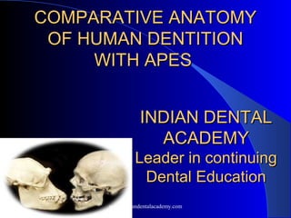
- 1. COMPARATIVE ANATOMYCOMPARATIVE ANATOMY OF HUMAN DENTITIONOF HUMAN DENTITION WITH APESWITH APES INDIAN DENTALINDIAN DENTAL ACADEMYACADEMY Leader in continuingLeader in continuing Dental EducationDental Education www.indiandentalacademy.com
- 2. CONTENTS Terminology Attachments of teeth Evolution of dental occlusion Evolution of teeth Comparisons with : - old world monkeys - new world monkeys - lesser apes - greater apes - gorilla Bibliography www.indiandentalacademy.com
- 3. TERMS ON THE BASIS OFTERMS ON THE BASIS OF TEETHTEETH Homodont: - Teeth all alike e.g. Dolphin , Crocodile. Heterodont: - Teeth differ in form. E.g. Man Monophyodont: - Having only set of teeth. E.g. Rodents Diphyodont: - Having two set of teeth. E.g. Man www.indiandentalacademy.com
- 4. Polyphyodont: - Having endless succession of teeth e.g. most of Fishes, Amphibians. Acrodonts: - Anchylosed teeth, the base of which are wholly Situated upon the bones of attachment e.g. Sphenodon. Plearodont: - Anchylosed teeth, outer side of bone of Attachment e.g. Mackerel. Lophodont: - Ridged teeth (transversely) e.g. Tapir and Elephant. www.indiandentalacademy.com
- 5. Bilophodont: - Teeth having two ridge e.g. Tapir . Polylophodont: - Teeth having more than two ridges e.g. Elephant. Bunodont: - Tooth crown supporting tubercles or cones e.g. pig Selenodont: - Bicrescentic teeth elongated before backward e.g. Ox. www.indiandentalacademy.com
- 6. Brachyodont: - Teeth having short crown and long root e.g. Man Hypsodont: - Teeth having long crown and short root e.g. Horse and Camel Haplodont: - Simple conical tooth having simple crown and roots e.g. Dolphin . www.indiandentalacademy.com
- 7. Thecodont: - Contain a bony socket, which persist for successional tooth . Carnassial Teeth: - Teeth special for tearing flesh e.g. Carnivores and Rodents Cynodontism: - The condition in which the pulp cavity is confined to crown. The tooth having large root or roots. Secodont: - Sectorial or cutting teeth e.g. cat family. www.indiandentalacademy.com
- 8. Polyprotodont: - Numerous Incisors - Having two lower Incisors Ciliiform / Setiform: - Closely set and very fine teeth e.g. some fishes . Tusk: - Incisors or canines of persistent growth, which protrude beyond Lip, when mouth is closed. www.indiandentalacademy.com
- 9. ATTACHMENT OF TEETHATTACHMENT OF TEETH The attachment of teeth with underlying structures also shows the variation in different species. There are four methods of tooth attachment in animal kingdom - 1. Fibrous attachment 2. Hinged attachment 3. Anchylosis 4. Gomphosis www.indiandentalacademy.com
- 10. FIBROUS ATTACHMENT : Well illustrated in Sharks and Rays. Skeleton is cartilaginous, teeth is not attached directly to cartilaginous Jaw. Here the one end of fibers (unmineralized) is embedded at one end into the dentine, and the other end into the bone of attachment. In this group the fiber anchors the teeth to a sheet of outer fiber, which run over the surface of jaw cartilage underneath the tooth rows. www.indiandentalacademy.com
- 11. HINGED ATTACHMENT : Mobile teeth are found in large number of Telosts e.g. Eels and Cod. The teeth are some time capable of a certain amount of movement in all directions, but usually movement is more or less restricted to labiolingual plane. www.indiandentalacademy.com
- 12. Considerably more mobile teeth are found in Hake, Pig and Angle fish. Inward tilting of tooth around its hinge aids the ingestion of prey easier into the oral cavity. Where as outward movement erect the teeth there by trapping the prey. In HAKE the teeth can be depressed lingually almost 90° angle by gentle pressure www.indiandentalacademy.com
- 13. ANCHYLOSIS : Found in Python, Frog, Eel, etc. In a rigidly attached tooth the region of union with the bone is completely mineralized. This condition is referred to ANCHYLOSIS. Where teeth anchylosed, the stress of biting must be take a byte hard tissue and the shape of tooth may be modified in such a way as to absorb stress more efficiently. www.indiandentalacademy.com
- 14. GOMPHOSIS : Attachment occurs in teeth of man, mammalians, in some reptiles and in some fishes (e.g. sow - fish) The presence of Periodontal ligament is characteristic feature of this type of attachment. Most advance type of attachment. www.indiandentalacademy.com
- 15. Evolution of Dental OcclusionEvolution of Dental Occlusion Study of the evolution of dental occlusion involves the study of the complex of general skeleton, which includes jaws and hence teeth. It is believed truly, from the study of Patten, that, vertebrates were closely related to Palaeozoic Eurypterids. The jaws of vertebrates were derived by the appendages of Trilobites. www.indiandentalacademy.com
- 16. The earlier trilobites were well equipped with numerous appendages, which combined the function of oars and gills. In the eurypterids and their modern relative, Limulus, the appendages around the mouth had become specialized in connection with the finding and prehension of food. www.indiandentalacademy.com
- 17. Chordates, shows direct evidence that in the ostracoderms the cavity of the mouth was in series with those of the primitive gill pouches and that the floor of the mouth could be moved up and down, somewhat like that of a frog. In certain ostracoderms the slit-like mouth was bordered by exoskeletal plates. www.indiandentalacademy.com
- 18. The beginning of jaws: An Ostracoderm , showing the position of the slit like mouth. Head shield, seen from below, showing the jaw – like rims of the mouth and mosaic of bony plates on the floor of the mouth. The black spots represent the opening of the gill cavities. www.indiandentalacademy.com
- 19. The ostraco-derms, together with their highly specialized descendants, are classed as Agnathi ( jawless) because they did not possess internal jaws of the shark type. In the sharks the underlying oralo- branchial arches became enlarged into a great fish-trap, while the surface layers of the dermal plates disintegrated into shark teeth which rest on "cartilage jaws"; these are serially homologous with the gills arches. www.indiandentalacademy.com
- 20. Sharks have probably lost the primitive bony dermal jaw plates and have greatly enlarged and specialized the inner or gill arch jaws. The upper bony plate (corresponding to the maxilla) was fixed and toothless. The lower or mandibular plate bore a series of vertical rows of small teeth arranged along the upper margin. www.indiandentalacademy.com
- 21. In the lobe-finned, air- breathing fishes, both the upper and lower jaws were of complex type, consisting of an inner core corresponding respectively with the Meckel's cartilage in the lower and the palatoquadrate in the upper, covered by a number of bony plates. www.indiandentalacademy.com
- 22. The jaw muscles arose as specialized gill-arch muscles. In the higher mammal- like reptiles, however, there was begun a movement to emancipate the jaw muscles proper from their hyoid and branchial companions. www.indiandentalacademy.com
- 23. This was carried to an extreme in the mammals, and meanwhile the old fulcrum at the back of the compound jaws began to diminish, while a totally new joint was established between the ascending ramus of the dentary plate and the squamosal plate. This mandibulo-temporal joint, together with the increasing functional differentiation of the jaw musculature from that of the hyobranchial complex, determines all types of dental occlusion found in the mammals. www.indiandentalacademy.com
- 24. Evolution of the teethEvolution of the teeth The earliest known acanthodians, the denticles around the mouth were loosely attached on or near the surface of the lower jaw. In the air-breathing, lobe-finned fishes of late Mesozoic time there were, two classes of teeth, comprising a row of numerous smaller teeth on the margins and a few much larger sabre-like tusks, forming a widely spaced inner row on the roof of the mouth, and inner sides of the lower jaw. www.indiandentalacademy.com
- 25. This labyrinthodont type of attachment was transmitted to the early amphibians. By the time of the earlier reptiles the pits in which the labyrinthodont teeth were sunk at the base and gradually changed into sockets and the labyrinthodont folds were gradually lost. These simple laniariform socketed teeth, which are seen in the earliest mammal-like reptiles, were set in jaws of a simple scissors-like type, in which the upper teeth overhung the lower teeth and sheared past them in simple vertical arcs. www.indiandentalacademy.com
- 26. Comparison of DentitionComparison of Dentition Marmosets and Tamarins (Callithricidae) : The most primitive of the latin American monkeys. The V-shaped mandible has a slender body and somewhat square ramus, the coronoid process and angle vary in size but the latter is usually well developed. The marmosets can be distinguish from other New World monkeys by the shape of the skull and the number or molar). www.indiandentalacademy.com
- 27. All the group have two molars except Callimico (Goeldi's marmoset) which retains a very much reduced third molar. Marmosets are described as 'shorttusked' (where the incisors are as large as the canines) or 'longtusked' (the canines project above the occlusal plane). The upper molars have only the smallest suggestion of a hypocone, the lower molars are fourcusped. The last molar is always the smallest and the first is the largest of the series. www.indiandentalacademy.com
- 28. NEW WORLD MONKEYSNEW WORLD MONKEYS (CEBIDAE)(CEBIDAE) All the cebids have forward facing orbits which are complete posteriorly, a tympanic bulla with an external tympanic ring fused to the bulla and three premolars in each jaw quadrant. They show Massive laryngeal expansion and modification of the shape of both the skull and lower jaw. www.indiandentalacademy.com
- 29. Aotus (night monkey), CebusAotus (night monkey), Cebus (capuchin), Saimiri (squirrel(capuchin), Saimiri (squirrel monkey)monkey),,CallicebusCallicebus The first three are short-faced and have an anthropoid mandible; the lower dental arcade is V-shaped and the ramus rectangular with a moderately developed coronoid process and a slightly expanded angle. In contrast, the laryngeal enlargement found in Callicebus and Aloriarta. The coronoid process is small, the condyle is high above the tooth row and the angle is greatly expanded. www.indiandentalacademy.com
- 30. Atele, Brachyteles andAtele, Brachyteles and LagothrixLagothrix The members of this group have fairly long faces with well-developed jaws. The mandible has a large angle (biggest in Brachyteles). The dentition is similar to that of the first group, but a hypoconulid is often developed on molar and although upper third molar is the smallest of the molars, all these teeth are of similar size. www.indiandentalacademy.com
- 31. CCacajao (uakari), Pitheciaacajao (uakari), Pithecia (( saki) alltisaki) allti Chirpotes (bearded saki)Chirpotes (bearded saki) These monkeys have a highly specialized anterior dentition. The upper incisors are spatulate and project forwards so sharply as to be almost horizontal. Both upper and lower incisors are separated from the tusk-like laterally flaring canines by a large diastema. The post-canine teeth conform to the general cebid pattern of bicuspid premolars and four-cusped molars but appear small in comparison to the anterior dentition. www.indiandentalacademy.com
- 32. Terrestrial groupTerrestrial group :: Erythrocebus (patasErythrocebus (patas monkeys):monkeys): MandrillusMandrillus (( mandrills),mandrills), Papio(baboons), TheropithecusPapio(baboons), Theropithecus This are the ground- living monkeys, and all shows marked sexual dimorphism of body size: the adult female weighs approximately half as much as the male. This difference extends to the skull and dentition. www.indiandentalacademy.com
- 33. The canines, large in both sexes, become long and dagger-like in the males. The molars are bilophodont and the third molars are large in size. In Mandrills, the anterior teeth are very large and the cheek teeth are unusually small. www.indiandentalacademy.com
- 34. Colobus (guerezas), Presbytis ( langurs), Pygathrix ( Douc langurs ), Rhinopithecus (snub nosed langurs) This are 'leaf-eating monkeys’. The distinctive features of these monkeys, including a sacculated stomach, have been associated with a herbivorous diet. www.indiandentalacademy.com
- 35. For example, the mandibular ramus is high, with corresponding distance between the level of the mandibular joint and that of the occlusal plane. The lower incisors occlude in front of the uppers. This condition is called 'underbite' and may be an adaptation for the more efficient ingestion of leaves. www.indiandentalacademy.com
- 36. GIBBON AND SIAMANGGIBBON AND SIAMANG HYLOBATIDAE (LESSER APES)HYLOBATIDAE (LESSER APES) Found in the tropical rain and forest of south east Asia. The palate in gibbons is exceptionally long (reaching 50- 54% of total skull length) and broad. No distinct condylar neck, , the posterior border of the mandibular notch merges into the condyle proper. The fossa has no distinct articular eminence although there is a post- glenoid process. www.indiandentalacademy.com
- 37. The incisors are heteromorphic: the upper centrals are weak and spatulate, the lateral small and often pointed. The lower incisors are fanshaped. The lower first premolar is sectorial, the remainder are bicuspid although the lower second is expanded distally. The upper molars are simple fourcusped teeth with an oblique ridge, the lower, have the 'Dryopithecus' pattern or Y5 pattern. www.indiandentalacademy.com
- 38. GREAT APES (PONGIDAE)GREAT APES (PONGIDAE) There are three great apes: – The Chimpanzee (Pan), – The OrangUtan (Pongo) and – The Gorilla (Gorilla). They have different distributions, habits, locomotor patterns and skulls but very similar dentitions. The general form of the dentition is the same in all three great apes. 2123 2123 www.indiandentalacademy.com
- 39. The juvenile skuII and deciduousThe juvenile skuII and deciduous dentitiondentition The juvenile pongid skull is much more 'human' in appearance than that of the adult This is due to the early development of the brain and neurocranium and the much later growth of the face. The first molar is the first tooth in the permanent dentition to erupt followed by the incisors, second molars, premolars and then the canine. As in man the shape of the second deciduous molar closely resembles that of the first permanent molar. www.indiandentalacademy.com
- 40. Chimpanzee ( pan )Chimpanzee ( pan ) There are two species of chimpanzee, Pan trog lodytes which is widely distributed across equa torial Africa and Pan paniscus, the pygmy chimpanzee, which is restricted to the area between the Congo and Lualaba Rivers of central West Africa. Both live in rain forest www.indiandentalacademy.com
- 41. The face is prognathic with a long premaxilla. The mandibular body is pressed near the canine region. The ramus is rectangular, with large areas for the attachment of masseter and temporalis, although the coronoid process is short and the mandibular notch shallow. The gently rounded condyle articulate with an almost flat glenoid fossa. www.indiandentalacademy.com
- 42. Orang-Utan (Pongo)Orang-Utan (Pongo) The cranium of the orang is comparatively short and domed. The dentition has the same general form as in the chimpanzee. The upper canine is much larger in the male. In both jaws the second molar is normally the largest and the third the smallest. Supernumerary molars are common. www.indiandentalacademy.com
- 43. GorillaGorilla Gorillas are found in western (lowland gorilla) and eastern equatorial Africa (highland gorilla). Both the neurocranium and facial skeleton are large. The very prognathic face has a rectangular hard palate extending backwards behind the third molars. www.indiandentalacademy.com
- 44. The lower jaw is strong and heavy with a sloping symphysis braced by a simian shelf. The rectangular ramus often shows strong muscle markings. The upper molars have an oblique ridge and the lowers have the Dryopirheclis' pattern but both have very sharply pointed cusps when newly erupted. www.indiandentalacademy.com
- 45. ReferencesReferences 1. J.W.OSBORN, Dental anatomy and embryology, Blackwell publications, vol.1 pg no. 401455. 2. William K.Gregory, evolution of dental occlusion from fish to man, Angle Orthod 1941:3. 3. Dental anatomy, histology and development; comparative anatomy, ed 1st .pg no 179215. www.indiandentalacademy.com