Fe male reproductive system 2
•Transferir como PPTX, PDF•
5 gostaram•1,016 visualizações


Recomendados
Mais conteúdo relacionado
Mais procurados
Mais procurados (20)
Semelhante a Fe male reproductive system 2
Semelhante a Fe male reproductive system 2 (20)
Mais de DrChintansinh Parmar
Mais de DrChintansinh Parmar (20)
Último
Último (20)
Fe male reproductive system 2
- 1. Female Reproductive System - Dr. Chintan
- 2. Anovulation hormones - hyposecretion of gonadotropic Abnormal ovaries – thick capsule - cyst Tests - to analyze the urine for a surge in pregnanediol, the end product of progesterone metabolism, during the latter half of the sexual cycle; the lack of this substance indicates failure of ovulation Body temp. chart - Secretion of progesterone during the latter half of the cycle raises the body temperature about 0.5°F, with the temperature rise coming abruptly at the time of ovulation.
- 5. Human chorionic gonadotropin, a hormone that is extracted from the human placenta has almost the same effects as LH and is therefore a powerful stimulator of ovulation. excess use of this hormone can cause ovulation from many follicles simultaneously; this results in multiple births, an effect that has caused as many as eight babies (mostly stillborn) to be born to mothers treated for infertility with this hormone. Endometriosis – endometrial tissue grows and even menstruates in the pelvic cavity surrounding the uterus, fallopian tubes, and ovaries – fibrosis – ovum cant be released – tube blockage.
- 6. Salpingitis -gonococcal infection - inflammation of the fallopian tubes; this causes fibrosis in the tubes, thereby occluding them. secretion of abnormal mucus by the uterine cervix at the time of ovulation, estrogen causes the secretion of mucus with special characteristics that allow rapid mobility of sperm into the uterus and actually guide the sperm up along mucous ―threads.‖ low-grade infection or inflammation, or abnormal hormonal stimulation of the cervix can lead to a viscous mucous plug that prevents fertilization.
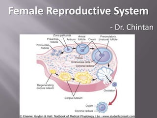
- 8. While still in the ovary, the ovum is in the primary oocyte stage. Shortly before it is released from the ovarian follicle, its nucleus divides by meiosis and a first polar body is expelled from the nucleus of the oocyte. The primary oocyte then becomes the secondary oocyte. In this process, each of the 23 pairs of chromosomes loses one of its partners, which becomes incorporated in a polar body that is expelled. This leaves 23 unpaired chromosomes in the secondary oocyte. It is at this time that the ovum, still in the secondary oocyte stage, is ovulated into the abdominal cavity. Then, almost immediately, it enters the fimbriated end of one of the fallopian tubes.
- 9. When ovulation occurs, the ovum, along with a hundred or more attached granulosa cells that constitute the corona radiata, is expelled directly into the peritoneal cavity. As many as 98 % succeed in entering fallopian tube. Women with one ovary removed and the opposite fallopian tube removed have had several children with relative ease of conception, thus demonstrating that ova can even enter the opposite fallopian tube.
- 10. After the male ejaculates semen into the vagina during intercourse, a few sperm are transported within 5 to 10 minutes upward from the vagina and through the uterus and fallopian tubes to the ampullae of the fallopian tubes. This transport of the sperm is aided by contractions of the uterus and fallopian tubes stimulated by prostaglandins in the male seminal fluid and also by oxytocin released from the posterior pituitary gland of the female. Of the almost half a billion sperm deposited in the vagina, a few thousand succeed in reaching each ampulla. Chemoattraction
- 11. Sperm penetration – acrosomal reaction Once a sperm has entered the ovum (secondary oocyte), the oocyte divides again to form the mature ovum plus a second polar body that is expelled. The mature ovum still carries in its nucleus (female pronucleus) 23 chromosomes. On entering the ovum, sperm head swells to form a male pronucleus. Later, the 23 unpaired chromosomes of the male pronucleus and the 23 unpaired chromosomes of the female pronucleus align themselves to re-form a complete complement of 46 chromosomes (23 pairs).
- 13. Weak contractions of the fallopian tube + beating of cilia towards uterus – 3 to 5 days The isthmus of the fallopian tube remains spastically contracted for about the first 3 days after ovulation. After this time, the rapidly increasing progesterone secreted by the ovarian corpus luteum, exerts a tubular relaxing effect that allows entry of the ovum into the uterus. Blastocyst (100 cells) – nutrition by FT secretion IVF – test tube babies – 5 to 10 %
- 14. After reaching the uterus, the developing blastocyst usually remains in the uterine cavity an additional 1 to 3 days before it implants in the endometrium – uterine milk Implantation results from the action of trophoblast cells that develop over the surface of the blastocyst. These cells secrete proteolytic enzymes that digest and liquefy the adjacent cells of the uterine endometrium – dorsal wall Once implantation has taken place, the trophoblast cells and other adjacent cells proliferate rapidly, forming the placenta and the various membranes of pregnancy.
- 16. Corpus luteum – progesterone - endometrial stromal cells - extra quantities of glycogen, proteins, lipids and some minerals After implantation, uterine endometrium stromal cells swell – decidual reaction - the decidua As the trophoblast cells invade the decidua, digesting and absorbing it, the stored nutrients in the decidua are used by the embryo for growth and development. During the first week after implantation, this is the only means by which the embryo can obtain nutrients – 8 weeks The placenta also begins to provide nutrition 1 week after implantation.
- 18. While the trophoblastic cords from the blastocyst are attaching to the uterus, blood capillaries grow into the cords from the vascular system of the newly forming embryo. By the 16th day after fertilization, blood also begins to be pumped by the heart of the embryo itself. Simultaneously, blood sinuses supplied with blood from the mother develop around the outsides of the trophoblastic cords. The trophoblast cells send out more and more projections, which become placental villi into which fetal capillaries grow. Thus, the villi, carrying fetal blood, are surrounded by sinuses that contain maternal blood.
- 19. Final structure - the fetus’s blood flows through two umbilical arteries, then into the capillaries of the villi, and finally back through a single umbilical vein into the fetus. The mother’s blood flows from her uterine arteries into large maternal sinuses that surround the villi and then back into the uterine veins of the mother. Nutrients and other substances pass through the placental membrane mainly by diffusion in much the same manner that diffusion occurs through the alveolar membranes of the lungs and the capillary membranes.
- 21. The major function of the placenta is to provide for diffusion of foodstuffs and oxygen from the mother’s blood into the fetus’s blood and diffusion of excretory products from the fetus back into the mother. Early months of pregnancy – not developed thick membrane – less permeability – not grown small surface area – less diffusion – later months vice versa Rarely, ―breaks‖ occur
- 22. The dissolved oxygen in the blood of the large maternal sinuses passes into the fetal blood by simple diffusion, driven by an oxygen pressure gradient from the mother’s blood to the fetus’ blood. 50 – 30 = 20 mmHg How it is possible for a fetus to obtain sufficient oxygen when the fetal blood leaving the placenta has a PO2 of only 30 mm Hg ???
- 23. The curve for fetal hemoglobin is shifted to the left of that for maternal hemoglobin. This means that at the low PO2 levels in fetal blood, the fetal hemoglobin can carry 20 to 50 per cent more oxygen than maternal hemoglobin can. The hemoglobin concentration of fetal blood is about 50 per cent greater than that of the mother – less affinity for 2,3 DPG The Bohr effect, hemoglobin can carry more oxygen at a low PCO2 than it can at a high PCO2. The fetal blood entering the placenta carries large amounts of CO2, but much of this CO2 diffuses from the fetal blood into the maternal blood.
- 25. Double Bohr effect The total diffusing capacity of the entire placenta for oxygen at term is about 1.2 milliliters of oxygen/minute/mmHg O2 pressure difference across the membrane. The PCO2 of the fetal blood is 2 to 3 mm Hg higher than that of the maternal blood. This small pressure gradient for carbon dioxide across the membrane is more than sufficient to allow adequate diffusion of carbon dioxide
- 26. Late stages of pregnancy, the fetus often uses as much glucose as the entire body of the mother uses. The glucose is transported by carrier molecule facilitated diffusion through the placental membrane. High solubility of fatty acids in cell membranes, these also diffuse from the maternal blood into the fetal blood, but more slowly than glucose, so that glucose is used more easily by the fetus for nutrition. Ketone bodies and potassium, sodium, and chloride ions diffuse with relative ease from the maternal blood into the fetal blood.
- 27. Same as CO2 NPN such as urea, uric acid and creatinine. Urea conc. slightly greater Creatinine considerably higher diffusion gradients across the placental membrane
- 28. hCG, E, P, hCS With the development of the trophoblast cells from the early fertilized ovum, the hormone hCG is secreted by the syncytial trophoblast cells into the fluids of the mother. The secretion of this hormone can first be measured in the blood 8 to 9 days after ovulation, shortly after the blastocyst implants in the endometrium. Then the rate of secretion rises rapidly to reach a maximum at about 10 to 12 weeks of pregnancy and decreases back to a lower value by 16 to 20 weeks. It continues at this level for the remainder of pregnancy.
- 29. It can be measured by radioimmunoassay and detected in the blood as early as 6 days after conception. Its presence in the urine in early pregnancy is the basis of the various laboratory tests for pregnancy, and it can sometimes be detected in the urine as early as 14 days after conception – it disappears if fetus dies hCG is not absolutely specific for pregnancy. Small amounts are secreted by a variety of GIT and other tumors in both sexes, and hCG has been measured in individuals with suspected tumors as a "tumor marker." It also appears that the fetal liver and kidney normally produce small amounts of hCG.
- 30. Glycoprotein - molecular weight of about 39,000 same molecular structure and function as LH – fetal testis to prevent involution of the corpus luteum at the end of the monthly female sexual cycle It causes the corpus luteum to secrete larger quantities of progesterone and estrogens for the next few months. E,P prevent menstruation and cause the endometrium to continue to grow and store large amounts of nutrients rather than being shed in the menstruation.
- 31. decidual cells—greatly swollen and nutritious—at about the time that the blastocyst implants. corpus luteum in the mother’s ovary grows to about twice its initial size by a month or so after pregnancy begins, and its continued secretion of E, P maintains the decidual nature of the uterine endometrium If the corpus luteum is removed before approximately the 7th week of pregnancy, spontaneous abortion almost always occurs, sometimes even up to the 12th week. The corpus luteum involutes slowly after the 13th to 17th week of gestation.