This document reports a rare case of inflammatory pseudotumor (IPT) of the stomach in a 65-year-old female patient. IPT is a benign tumor that can occur in any part of the body. The patient presented with abdominal discomfort, constipation, and abdominal distension for a year. Imaging showed a large cystic mass in her stomach. She underwent surgery to remove the mass, which was diagnosed as IPT based on pathological examination. IPT of the stomach is very rare, and this case highlights the difficulty in diagnosis due to non-specific presentations that can mimic other tumors.
1. ORIGINAL ARTICLE
Inflammatory Pseudotumour of Stomach in an
old lady: a rare case report
Azhar AHa, Pasha MAa, Hassan Sa, Zainal Ma and Rashidi Ab
a
Department of Surgery and b Department Of Emergency Medicine, School of Medical Sciences, Health Cam-
pus, Universiti Sains Malaysia, Kubang Kerian, Kelantan, Malaysia
ABSTRACT
Inflammatory pseudotumour (IPT) is a rare benign solid tumor in adults and children. The prevalence, etiology
CASE REPORT
and pathogenesis of this condition are still uncertain. Despite the use of modern laboratory techniques and
imaging, it is often difficult to make the diagnosis of IPT. Besides, occasionally the nonspecific morphological
appearance and clinical presentation of the mass may mimic other more common primary or secondary
neoplasms. IPT is commonly encountered in the lung and mediastinum. Other sites include abdomen (liver,
pancreas, stomach, omentum), retroperitoneum, pelvis (bladder) and extremities in children. We report a
rare case of gastric inflammatory pseudotumour in a 65-year-old female patient. Clinical presentations and
its management along with review of literatures are presented.
KEYWORDS: Inflammatory pseudotumour of stomach, Elderly, Rare
INTRODUCTION
Inflammatory pseudotumour (IPT) is a rare benign le- measuring 20 x 15 cm in size. The mass had a smooth
sion in adults and children and it can occur in any surface and well-defined margins. The mass was hard
site of the body.1,2,3 Other synonyms for IPT include in consistency and moved in both vertical and hori-
inflammatory fibromyoblastic pseudotumor, plasma zontal directions during the palpation. There were
cell granuloma, plasma cell pseudotumour, omental no stigmata of chronic liver disease and no hepat-
mesentreric myxoid hamartoma and inflammatory fi- osplenomegaly. Other physical examinations were
brosarcoma.4 However, the term IPT being the most normal. Blood, renal and hepatic parameters were
descriptive is widely used in the literature. normal. Hepatitis serology of HbsAg and anti-HCV,
a-fetoprotein, and carcinoembryonic antigen were
Gastric IPT in adults is a very rare disease. Only five negative.
reported cases of gastric IPT in adults exist in the
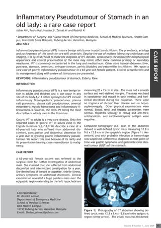
English literature.5,6,7,8,9 We describe a case of a Computed tomography (CT) scan of the abdomen
65-year-old lady who suffered from abdominal dis- showed a well-defined cystic mass measuring 12.8 x
comfort, constipation and abdominal distension for 9.6 x 12.8 cm in the epigastric region (Figure 1). Me-
a year due to growing gastric inflammatory pseudo- senteric cyst with probable infection or hemorrhage
tumour. We report this case because of its rarity and was suspected. Differential diagnosis at that point of
its presentation bearing close resemblance to malig- time was gastric lymphoma and gastrointestinal stro-
nancy. mal tumour (GIST) of the stomach.
CASE REPORT
A 65-year-old female patient was referred to the
surgical clinic for further investigation of abdominal
mass. She claimed that she suffered from abdominal
discomfort and intermittent constipation for a year.
She denied loss of weight or appetite, febrile illness,
urinary symptoms or abdominal distension. Clinical
examination revealed a huge painless mass over the
epigastric region extending to the left hypochodrium
Correspondence:
Dr. Rashidi Ahmad
Department of Emergency Medicine
School of Medical Sciences
USM Health Campus
16150 Kubang Kerian, Kelantan Malaysia Figure 1: Photography of CT abdomen showing de-
Email: Shidee_ahmad@yahoo.com fined cystic mass 12.8 x 9.6 x 12.8 cm in the epigastric
region (white arrow). The cystic mass has thickened
Volume 8 Number 1, June 2009 45
2. THE INTERNATIONAL MEDICAL JOURNAL
wall and contained protein-like material. an unusual tissue response to injury, bacterial, viral
and fungal infection and autoimmune causes.10 There
The patient underwent explorative laparatomy, com- are many reports which associate IPT with Mycobacte-
plete excision of the mass and subtotal gastrectomy rium avium intracellulare, Campylobacter jejuni, Ba-
with gastrojujenostomy. Macroscopically, the tumour cillus sphaericus, Coxiella burnetii, Escherichia coli,
was solid and lobulated measuring 20 x 14 x 12 cm Epstein-Barr Virus and acute retroviral syndrome.11
in size arising from the greater curvature of stom- Association of IPT and radiotherapy, steroid usage and
ach. Histopathological study of the excised specimen some genetic factors has also been reported.12
showed diffuse infiltration of plasma cells in all the
specimens, walled by dense hyalinised fibrous colla- There are still two controversial issues regarding IPT.
genous tissue (Figure 2). Giant cells of foreign body Is IPT purely an inflammatory entity or neoplastic in
type and fibroblastic reaction were noted. There was its origin? If it is neoplatic, is IPT benign or malignant
no evidence of dysplasia or metaplasia from the speci- tumour? Few genetic studies on cytogenetic abnor-
men. Histopathological diagnosis was IPT of stomach. malities such as rearrangements of the ALK gene on
chromosome 2p23, clonal chromosome abnormalities
Figure 2: Photomicrograph showing diffuse infiltra- and DNA aneuploidy, and the role of oncogenic viruses
tion of plasma cells, that walled by dense hyalinised suggest that IPT is a true neoplasm.13-20 According to
fibrous collagenous. (Arrow head - stomach wall, the current classification of the World Health Organi-
zation (WHO), IPT is a neoplasm with a tendency for
local recurrence and a very low rate of metastasis.21
Hussong et al. confirmed that IPT tumors testing posi-
tive for p53 showed recurrence or malignant transfor-
mation.22 However, Yamamoto et al. do not support
the theory that p53 plays a major role in the patho-
genesis of IPT.23
Most IPTs require surgery to obtain definite diagnosis
and cure. Complete resection is the preferred option,
because incomplete excision has been shown to be a
risk factor for recurrence.2 According to Hakozi et al,
pronounced inflammatory reaction is the main char-
acteristic of IPT. Therefore, the logical option is an-
ti-inflammatory treatment. In fact the trial of NSAID
treatment may both confirm the diagnosis of IPT and
at the same time successfully treating the tumor.24
black arrow - inflammatory plasma cells, white arrow There is no uniform recommendation in the literature
hyalinised fibrous collagenous matrix). regarding when to use additional therapy.25 Steroid
therapy has been tried for cases in whom diagnosis is
Postoperative course was uneventful. At follow up made preoperatively, but the outcome is not signifi-
nine months later, she was well, with no evidence of cant.26 Other adjuvant therapy like radiotherapy and
recurrence. chemotherapy can also be tried. However, additional
therapy for IPT that show a high risk of recurrence
indicated by ill-defined margins or intra-abdominal,
mesenteric, omental, and retroperitoneal localiza-
tions is strongly recommended.1
DISCUSSION
Recurrences are documented in 18-40% of the cases
We highlight a rare case of IPT in elderly patient who and appear to be more frequent in the extrapulmonary
presented with vague abdominal pain, change of lesions, which are larger than 8 cm and locally inva-
bowel habit and fast growing abdominal mass within sive.27 Unfortunately, there are no definitive clinical,
a year. Despite the use of modern laboratory tech- histopathological, or genetic features to predict re-
niques and imaging, making a definite diagnosis of IPT currence or metastasis though one of the recent stud-
is often difficult. Our patient had non-specific clinical ies suggests that reactivity of ALK to be a favorable
presentation and the mass also had non-specific mor- prognostic indicator.3 Novosel et al suggest a palette
phological appearances that mimic other lesions, such of immunohistochemical agents with p53 or ALK in ad-
as primary or secondary neoplasm. dition to standard immunohistochemical procedures
for prognostic stratification. Negative p53 expression
The IPT is characterized pathologically by a diffuse or positive ALK expression should be considered as a
infiltration of inflammatory cells with fibromyoblastic favorable IPT diagnosis.10
proliferation. The predominant cells may be plasma
cells, neutrophils or lymphocytes.3 The prevalence, A year after surgical IPT resection, our patient is well,
etiology and pathogenesis of this condition are still with no signs of recurrence. We suggest long term fol-
uncertain. The condition is thought to be related to low up for this patient.
46 Volume 8 Number 1, June 2009
3. ORIGINAL ARTICLE
REFERENCES 12:424- 6
1. Coffin CM, Watterson J, Priest JR, Dehner LP. 12. Karnak I, Senocak ME, Ciftci AO, et al.
Extrapulmonary inflammatory myofibroblas Inflammatory myofibroblastic tumor in
tic tumor (inflammatory pseudotumor). children: diagnosis and treatment. J
A clinicopathologic and immunohistochemical Pediatr Surg 2001; 36:908-12
study of 84 cases. Am J Surg Pathol 1995;
19:859-72 13. Coffin CM, Patel A, Perkins S, Elenitoba-
Johnson KS, Perlman E, Griffin CA. ALK1 and
2. Souid AK, Ziemba MC, Dubansky AS, et al. p80 expression and chromosomal rearrange
Inflammatory myofi broblastic tumor in ments involving 2p23 in inflammatory
children. Cancer 1993; 72:2042-8 myofi broblastic tumor. Mod Pathol 2001;
CASE REPORT
14:569-76
3. Coffin CM, Hornick JL, Fletcher CD.
Inflammatory myofibroblastic tumor: 14. Yousem SA, Shaw H, Cieply K. Involvement
comparison of clinicopathologic, histologic, of
and immunohistochemical features including 2p23 in pulmonary inflammatory pseudotu
ALK expression in atypical and aggressive mors. Hum Pathol 2001; 32:428-33
cases. Am J Surg Pathol 2007; 31:509-20
15. Su LD, Atayde-Perez A, Sheldon S, Fletcher
4. Day DL, Sane S, Dehner LP. Inflammatory JA, Weiss SW. Inflammatory myofibroblastic
pseudotumor of the mesentery and small tumor: cytogenetic evidence supporting
intestine. Pediatr Radiol 1986; 16:210-5 clonal origin. Mod Pathol 1998; 11:364-8
5. Kim KA, Park CM, Lee JH, et al. Inflammatory 16. Snyder CS, Dell’Aquila M, Haghighi P, Baer-
myofibroblastic tumor of the stomach with gen
peritoneal dissemination in a young adult: RN, Suh YK, Yi ES. Clonal changes in
imaging findings. Abdom Imaging 2004; 29: inflammatory pseudotumor of the lung: a
9-11 case report. Cancer 1995; 76:1545-9
6. Al-Taie OH, Mork H, Jenett M, Klein D, Muller 17. Treissman SP, Gillis DA, Lee CL, Giacomanto
JG, Scheurlen M. Fast-growing gastric nio M, Resch L. Omental-mesenteric infl
inflammatory pseudotumor: a rare manifesta ammatory pseudotumor. Cytogenetic
tion of peptic ulcer disease. Endoscopy 2002; demonstration of genetic changes and
34:239-41 monoclonality in one tumor. Cancer 1994;
73:1433-7
7. Leon CJ, Castillo J, Mebold J, Cortez L,
Felmer R. Inflamatory myofibroblastic tumor 18. Biselli R, Ferlini C, Fattorossi A, Boldrini R,
of the stomach: an unusual complication Bosman C. Inflammatory myofibroblastic
after gastrectomy. Gastrointest Endosc 2006; tumor (inflammatory pseudotumor): DNA
63:347-9 flow
cytometric analysis of nine pediatric cases.
8. Kojimahara K, Mukai M, Yamazaki K, et al. Cancer 1996; 77:778-84
Inflammatory pseudotumor of the stomach:
report of a highly infiltrative case with 19. Lewis JT, Gaffney RL, Casey MB, Farrell MA,
electron microscopic and immunohistochemi Morice WG, Macon WR. Inflammatory
cal studies. Acta Pathol Jpn 1993; 43:65-70 pseudotumor of the spleen associated with
a clonal Epstein-Barr virus genome. Case
9. Park SH, Kim JH, Min BW, Song TJ, Son GS. report and review of the literature. Am J
Exophytic inflammatory myofibroblastic Clin
tumor of the stomach in an adult woman: Pathol 2003; 120: 56-61
A rare cause of hemoperitoneum. World J
Gastroenterol 2008; 14:136-9 20. Gomez-Roman JJ, Sanchez-Velasco P,
Ocejo-Vinyals G, Hernandez-Nieto E,
10. Novosel I, Babiæ D, Iliæ J, Seiwerth S. Leyva-Cobian F, Val-Bernal JF. Human
Inflammatory pseudotumour of the cervix: herpesvirus-8 genes are expressed in
case report and review of the literature. pulmonary inflammatory myofibroblastic
Acta Clin Croat 2007; 4:265-70 tumor (inflammatory pseudotumor).
Am J Surg Pathol 2001; 25:624-9
11. Navai N, Yap RL, Guptar R, Fraser TG,
Gonzales CM. inflammatory pseudotumour of 21. Coffin CM, Fletcher JA. Inflammatory myofi
the testis: a novel presentation of acute broblastic tumour. In: Fletcher CDM, Unni
retroviral syndrome. Int J Urol 2005; KK, Mertens F. Pathology and genetics of
Volume 8 Number 1, June 2009 47
4. THE INTERNATIONAL MEDICAL JOURNAL
tumours of soft tissue and bone. World
Health Organization Classification of
Tumours. Lyon: IARC Press, 2002: 91-3
22. Hussong Jw, Brown M, Perkins SL, Dehner LP,
Coffin CM. Comparison of DNA ploidy,
histologic, and immunohistochemical findings
with clinical outcome in inflammatory myofi
broblastic tumors. Mod Pathol 1999; 12:279-
86
23. Yamamoto H, Oda Y, Saito T, et al. p53
Mutation and MDM 2 amplification in
inflammatory myofibroblastic tumours.
Histopathology 2003; 42:431-9
24. Hakozaki Y, Katou M, Nakagawa K,
Shirahama T, Matsumoto T. Improvement of
Inflammatory pseudotumour of the liver after
nonsteroidal anti-inflammatory agent
therapy. Am J Gastroenterol 1993; 88:1121-2
25. Shah MD, Mcclain KL. Intracranial plasma cell
granuloma: case report and treatment of
recurrence with methotrexate and 6-mercap
topurine. J Pediatr Hematol Oncol 2005;
27:599-603
26. Messineo A, Mognato G, D’Amore ES, Anton
iello L, Guglielmi M, Cecchetto G. Inflamma
tory pseudotumors of the lung in children:
Conservative or aggressive approach?
Med Pediatr Oncol 1998; 31:100-4
27. Sanders BM, West KW, Gingalewski C,
Engum S, Davis M, Grosfeld JL. Inflammatory
pseudotumor of the alimentary tract: Clinical
and surgical experience. J Pediatr Surg 2001;
36:169-73
48 Volume 8 Number 1, June 2009