SlideShare uma empresa Scribd logo
1 de 189
Baixar para ler offline
Followyour heart ... but ... takeyour brain with you ...
Giossitis
+ ve Babinski
M~a/ol,lastlcantmla
Sherif EL Hawary,
glove and stocking
hyposthesia
MD
NEUROLOGY
Sherif EL Hawary, MD
Copyright © 2019, Sherif EL Hawary, MD
Text copyright © 2019, Sherif EL Hawary, MD
Figures, tables, illustrations and images copyright © 2019, Sherif EL Hawary, MD
All rights reserved. No part ofthis book may be translated, reprinted, used or reproduced
in any form or by any electronic, mechanical, or other means, now known or hereafter
invented, including photocopying and recording, or in any information storage or
retrieval system, without permission in writing from the author.
INDEX
CENTRAL NERVOUS SYSTEM----------------------------------
THE MOTOR SYSTEM---------------------------------------------
Differentiation between UMNL & LMNL -------------------------
THE SENSORY SYSTEM ------------------------------------------
AREAS OF THE CEREBRAL CORTEX-----------------------
SPEECH DISORDERS -----------------------------------------------
THE CRANIAL NERYES -------------------------------------------
BELL'S PALSY -------------------------------------------------------
VERTIGO---------------------------------------------------------------
BULBAR PALSY -----------------------------------------------------
CEREBRO - VASCULAR DISEASE ------------------------------
STROKE-----------------------------------------------------------------
TRANSIENT ISCHEMIC ATTACKS (TIAs) -------------------
HEMIPLEGIA ----------------------------------------------------------
BLOOD SUPPLY OF THE BRAIN------------------------------
NEUROGENIC BLADDER-----------------------------------------
PARAPLEGIA ----------------------------------------------------------
SPECIAL FORMS OF PARAPLEGIA ---------------------------
CAUDA EQUINA -----------------------------------------------------
SPONDYLOSIS--------------------------------------------------------
SCIATICA---------------------------------------------------------------
MOTOR NEURONE DISEASE------------------------------------
DD ofwasting ofthe small muscles ofthe hand -------------------
THE EXTRAPYRAMIDAL SYSTEM ----------------------------
THE CEREBELLUM-------------------------------------------------
PERIPHERAL NEURITIS -------------------------------------------
MYASTHENIAS -------------------------------------------------------
MYOPATHIES ---------------------------------------------------------
BRAIN TUMOURS---------------------------------------------------
EPILEPSY ---------------------------------------------------------------
MULTIPLE SCLEROSIS --------------------------------------------
HEADACHE ------------------------------------------------------------
MIGRAINE --------------------------------------------------------------
SUBARACHNOID HEMORRHAGE ------------------------------
MENINGITIS-----------------------------------------------------------
ENCEPHALITIS -------------------------------------------------------
COMA--------------------------------------------------------------------
1
4
10
11
15
21
24
41
43
45
46
46
57
61
69
78
79
88
92
95
98
100
102
103
108
111
123
126
130
136
142
150
151
155
163
170
172
A wise man once said .........
" Everyone has 2 cerebral hemispheres,
right & left....
In the left: nothing is right........ and...
In the right: nothing is left....."
CENTRAL NERVOUS SYSTEM
The CNS is formed of 2 parts:
I. INTRACRANIAL PART
1. Two cerebral hemispheres.
2. Basal ganglia.
3. Brain stem.
4. Cerebellum.
1. Two cerebral hemispheres:
• CONNECTION: They are connected to each other by the CORPUS CALLOSUM.
• SURFACE: The surface ofeach hemisphere is divided into 4 LOBES:
1. Frontal 2. Parietal. 3. Temporal. 4. Occipital.
• CEREBRAL LOBES:
1. Outer gray matter:
o It is composed of:
(cerebral cortex)
NERVE CELLS.
o It contains certain AREAS that control specific functions.
2. Inner white matter: (depth ofthe cerebral hemisphere)
o It is composed of: NERVE FIBRES.
o It CONDUCTS IMPULSES to & from the cerebral cortex.
2. Basal nuclei:
- At the base ofeach cerebral hemisphere (At various levels deep in the white matter):
• BASAL GANGLIA: Caudate, Putamen, Globuspallidus.
• THALAMUS, SUBTHALAMUS, HYPOTHALAMUS.
3. Brain stem: "3 parts from above downwards"
• MIDBRAIN: contains the motor nuclei of the cranial nerves 3 & 4.
• PONS: contains the motor nuclei of the cranial nerves 5, 6, 7.
• MEDULLA: contains the motor nuclei ofthe cranial nerves 9, 10, 11, 12.
• The 1st, 2
nd
& 8
th
cranial nerves have no motor nuclei.
• They are SENSORY NERVES concerned with special sensations.
4. Cerebellum:
- It lies at the BACK & the BOTTOM ofthe cranium behind the brain stem,
in the posterior cranial fossa.
1
l
2
2
II. SPINAL PART
1. Spinal Cord.
2. Cauda Equina.
1. Spinal Cord:
o It lies in the spinal canal.
o It ends at the lower border ofthe 1
st
lumbar vertebra (Lt).
Segments of the spinal cord:
• Cervical segments: 8 segments.
• Thoracic segments: 12 segments.
• Lumbar segments: 5 segments.
• Sacral segments: 5 segments.
o CONUS MEDULLARIS: The lowermost 3 segments ofthe spinal cord (S3,4, 5).
o EPICONUS: The 4 segments above the conus medullaris (L4, 5, S1, 2).
Section in the spinal cord:
- It contains: gray matter (cells) surrounded by white matter (fibres).
- The GRAY MATTER: H - shaped in a transverse section & is formed of:
• 2 anterior horns: MOTOR function.
• 2 posterior horns: SENSORY function.
- The WIDTE MATTER: contains nerve fibres arranged into tracts:
1. The important ascending tracts are:
• Lateral & ventral spinothalamic:
• Posterior column:
• Spinocerebellar:
2. The important descending tracts are:
for superficial sensations.
for deep sensations.
for cerebellar information.
• The pyramidal tract (corticospinal tract).
• The extrapyramidal tracts.
• The cerebello-spinal tracts.
2. Cauda Eguina: " Collection of Lumbo - sacral roots "
o It fills the lower part ofthe spinal canal.
o It starts at the lower border ofthe 1
st
lumbar vertebra (Lt).
C2 cord segment
C. nerve root
Anterior
S, cord segn1ent
~a..-"r' )..r-:~ ;;:.__ Spinous process T 1
vertebra
Posterior
Spinous process 1..1
vertebra
Cavda equ;na
51nerve root - -----
THE SPlNAL CORD, LATERAL VIEW
3
3
4
4
THE MOTOR SYSTEM
1. The Pyramidal System (UMN)
• ORIGIN: in the cerebral cortex (motor area" 4" & premotor area" 6").
• TERMINATION: at the AHCs of the different levels ofthe spinal cord.
• CONTROL: it controls the opposite side ofthe body.
• FUNCTIONS:
o /NIT/ATION ofthe voluntary motor activity.
o Inhibition ofthe deep r~fiexes.
o Inhibition ofthe muscle tone.
2. The Extrapyramidal System
• ORIGIN: from the basal ganglia.
• TERMINATION: at the AHCs of the different levels ofthe spinal cord.
• CONTROL: it controls the opposite side of the body.
• FUNCTIONS:
o REGULATJON ofthe voluntary motor activity.
o Regulation ofthe emotional & associated movements.
o Inhibition ofthe muscle tone.
3. The Cerebellar System
• ORIGIN: from the cerebellum.
• TERMINATION: at the AHCs ofthe different levels of the spinal cord.
• CONTROL: it controls the same side of the body.
• FUNCTIONS:
o Co-ordination ofthe voluntmy motor activity initiated by the pyramidal system.
o Maintenance ofequilibrium.
4. The Lower Motor Neurone (LMN)
• ORIGIN:
• TERMINATION:
• COMPONENTS:
• FUNCTIONS:
in the AHCs of the different levels of the spinal cord.
at the voluntary muscles.
AHCs, PN, NMJ & Voluntary muscles.
o Transmission ofthe motor impulsefrom the AHCs to the voluntary muscles.
THE MOTOR
The Pyramidal System
"Controls opposite side"
The Extra Pyramidal System
"Controls opposite side"
I LMN
AHC
SYSTEM
The Cerebellar System
"Controls same side"
5
5
6
6
MOTOR PATHWAY
• ORIGIN: the order starts in the Cerebral cortex.
• TERMINATION: on the Voluntar muscle which will respond by movement.
From the origin to the termination, the impulse passes through 2 neurones:
1. Upper Motor Neurone ( UMN ): PYRAMIDAL TRACT.
2. Lower Motor Neurone ( LMN ): ARCS, Nerves, NMJ, Muscles.
1. UPPER MOTOR NEURONE (UMN)
1. MOTOR AREA " 4 " " CEREBRAL CORTEX "
The voluntary motor impulse originates mainly in the large pyramidal cells (Betz cells)
ofthe motor area" 4" & to a lesser extent in the cells ofthe premotor area" 6 ".
2. INTERNAL CAPSULE
The axons of these cells (canying the impulse) descend in the depth of the
Cerebral hemisphere in the Corona radiata to pass in the Internal capsule, and,
continue their descent in the brain stem (midbrain, pons & medulla).
3. CORTICOBULBAR TRACT "BRAIN STEM"
In the Brain Stem, some of the descending fibres separate to supply the motor nuclei
of the cranial nerves of BOTH sides except the lower ½ ofthe facial nucleus and
all the bypoglossal nucleus which are supplied only from the opposite pyramidal tract.
These fibres are known as the corticobulbar tract as they do not reach the spinal cord.
The pathway from the cerebral cortex down to the cranial nuclei
in the brain stem is known as the " Corticobulbar Tract ".
4. CORT/CO SPINAL TRACT "SPINAL CORD"
In the lower medulla:
- 85 % of fibres cross (decussate) to descend in the white matter ofthe
opposite side of the spinal cord,
- 15 % ofthe fibres descend directly in the white matter ofthe
same side of the spinal cord.
The fibres ofthe pyramidal tract tenninate at different levels of the AHCs of
the spinal cord (corticospinal tract).
The pathway from the cerebral cm1ex down to the AHCs
in the spinal cord is known as the " Corticospinal tract ".
I Pathway of the voluntary motor impulse (UMN & LMN)I
, - - - - - - Visual fibres
Sensory fibres
Internal capsule
posterior hmb
~'c'--------- F bres for lower limb
Internal capsule
- - - Fibres to motor nuclei of
Mid-brain other half or mid-brain
Crus cerebri
Fibres for lower imb
,,___ ____ 6th nerve nucleus
Pons - - - - 7th nerve nucleus
Corticospinal ----w,
(pyramidal) tract
Medulla - - -- 'Fibres to motor nuclei of
other half of medulla
V. muscle
Spinal cord
Antenor (direct) ______::::rr•........-
cort,cospinal tract
- --- Lateral {indirect)
corticospinal tract
-.. To anterior hom
7
7
8
8
2. LOWER MOTOR NEURONE
I. AHCs
(LMN)
• They are: special type ofnerve cells situated in the anterior horns ofthe:
"H-shaped gray matter o{the spinal cord".
• They receive the voluntary motor impulse from the corticospinal pyramidal tract.
• Their axons exit from the spinal cord as the anterior roots.
2. PERIPHERAL MOTOR NERVES
• They carry the motor impulse from the AHCs to the voluntary muscles.
3. NEUROMUSCULAR JUNCTION.
4. VOLUNTARY MUSCLES.
!Muscle Tonel
1. This is a SPONTANEOUS local axon stretch reflex.
2. It is spontaneous because: the length of any skeletal muscle is shorter than
the distance between the origin and the insertion. So any muscle is always
in a state of: Persistent spontaneous slight stretch.
• This spontaneous persistent stretch will persistently activate the local axon reflex.
• This will result in ersistent (maintained) contraction of the muscle (tone).
3. The Muscle tone receives inhibition from the Pyramidal & Extrapyramidal systems:
a) UMNL results in loss of inhibition on the muscle tone, leading to:
Increased muscle tone (spasticity) below the level ofthe lesion.
b) LMNL results in inten-uption ofthe reflex arc, leading to:
Decreased muscle tone (flaccidity) at the level ofthe lesion.
lneep Reflex (Tendon Jerk)I
1. This is an INDUCED local axon stretch reflex.
2. It is induced by: sudden stretch of the muscle by tapping the tendon with a hammer.
• This induced sudden stretch will suddenly & temporarily activate the local axon reflex.
• This will result in sudden transient contraction ofthe muscle Uerk).
3. The deep reflex receives inhibition from the Pyramidal system:
a) UMNL results in loss of inhibition on the deep reflex, leading to:
Increased deep reflex (hyperreflexia) below the level ofthe lesion.
b) LMNL results in inte1ruption ofthe reflex arc, leading to:
Decreased deep reflex (hyporeflexia) at the level ofthe lesion.
STRETCH REFLEX
(Muscle tone & Deep reflex)
Higher control
"mainly inhibitory"
Inhibitoryintemeuron Posterior
root
"~I
lc1onusl
- Sudden sustained stretch ofthe muscle tendon, results in:
Rapid, Rhythmic, Regular contractions.
1. It indicates: Severe pyramidal lesion due to:
loss of inhibition on the stretch reflex.
2. It is elicited in the: ankle, patella, wrist.
3. It stops when: the stretch is stopped.
9
9
10
How would you clinically differentiate between:
UMNL & LMNL ??
UMNL LMNL
1. Muscle power Paralysis or weakness below the Paralysis or weakness at the
level ofthe lesion. level of the lesion.
2. Muscle wasting No wasting, & if present it is late Early & marked wasting due
and due to disuse atrophy. to loss ofmuscle tone.
3. Muscle tone Hypertonia (spasticity) below Hypotonia (flaccidity) at
the level oftbe lesion. the level of the lesion.
4. Deep reflexes Hyperreflexia below the level. Hyporeflexia at the level.
5. Pathological May be present. Absent.
deep reflexes
6. Clonus May be present. Absent.
7. Superficial Lost ifthe lesion is above the Lost if the lesion involves
reflexes segmental supply of the reflex. the supply of the reflex.
8. Plantar reflex Positive, i.e. dorsiflexion of the Plantar flexion ofthe toes, or
(Babinski) *
big toe ± fanning of the other toes. no response.
9. Fasciculations Absent May be present in irritative
lesion of the AHCs.
· Joseph Babinski (1857-1932) was a French neLtrologist ofPolish origin, he described the plantar reflex.
10
11
THE SENSORY SYSTEM
- There are 3 types of sensations in the body:
I. SOMATIC SENSATIONS:
* They are conducted to the CNS via the " somatic nerves."
* They include:
1. Superficial sensations:
• Pain.
• Temperature.
• Touch.
2. Deep sensations (proprioceptive):
• Vibration sense.
• Joint sense.
• Muscle sense.
• Nerve sense.
3. Cortical sensations:
• Tactile localization.
• Two points discrimjnation.
• Stereognosis.
• Graphosthesia.
II. VISCERAL SENSATIONS:
* They are conducted to the CNS via the "autonomic nerves."
* They include all sensations coming from the internal viscera,
e.g. heart & intestines.
Ill. SPECIAL SENSATIONS:
* They are conducted to the CNS via the " cranial nerves. "
* They include: Smell, Vision, Hearing.
11
12
!somatic Sensations!
- Allsomatic sensations, whether supe1ficial or deep pass through 3 ORDER NEURONES
from receptors in the skin & deep structures to reach the cortical sensmy area ofthe opposite side.
I. The cell of 1st
order neurone is: always in the posterior root ganglion ofthe same side.
2. The cell of3rd
order neurone is: always in the thalamus ofthe opposite side.
3. The cell of2nd
order neurone: varies according to the type ofsensation.
A) PATHWAY OF SUPERFICIAL SENSATIONS
I. PathwaY- of Pain & Temperature:
12
1. The P' order neurone:
• The 1st
order neurone is the cell of the posterior root ganglion (PRG).
• The afferent sensory nerve relays in the PRG.
• The efferent sensory nerve enters the spinal cord in Lissauer's tract to relay
in the cells ofthe Substantia Gelatinosa ofRolandi (SGR),
at the posterior horn ofthe gray matter.
2. The 21/(1
order neurone:
• The 2nd
order neurone is the cell of SGR & its axon.
• This axon crosses to the OPPOSITE side & ascends in the:
Lateral Spinothalamic tract of the spinal cord, then in the:
Lateral Lemniscus ofthe brain stem, to relay in the thalamus.
3. The 3
rd
order neurone:
• The 3rd
order neurone starts in the cell ofthe thalamus.
• Its axon ascends to pass through the internal capsule conducting
the impulse to the cortical sensory area in the parietal lobe.
II. Pathway of Touch:
1. Crude touch: has the same pathway as pain & temperature, BUT:
In the 2nd
order neurone: it ascends in the Ventral Spinothalamic
tract of the OPPOSITE side ofthe spinal cord.
2. Fine touch: has the same pathway as deep sensations.
IPathway of Superficial & Deep Sensationsl
Arm
area
Face
area
Thigh area
Parietal lobe
{Cortical sensory area)
(1,2,3)
Thalamus
(3rd order neurone)
' ·
~ - - - - Medial lemnlscus
Lower medulla
Lateral lemniscus
Spino-thalamic T
- Pain & Temp.
- Crude touch
Spinal cord
(Thoracic)
Spinal cord
(Lumbar)
t
PHC, SGR (2nd order neurone)
Gracile
&
Cuneate
nuclei
Posterior column
- Position & Vibration
- Fine touch
(Gracile & Cuneate tracts)
13
13
14
B) PATHWAY OF DEEP SENSATIONS
14
1. The I51
order neurone:
• The 1st
order neurone is the cell of the posterior root ganglion (PRG).
• The afferent sensory nerve relays in the PRG.
• The efferent sensory nerve enters the spinal cord & ascends in
Gracile & Cuneate tracts within the posterior column ofthe SAME side
(along with fibres carrying fine touch) to relay in Gracile & Cuneate
nuclei in the medulla:
- Gracile Tract: canies fibres from lower ½ ofbody & lies medially.
- Cuneate Tract: canies fibres from upper ½ ofbody & lies laterally.
2. The 211d
order neurone:
• The 2nd
order neurone is the cell of Gracile & Cuneate nuclei & its axon.
• This axon crosses to the OPPOSITE side & ascends in the
Medial Lemniscus through the brain stem, to relay in the thalamus.
3. The 3
rd
order neurone:
• The 3rd
order neurone starts in the cell ofthe thalamus.
• Its axon ascends to pass through the internal capsule conducting
the in1pulse to the cortical sensory area in the parietal lobe.
C) PATHWAY OF CORTICAL SENSATIONS
• These are a mixture ofrefined superficial & deep sensations arriving to
the thalamus via the Ist
& 2nd
order neurones & conducted from the
thalamus to the cortical sensory area (1, 2, 3) in the parietal lobe.
jAreas of the Cerebral Cortexl
The Human Cerebral Cortex
Primary motor area
Premotor
Primary somatosensory area
4 Supramarginal Gyn1s
Prefrontal
association
I
I f ,C:
I -f~l I
--- I 1
area
(personality)
' 1
~ 44 ~
 ,, ...,_,...,._
, ,... 1 '
, I .,._,.__,,.
Limbic .~~,,J; ~'
association K' I
Angular Gy111s
Secondary visual area
17 Primary visual area
I
area
,"'-..,
Secondary auditory area
Pr,imary auditory area
The surface of each cerebral hemisphere is divided into 4 LOBES:
1. Frontal. 2. Parietal. 3. Temporal.
Each lobe consists of: MANY AREAS.
Each area is responsible for a specificfunction.
4. Occipital.
15
15
16
!
FRONTAL LOBEi
1 . MOTOR AREA (4)
• Function:
- Initiation ofthe voluntary motor activity ofthe OPPOSITE halfofthe body.
- In this area the body is represented upside down.
• Lesion:
- Irritative:
1. SITE:
" Contralateral Motor Jacksonian Fits "
Convulsions that involve the muscles of one side ofthe body.
2. ONSET: Focal onset, they start either in:
• The thumb or angle ofthe mouth (if the lesion starts in the lower part).
• The big toe ( ifthe lesion starts in the upper part).
3. SPREAD: (Special march)
• e.g. Thumb ~ arm ~ shoulder ~ trunk ~ LL.
4. OFFSET: Followed by transient paralysis due to fatigue ofBetz cells:
• Todd's paralysis.
- Destructive: " Contralateral Motor Paralysis"
2. PREMOTOR AREA (6)
• Function:
- Partly supplies the pyramidal tract.
- Partly supplies the extra-pyramidal tract.
• Lesion:
- Contralateral Hypertonia & Hyperreflexia.
16
3. AREA OF CONJUGATE EYE DEVIATION (8)
• Function:
- Voluntary conjugate eye deviation to the OPPOSITE side.
• Lesion:
- Irritative:
Attacks of conjugate eye deviation to the OPPOSITE side ofthe lesion.
- Destructive:
Loss ofconjugate eye deviation to the OPPOSITE side ofthe lesion.
4. BROCA'S AREA (44)
• Function:
- It is the Motor center for speech.
"In The Dominant Hemisphere Only''
- It is responsible for fonnulation ofspoken words.
- It is present in the left cerebral cortex in right-handed persons & vice versa.
• Lesion: "Motor Aphasia (Verbal Aphasia)"
- The patient cannot express himself in spoken words.
5. EXNER'S AREA (45) "In The Dominant Hemisphere Only''
• Function:
- It is the motor center for writing.
- It is responsible for formulation ofwritten words.
- It is present in the left cerebral cortex in right-handed persons, and vice versa.
• Lesion: "Writing Aphasia (Agraphia)"
- The patient cannot express himselfin written words.
6. PARACENTRAL LOBULE
• Function:
- Cortical inhibition (conh·ol) ofbladder voiding & bowel evacuation.
• Lesion:
- Incontinence ofurine & stools.
17
17
18
7. PREFRONTAL AREA
• Function:
- It is the higher center for:
o Mentality (Memory & Intelligence).
o Personality (Behaviour, Planning, Problem - solving).
- It inhibits the primitive reflexes present in the newborn, e.g. grasp reflex.
• Lesion:
- Disturbances in: Mentality & Personality -+ DEMENTIA.
- Reappearance of the primitive reflexes, e.g. grasp reflex.
[PARIETAL LOBEi
18
1. CORTICAL SENSORY AREA ( 1, 2 , 3)
• Function:
- Perception of c01iical sensations from the OPPOSITE halfofthe body.
- In this area the body is represented upside down.
• Lesion:
- Irritative: " Contralateral Sensory Jacksonian Fits"
1. SITE: Sensory fits (numbness or tingling) in one side ofthe body.
2. ONSET: Focal onset.
3. SPREAD: Special march.
- Destructive: " Contralateral Cortical Sensory Loss "
2. ANGULAR G YRUS (3 9) "In The Dominant Hemisphere Only''
• Function:
- It is responsible for Reading, (Recognition & Recall of letters & numbers).
• Lesion: "Alexia (Visual Aphasia, Word Blindness)"
- The patient is able to see the letters & numbers, BUT,
He is not able to recognize (understand) them,
He is not able to read.
THEREFORE,
3. SUPRAMARGINAL GYRUS (37) "In The Dominant Hemisphere Only'
• Function:
- It is responsible for: Storage & Recall of:
1. IDEAS of speech.
2. IDEAS ofcomplex voluntary motor activity.
• Lesion:
1. Jargon's Aphasia (Word salad).
2. Apraxia: inability to perform complex voluntary motor activity,
in absence of: paralysis, sensory loss or incoordination.
[TEMPORAL LOBEi
1. PRIMARY AUDITORY AREA (41, 42)
• Function:
- It is the center for HEARING.
• Lesion:
- Irritative:
o Auditory Hallucinations.
- Destructive:
o Slight impairment of HEARING.
o Never complete deafness because hearing is bilaterally represented.
2. SECONDARY AUDITORY AREA (22) "In The DominantHemisphere Only'
• Function:
- Recognition & Recall of sounds & heard words.
• Lesion: "Auditory Agnosia"
- The patient is able to hear the sounds, BUT,
He is not able to recognize (understand) them.
19
19
20
3. LIMBIC SYSTEM (UNCUS)
• Function:
- It is the center for SMELL.
• Lesion:
- Irritative:
o Olfactory Hallucinations.
- Destructive:
o Slight impairment of SMELL.
o Never complete anosmia because smell is bilaterally represented.
I
OCCIPITAL LOBEi
1. PRIMARY VISUAL AREA ( 17)
• Function:
- It is the center for VISION.
• Lesion:
- Irritative:
o Visual Hallucinations.
- Destructive:
o Contralateral Homonymous Hemianopia (bilateral halfblindness).
o Never bilateral complete blindness because vision is bilaterally represented.
2. SECONDARY VISUAL AREA ( 18, 19) "In The Dominant Hemisphere Only''
• Function:
- R ecognition & Recall of images.
20
• Lesion: " Visual Agnosia "
The patient is able to see the images, BUT,
He is not able to recognize (understand) them.
SPEECH DISORDERS
- Normal speech involves 2 stages:
1. I
Formulationl whether Spoken or Written, .... This needs:
A) Sensory System:
1. Visual areas: 17, 18, 19,
2. Auditory areas: 41, 42
B) Motor S stem:
1. Broca's area: 44.
2. Exner's area: 45.
C) Associative S stem:
- Supramarginal gyrus: 37.
Areas 17, 41, 42 in BOTH hemispheres.
and 39.
and 22.
Areas 18, 19, 39, 22, 44, 45, 37 in DOMINANT hemisphere ONLY.
2. [Articulation! This needs:
A) UMN: Pyramidal tracts.
B) LMN:
• Cranial Nuclei (5, 7, 10, 12).
• Nerves.
• NMJ
• Muscles.
C) Cerebellum: for co-ordination ofthe muscles ofspeech.
D) Extrapyramidal system: for making the speech expressive.
21
21
22
22
- Disorders of speech include:
[APHASIA!
Definition
- Difficulty or inability ofthe fon1mlation of speech:
o In the absence oflesions ofthe sense organs (e.g. Vision or Hearing).
o In the absence ofmental defect.
Types
1. Sensory Aphasia: " due to defect in comprehension "
a) Visual:
- Visual agnosia: (lesion in area 18, 19)
The patient sees objects but does not recognize them.
- Alexia: (lesion in area 39)
The patient cannot read because he does not understand the
letters & numbers (word blindness).
b) Auditory:
- Auditory agnosia (lesion in area 22)
The patient hears sounds but does not understand them.
2. Motor Aphasia:
a) Verbal aphasia:
" due to defect in expression "
(lesion in Broca's area)
The patient cannot express himself in spoken words;
(although he can understand visual & auditory stimuli).
b) Agraphia: (lesion in Exner's area)
The patient cannot express himself in written words
(although he can understand letters & numbers).
3. Jargon's Aphasia (Word salad): "due to defect in association"
- It is due to lesion in the supramarginal gyrus (area 37).
- The patient can speak, BUT, the words are meaningless & not related.
- This condition is usually associated with apraxia.
JDYSARTHRIAI
Definition
- Difficulty in articulation of speech (with normal formulation of speech).
Types
1. Slurred Speech:
- Disturbance in the roduction of consonants es eciall _:
o Labials (e.g. B, M).
o Dentals (e.g. D, T).
- Causes include:
a) Bilateral pyramidal tract lesion (Cortico-bulbar lesion), as in :
o Pseudobulbar palsy.
b) LMNL ofthe cranial nerves concerned with speech, e.g.:
o Nuclear: True bulbar palsy.
o Nerve:
o NMJ:
o Muscle:
2. Staccato Speech:
Facial nerve palsy.
Myasthenia.
Myopathy (Facio-scapulo-humeral myopathy).
- The speech is explosive with separation ofthe syllables.
- Causes include: Cerebellar lesions:
o Hereditary ataxias.
o DS.
3. Scanning Speech: (Slurred Staccato)
- It occurs also in: Cerebellar lesions.
4. Monotonus Speech:
- The s eech is ex ressionless & monotonus.
- Causes include: Extrapyramidal lesions:
o Parkinsonism.
23
23
24
24
THE CRANIAL NERVES
It. OLFACTORY NERVE!
• Function
- Sense ofsmell.
• Lesion
1. Anosmia:
a) Bilateral anosmia:
ENT causes,
Congenital.
b) Unilateral anosmia:
Traumatic:
Inflammatory:
Neoplastic:
" loss ofsense ofsmell"
causes:
e.g. atrophic rhinitis.
causes:
fracture cribrifonn plate ( in fracture base ofskull ).
basal meningitis.
meningioma ofthe olfactory groove.
2. Olfactory Hallucinations: "f
alse perception ofbad smell "
It occurs in irritative lesions ofthe limbic system in the
temporal lobe.
1
2. OPTIC NERVE!
• Pathway
- From the retina, the nerve fibres converge to form the optic nerve.
- The nasal fibres of the nerve (which carry the temporal field of vision),
decussate at the optic chiasma to pass in the optic tract ofthe QJ2posite side.
- The tern oral fibres ofthe nerve ( which carry the nasal field ofvision),
pass in the optic tract of the same side.
- The fibres relay in the lateral geniculate body, where new fibres alise & pass in
the posterior limb ofthe internal capsule.
- The fibres then pass in the optic radiation ( in the parietal & temporal lobes).
- The fibres of the optic radiation finally terminate in area 17 of the occipital lobe
(visual sensory area), where vision is perceived.
- The neighbouring areas 18 and 19 (visual associative areas) are responsible
for the recognition & recall ofimages.
• Function
- Sense ofvision.
• Lesion differs according to the site in the pathway:
1. Lesion in the O tic Nerve:
- Ipsilateral loss of vision.
- Loss ofdirect & consensual. light reflex.
2. Lesion in the O tic Chiasma:
- Bitemporal Hemianopia.
3. Lesion in the O tic Tract:
- Contralateral Homonymous Hemianopia (bilateral half blindness).
4. Lesion in the O tic Radiation (lower fibres):
- Upper quadrantic Contralateral Homonymous Hemianopia.
- Preservation of the light reflex.
5. Lesion in the O tic Radiation (upper fibres):
- Lower quadrantic Contralateral Homonymous Hemianopia.
- Preservation of the light reflex.
In complete lesion of the Optic Radiation, there will be:
- Contralateral Homonymous Hemianopia (bilateral halfblindness).
- Preservation ofthe light reflex.
6. Lesion in the Occi ital Lobe:
- Contralateral Homonymous Hemianopia (bilateral halfblindness).
- Preservation of the light reflex.
- Preservation ofthe macular vis.ion (due to double blood supply).
Vlsual field
defects
L A
1co
2
Visual
L/fields~A
- - - - Retina
- - -- -- Optic nerve
,:,-- - - - - - Optic chiasma
' '
' '
•~:-•,:-,,----- Optic tract
,.... "
<lm)___ Lateral geniculate
.. .. .... body
',:,:·'..~,, Optic radiation
'.  '.-;--- Lower fibres in
:i -); temporal lobe
,:',',,,: ' Upper fibres in
,:::(:>' anterior parietal Jobe
,;:::~
, (.,
-.(;';:
,?"----..:;r--- All fibres in posterior
...__._.._
, ..-- parietal lobe
~ Occipital cortex
25
25
26
oc;1GHT REFLExj
Definition
- Exposure ofone eye to bright light results in :
1. Constriction ofthe same side (direct reflex).
2. Constriction ofthe opposite side (consensual reflex).
Pathway
- Impulses pass in the optic nerve, chiasma & then optic tract.
- Fibres do not reach the lateral geniculate body, but pass to the Edinger-Westphal
nuclei (EW nuclei) ofboth sides in the midbrain.
- Fibres then pass from the EW nuclei ofboth sides to the oculomotor nerves.
- Fibres finally terminate in the constrictor pupillae muscles ofboth eyes.
Cranial nerve 3
EW nucleus
Pathway oflight reflex
26
~- OCULOMOTOR NERVEI
• Function
- Extraocular movements.
- Elevation of the upper eyelid.
- Pupillary constriction.
• Lesion
A) External 0
1. Ptosis.
2. Divergent paralytic squint ( the eye looks out & down due to the unopposed action of
the lateral rectus " Cr N 6 " & the superior oblique " Cr N 4 " ).
3. Diplopia.
B) Internal Ophthalmoplegia:
1. lpsilateral mydriasis.
2. !psilateral loss oflight reflex: loss ofdirect light reflex on the affected eye.
1
4. TROCHLEAR NERVEI
• Function
- Extra.ocular eye movement: inwards & downwards (superior oblique muscle).
• Lesion
1. Limitation ofeye movement on looking inwards & downwards.
2. Diplopia.
1
6. ABDUCENT NERVEI
• Function
- Extraocular eye movement: outwards (lateral rectus muscle).
• Lesion
1. Limitation ofeye movement on looking outwards.
2. Diplopia.
27
27
28
28
WUPILLARY DISORDERS!
A. Myosis:
1. Horner's syndrome ( due to sympathetic lesion ):
• Ptosis.
• Myosis.
• Enophthalmos.
• Anhydrosis.
2. Argyll-Robertson pupil ( due to neurosyphilis, encephalitis, DS, DM ):
• Myotic, irregular, eccentric pupil.
• Lost light reflex.
It is due to a lesion in the MB destroying:
• The sympathetic pupillodilator fibres.
• The fibres ofthe light reflex.
3. Pontine hemorrhage.
4. Morphine poisoning.
B. Mydriasis:
1. Oculomotor nerve lesion:
• Dilated fixed pupil.
}
2. Adie's pupil ( Heredo-familial ):
• Dilated pupil.
• Lost light reflex.
• Lost ankle reflex.
It is Heredo-familial:
• In females.
• Unilateral.
Pin-point pupiJ
I
s
. TRIGEMINAL NERVE!
PATHWAY OF THE TRIGEMINAL NERVE
MB
Pons
Med
Trigeminal
nerve

Cortex
Thalamus
Mesenchephalic n.
Spinal sensory n. (pain & temp)
{
- - ■ ■ ■ ■ ■ ■ • Motor (nucleus &
Pons A
v Main sensory n. (touch)
29
29
30
30
• Function
1- The Sensor Division:
- It conducts sensations from the face ( except the angle of the mandible,
supplied by C2 ), the anterior 2/3 s of the tongue & the buccal cavity.
- Jt is formed of3 branches: ophthalmic, maxilla,y , and mandibular.
- Pathway:
a) Fibres carrying pain & temperature sensations relay in the
spinal sensory nucleus in the medulla on the same side, where:
• The upper face is represented in the lower part of the nucleus & vice versa.
• The outer face is represented in the inner pait ofthe nucleus & vice versa.
b) Fibres catTying dee sensations relay in the
mesencephalic nucleus in the midbrain on the same side.
c) Fibres carrying touch relay in the main sensory neucleus in the pons
on the same side.
- From these 3 nuclei, new fibres cross to the opposite side, then ascend
with the medial & lateral lemnisci, to reach the thalamus.
- From the thalamus, fibres canying cortical sensations ofthe face ascend
to end in the cortical sensory area in the parietal lobe.
2- The Motor Division:
- It arises from the motor nucleus in the pons.
- It supplies the muscles ofmastication.
• Lesion
1. Sensor affection:
a) Peripheral lesion:
- Loss ofsensations on the same side of the face (sparing the angle of the
angle of the mandible).
- Loss of general sensations over the anterior 2/3 s of the tongue,
(in lesions ofthe mandibular division).
b) Central lesion: (mainly affects the spinal sensoty nucleus in the medulla):
- Ipsilateral dissociated sensory loss ofthe face, i.e. lost pain & temperature
with preservation of touch & deep sensations.
- The clinical picture varies according to the site of the lesion:
• Lesion starting from above (lower pontine tumour):
• Lesion starting from below (upper cervical lesions):
• Lesion starting from periphery (Tabes dorsalis):
• Lesion starting from midline (syringobulbia):
affects the lower part of face.
affects the upper part offace.
affects the central part of face.
affects the outer part of face.
2. Motor affection:
a) Weakness ofthe muscles ofmastication on the same side ofthe lesion.
b) Deviation ofthe jaw to the affected side due to the unopposed action
ofthe pterygoid muscles ofthe healthy side.
3. Reflex affection:
a) Ipsilateral loss ofthe corneal reflex (afferent 5, efferent 7).
b) Ipsilateral loss ofthe palatal reflex (afferent 5, efferent 10).
c) Appearance ofjaw reflex (afferent 5, efferent 5):
in cases ofbilateral UMNL above the pons.
~D OF FACIAL PAIN[
1. Trigeminal neuralgia.
2. Post-herpitic neuralgia:
- The pain usually affects the o hthalmic division.
- The pain is followed by herpetic skin eruption.
3. Sinusitis:
- The pain is worse in the morning, with tenderness over the involved sinus.
4. Ocular (glaucoma):
- The pain is behind the eye, with associated visual symptoms.
5. Dental:
- The pain is around the mouth.
6. Cluster headache:
- Refer to the chapter of "Migraine".
7. Costen's syndrome: (Temporo-mandibular joint dysfunction).
31
31
32
TRIGEMINAL NEURALGIA
(Tic douloureux)
EFINITION
- Severe attacks ofpain along one or more ofthe sensory branches ofthe
trigeminal nerve, usually the maxillar_ or mandibular.
ETIOLOGY
32
- The exact cause is UNKNOWN, but there are some related diseases:
o Nerve root compression (in the cerebello-pontine angle by):
- Tumour.
- Aneurysm.
- Vascular anomaly: Abnormal vascular course ofthe SCA.*
o DS & DM.
CLINICAL PICTURE
- The "Pain s ndrome" is dia nosed b the "Risto " alone:
• Site & reference:
- One or more branches of the TN (usually the maxilla or mandibular).
- Pain is unilateral (rarely bilateral).
• Character & duration:
- Severe agonizing: stabbing, burning, electric shocks.
- Several seconds to several minutes (rarely hours or even longer).
- Recurrent: but the patient is completely free in between the attacks.
* SCA: Superior cerebellar artery
33
• Precipitated by:
- Movements ofthe jaw: e.g. laughing & mastication.
• Relieved by:
- Spontaneously, or by potent analgesics.
• Associated symptoms:
- Pain provokes briefmuscle spasm ofthe facial muscles (tic).
TREATMENT
- Medical:
o Carbamazepine: 500 - 1000 mg/ day orally.
o Analgesics.
- Surgical: e.g.
o Section ofthe affected sensory root.
o MICROVASCULAR DECOMPRESSION.
33
34
1
7. FACIAL NERVE!
• Function
The facial nerve is a mixed nerve, containing: motor, sensory_ and autonomic fibres.
The motor part: supplies the muscles ofexpression of the face as well as 4 other muscles,
• Platysma. • Stapedius.
• Posterior belly ofthe digastric muscle. • Stylohyoid.
The sensory part: receives taste sensations from the anterior 2/3 s of the tongue.
The autonomic part: supplies the lacrimal, the submandibular & sublingual saliva1y glands.
• Anatomy
34
1. Anatomy of the motor part: " See figure "
The motor nucleus of the facial nerve Lies in the pons, near the 6th nerve nucleus.
It is controlled by 2 types ofsupranuclear fibres:
a) Pyramidal Fibres:
- The upper part of the nucleus is supplied from both pyramidal tracts.
- The Lower part of the nucleus is supplied from the opposite pyramidal tract only.
b) Extra-pyramidal Fibres:
- For emotional & associated movements.
From the nucleus, the motor fibres form a loop around the 6th nerve nucleus, then pass
laterally to emerge at the lower part of the pons.
The nerve runs laterally between the 6th and 8th cranial nerves, in the subarachnoid
space of the cerebeUopontine angle to enter, through the internal auditory meatus
(1AM), into the facial canal.
In the facial canal, the motor part of the facial nerve becomes adherent to its senso1y
and autonomic parts. It gives the nerve to Stapedius muscle.
Finally, the facial nerve leaves the canal through the stylomastoid foramen SF, passes
through the parotid gland to divide into its terminal branches to innervate the muscles
of the face.
2. Anatomy of the sensory and autonomic arts: "See figure"
- In the facial canal lies the geniculate ganglion.
This ganglion gives 2 branches, a peripheral branch & a central branch.
a. The ~ ~;....c.cc..~-'-"--'~"'-=..c..= runs laterally and divides into the greater superficial
petrosal nerve and the chorda tympani.
• The greater superficial petrosal GSP nerve passes forwards to relay in the
sphenopalatine ganglion where a new set offibres gives autonomic supply to the
lacrimal gland.
• The chorda tympani leaves the facial nerve before the stylomastoid foramen, to supply
the submaxillary and sublingual salivary glands and to carry taste sensations from the
anterior 2/3s ofthe tongue.
b. The central branch: passes centrally, joins the motor pait ofthe nerve, then
enters the cranial cavity through the internal auditory meatus as the nervus intermedius.
• This nervus intermedius then enters the brain stem to tenninate in the solitary nucleus
in the medulla.
• A new set offibres passes from the nucleus to the opposite side and runs upwards
to terminate in the lower part of the cortical sensory area, where taste sensation from
the anterior 2/3s of the tongue is perceived.
35
35
36
36
PATHWAY OF THE FACIAL NERVE
MB
Pons
Submand & subling
salivary glands
• Lesions of The Facial Nerve
The lesion may be:
1. UMNL: (Supra-nuclear lesions)
• Lesions ofthe cortico-bulbar tract ( pyramidal tract ) at any point from:
The motor area in the cortex ~ to the facial nucleus in the pons.
2. LMNL: (Nuclear & infra-nuclear lesions)
• Lesions that affect the facial motor nucleus or the nerve itself.
1. UMNL: (SUPRA-NUCLEAR LESIONS)
- Causes:
1. Vascular:
2. Traumatic:
3. Inflammatmy:
4. Neoplastic:
5. Demyelinating:
6. Degenerative:
- Clinical/f!atures:
Embolism, Thrombosis, Hemorrhage.
Head injury.
Post-encephalitic.
Glioma.
DS.
MND.
Features of UMN Facial paralysis:
37
Paralysis ofthe LOWER HALF ofthe face on the OPPOSITE SIDE ofthe lesion:
• Dropping ofthe angle ofthe mouth.
• Deviation of the mouth to the healthy side on showing the teeth.
• Dribbling ofsaliva.
• Flattening ofthe naso-labial fold.
• Inability to blow the cheek.
• Accumulation of food behind the cheek.
37
38
2. LMNL: {NUCLEAR & INFRA-NUCLEAR LESIONS)
- Causes:
1. Pontine Lesions. } Nuclear Lesions
2. Cerebello-Pontine Angle
3. Facial Canal Lesions.
4. Extracranial Lesions.
} Infra-nuclear Lesions
- Clinical features:
1. Features according to the level of the lesion *
(see later: ).
2. Features of LMN facial paralysis:
Paralysis of: ALL MUSCLES ofthe face on the SAME SIDE oftbe lesion:
• Dropping ofthe angle ofthe mouth.
• Deviation ofthe mouth to the healthy side on showing the teeth.
• Dribbling of saliva.
• Flattening ofthe naso-labial fold.
• Inability to blow the cheek.
• Accumulation of food behind the cheek.
PLUS
• Inability to raise the eye brows.
• Inability to close the eyes.
• Absence ofwrinkles ofthe forehead on looking upwards.
38
* !
LOCALISATION OF THE SITE OF LMNLI
SITE OF LESION
Pontine Lesions (Nuclear Lesions)
1. Paralysis of facial muscles.
2. No impaitment oftaste sensation.
3. No impairment of salivation or lacrimation.
4. May be other LMN cranial palsies on the
same side or hemiplegia on the opposite side.
Cerebello-Pontine Anf!le Lesion (ALL)
CAUSES
1. Vascular:
- Vetebro-basilar insufficiency.
- Millard-Gubler syndrome.
2. Infective:
- Encephalitis, Poliomyelitis.
3. Neoplastic:
- Glioma.
4. Demyelinating:
- DS.
1. Paralysis offacial muscles. 1. Infective:
2. l taste on anterior 2/3s oftongue. - Basal meningitis.
3. l salivary & lacrimal secretions.
4. Associated Cr. 5, 6 & 8 palsies on same side. 2· NeoplaStic:
- Acoustic neuroma, Meningioma.
5. Ipsilateral cerebellar ataxia.
Facial Canal Lesion (If)
1. Paralysis offacial muscles.
2. t taste on anterior 2/3s oftongue, and
t salivation if chorda tympani is involved.
3. Diminished lacrimation if the greater
superficial petrosal nerve is involved.
4. Hyperacusis ifthe nerve to Stapedius is
involved.
Extracranial Lesions
(After its exit from the stylomastoid foramen)
Paralysis offacial muscles only.
1. Traumatic:
- Fracture base.
2. Infective:
- Otitis media, Herpes zoster.
3. Neoplastic:
- Facial neuroma.
4. Bell's palsy.
1. Neuropathy: Diabetes, Leprosy.
2. Myopathy: Facioscapulohumeral.
3. Myasthenia.
4. Invasion by a tumour: (parotid).
5. Injury to the nerve during surgery.
39
39
40
tf>ifferentiation Between UMNL & LMNL of the Facial Paralysis!
UMNL LMNL
1. Affects the Pyramidal tract above the I. Affects the Facial Motor Nucleus, or,
Facial Nucleus. the Nerve itself.
2. Paralysis of the muscles of LOWER HALF of 2. Paralysis of ALL MUSCLES ofthe face
the face on the OPPOSITE SIDE of the lesion (upper & lower halves), on the SAME SIDE
(supplied from the opposite pyramidal only). of the lesion).
3. Paralysis involves the voluntaiy movement, 3. Paralysis involves the voluntary, emotional
BUT: spares the emotional & associated and associated movements.
movements (supplied from the
extra-pyramidal fibres ).
4. Paralysis is associated with Hypertonia 4. Paralysis is associated with Hypotonia
and Hyperreflexia. and Hyporeflexia.
5. There is associated Hemiplegia on the SAME 6. If there is Hemiplegia, it is on the
SIDE of the facial paralysis. OPPOSITE SIDE ofthe facial paralysis
(crossed Hemiplegia).
Lesion (Right) Lesion (Right)
40
Bell's Palsy
toEFINITIONI
- It is an acute LMN paralysis ofthe face, due to a non-suppurative inflammation and
oedema ofthe facial nerve in the FACIAL CANAL near the stylomastoid foramen.
- It is usually unilateral, may be recurrent & sometimes runs in families.
[ETIOLOGY]
- UNKNOWN:
1. Air drafts:
Many causes have been suggested:
Exposure to air drafts may lead to inflammation, oedema, & compression
ofthe nerve at the stylomastoid foramen.
2. Viral infection: It may be secondary to a neurotropic vims (Herpes zoster), Herpes simplex.
3. Autoimmune: There is evidence ofhigh levels ofimmunoglobulins in the patient's serum.
!CLINICAL PICTURE!
l. PAIN:
• The entire course ofthe disease may be PAINLESS, OR,
• The disease may also present with acute PAIN behind the ipsilateral ear.
2. PARALYSIS: "one or two days a(ter the pain "
• Complete paralysis of the facial muscles on the affected side of LMN features,
(features of FACIAL CANAL lesion).
I
COMPLICATIONSI
I. CROCODILE TEARS:
• Excessive tears:
• Explanation:
2. CORNEAL ULCERS.
flow from the affected eye during eating.
the regenerating fibres to the submandibular salivary glands become
misdirected and reach the lacrymal gland.
3. INCOMPLETE RECOVERY:
• Tbi.s may result in residual facial asymmetry.
41
41
42
ITREATMENTI
1. REASSURANCE.
2. PHYSIOTHERAPY:
a. Massage of the facial muscles.
b. Infrared inadiation to the face.
c. Galvanic stimulation to the facial muscles.
3. MEDICAL TREATMENT:
a. Corticosteroids: Oral prednisolone (1 mg / Kg / day): to ! inflammation & oedema.
b. Corneal protection: Eye ointment during sleep, sunglasses during daytime.
4. SURGICAL TREATMENT:
a. For Facial nerve palsy: Decompression of the facial nerve or Facial nerve grafting.
b. For Crocodile tears: Section of the regenerating fibres.
C. For Residual asymmetry: Plastic surgery.
IPROGNOSISI
• Most patients ( 80% ) recover in few weeks.
• The remaining ( 20%) need surgical interference.
42
I
s. cocHLEO-VESTIBULAR NERVEI
It has 2 divisions:
1. Cochlear division:
• Function:
- Hearing.
• Lesion:
- Irritative: Tinnitus.
- Destructive: Deafness.
2. Vestibular division:
• Function:
- Equilibrium.
• Lesion:
- Vertigo.
- Nystagmus.
- Incoordination in the same side ofthe body.
Vertigo
PEFINITIONI
- It is the sense of rotation of the body in steady surroundings or the reverse i.e.:
o The body may be felt to rotate or fall while the surroundings are steady, or
o The surroundings themselves appear to rotate around the body.
43
It is aggravated by movements of the head and it persists in all positions: Sitting, S tanding or Supine.
- It is usually associated with autonomic manifestations in the form of:
pallor, sweating.
Nausea, vomiting,
- It sbould be differentiated from dizziness, where the unsteadiness is not associated with
a sense ofrotation.
43
44
[ETIOLOGjj
1) Labvrintbioe:
a) Physiological:
b) Pathological:
2) Peri heral nerve:
sea sickness, car sickness.
labyrintbitis, Meniere's disease.
- Cerebello-pontine angle lesion as acoustic neuroma.
- Vestibular neuritis.
3) Brain stem:
- Vertebra-basilar artery insufficiency.
- Posterior inferior cerebellar artery occlusion: PICA.
- Disseminated sclerosis: OS.
- Encephalitis.
4) Cerebral:
- Increased intracranjaJ tension.
5) Psychogenic:
- It occurs in acute anxiety . It responds to tranquillisers.
6) Benign Positional Vertigo:
- It is usually idiopathic or due to inner ear disease.
- It is associated with nystagmus & is usually by changing the position such as turning in bed.
- [t is usually mild, transient, lasting less than 30 seconds.
ITREATMENjj
1) Treatment ofthe cause.
2) Cinnarizine (Stugeron) and Dramamine may be used.
44
Bulbar Palsy
loEFINITIONI
- Paralysis of the muscles supplied by tbe cranial nerves arising from the Medulla: 9 , IO , 12.
!CLINICAL PICTURE!
1. Dysarthria.
2. Dysphagia.
3. Hoarseness ofvoice.
4. Nasal regurgitation of food.
I
TYPESI
I. True Bulbar Pals
- Bilateral LMNL of the cranial nerves in the Medulla.
- Common causes are:
1. Poliomyelitis.
2. MND.
3. Syringobulbia.
II. Pseudo Bulbar Pals :
- Bilateral UMNL ofthe cranial nerves in the Medulla.
- Common causes are:
1. DS.
2. MND.
3. Brain stem tumours.
True Bulbar Palsy Pseudo Bulbar Palsy
1. Palatal reflex Absent Exaggerated
2. Pharyngeal reflex Absent Exaf.!:2erated
3. Jaw reflex Absent Present
4. Quadriple~ia Absent Present
5. To11gue - Wasting - No wasting
- Placidity - Spasticity
- Fasciculations - No fasciculations
45
45
46
46
CEREBRO-VASCULAR DISEASE
- Diseases ofthe BRAIN VESSELS include:
1. STROKE.
2. TRANSIENT ISCHEMIC ATTACKS ( TIAs ).
STROKE
"BRAIN ATTACK"
• ACUTE INJURY to the brain due to a VASCULAR problem.
• It produces neurological deficits:
- Acute in onset.
- Persistentfor more than 24 hours.
ETIOLOGY
1. Ischemic stroke:1
" The most common cause " 85 %
OCCLUSION ofa blood vessel to a part ofthe brain ~ Ischemia.
• Thrombosis:
• Embolism:
Acute occlusion.
Sudden occlusion.
2. Hemorrhagic stroke: "Less common cause " 15%
RUPTURE of a blood vessel in a part of the brain ~ Bleeding.
1. Thrombosis: "clot in situ resulting in cerebral infarction"
1. Vessel wall diseases: ~-
- Cerebral ATHEROSCLEROSIS: "THE MOST COMMON"
- Vasculitis: PAN & SLE.
2. H'i/l_ercoaaulability_: e.g.
- Polycythemia ( i RBCs).
- Leukemia ( i WBCs ).
- Thrombocytosis ( j Platelets ).
3. Slow circulation e.g.
- Heart failure.
- Systemic hypoperfusion, e.g. Dehydration & shock.
1
Lacunar infarct is the most common type ofstroke (Refer to Hypertension).
Sickle cell disease can also cause ischemic stroke.
2. EmboliSm: "wandering clot resulting in cerebral infarction"
1. Heart:
The source ofthe embolus may be:
" The most common source "
- MS with AF, Vegetations of SBE, Mural thrombus in AMI.
2. Distal vessels:
- Arterial: Detached atheromatous plaque from carotid vessels.
- Venous: DVT ~ detached thrombus ~ paradoxical embolism
through VSD or ASD.
3. Other (unusual): " very rare "
- Fat emboli, air emboli, parasitic emboli, malignant cell emboli.
3. Hemorrhage:
Types of lntracranial Hemorrhage
1. lntracerebral:
• The bleeding is in the brain substance & may leak into the ventricles.
• This is very serious as the blood may compress vital centres.
• The commonest artery causing intracerebral hemorrhage is the
"lenticulo-striate branch ofthe middle cerebral artery"
2. Subarachnoid: Bleeding is in the subarachnoid space.
3. Subdural or extradural: Bleeding often forms a hematoma.
Causes of lntracranial Hemorrha e
1. Hypertension:
The commonest cause of: Intracerebral haemorrhage.
2. Rupture of intracranial aneurysm:
The commonest cause of: SAH.
3. Head trauma:
The commonest cause of: Subdural haematoma & SAH.
1
4. Hemorrhagic blood diseases:
Purpura & Hemophilia.
5. Anticoagulants.
1
The commonest causes of SAH are: head trauma & ruptw-e ofan intracranial aneurysm
47
47
48
48
Blood clot stops Lhe
now of blood to an area
ofthe brain
lschemic Stroke
85 % of STROKES are ischemic
i .,
)(

Blood leaks into brain tissue
Hemorrhagic Stroke
Weakened/diseased
blood vessels rupture.
15 °/o of STROKES are hemorrhagic
1. Non - modifiable:
• Age:
• Sex:
• Type A personality:
• Genetic factors:
2. Modifiable:
a) High risk:
"For Stroke & TIAs"
old.
male: female = 3: 1.
nervous, intellectual.
positive family history.
• Hea1t diseases: especially valvular heart diseases & AF.
• Hyperlipidemia: j total cholesterol, j LDL, t HDL.
• HYPERTENSION: causes endothelial damage.
• Diabetes mellitus.
• Cigarette smoking.
b) Less risk:
• Obesity.
• Diet: rich in saturated fat & cholesterol.
• Physical inactivity.
• Psychological stress.
• Hyperuricemia.
• Homocysteinemia.
• Heavy alcohol intake.
• CCPs.
CLINICAL PICTURE
- Neurological deficits commonly roduced de end on the affected brain area:
1. HEMIPLEGIA.
2. HEMIANESTHESIA.
3. Speechproblems:
4. Vision problems_:
5. Ataxia:
6. Cranial nerve paralysis.
Aphasia or Dysarthria.
especially field defects.
Lack ofco-ordination or Lack of balance.
49
49
50
CLINICAL TYPES
1. Stroke in evolution: (Progressing stroke)
An enlarging brain infarct with neurologic deficits that worsen over 24 to 48 h.
2. Completed stroke: (Non - progressing stroke)
A stable brain infarct with neurologic deficits that do not worsen with time.
INVESTIGATIONS
~-~------------------'
I. BRAIN IMAGING
a) CT scan
• Image acquisition is faster with CT scanning than with MRI.
• CT is more available i11 emergencies than MRI.
• NonContrast CT (NCCT) scanning is the most commonly used form.
• CT also includes: CT angiography (CTA) & CT perfusion (CTP) scanning.
1. lschemic stroke:
• Infarctions may not appear until 24 hours or more after onset.
• Small infarctions may not appear at all.
2. Hemmorrhagic stroke:
•Bleeding appears immediately (even very small lesions).
• Therefore: CT scan can help to rule out a hemorrhagic stroke.
b) MRI
- Recent Brain MRI protocols reliably diagnose both types of stroke.
II. OTHER IMAGING
a) CARDIAC IMAGING: CXR, ECG, Echocardiography.
b) VASCULAR IMAGINMG: DOPPLER ultrasonic imaging.
III. LABORATORY TESTS
a) CBC.
b) PT & APTT.
C) Risk factors: e.g. blood glucose, lipid profile, UA, homocysteine.
d) !Jypercoagulability: e.g. protein C, protein S, Antithrombin III.
50
TREATMENT
I■ GENERAL CA~E: "For: Hemiplegic patient or a Comatosed patient"
" Stroke Unit "
1. Skin:
• Frequent change of the patient's position (every 2 hours), & ofthe bed sheets.
• Frequent wash of the back & pressure points by alcohol followed by powder.
2. Swallowin & nutrition:
• Tube feeding & IV fluids.
3. Balance of fluids:
• Controlled IV fluids.
4. Breathing:
• Suction of secretions.
• Oxygen inhalation.
5. Bladder:
• Catheterisation & urinary antiseptics.
6. Bowels:
• Daily enema.
II. SYMPTO~ATIC:
1. EARLY:
• Cerebral dehydrating agents (e.g. Mannitol): to l brain oedema.
• Prophylaxis against STRESS ULCER: "Refer to Cardiology".
2. LATE: PHYSIOTHERAPY.
51
51
52
III. SPECIFIC: " According to the type "
52
A) ISCHEMIC STROKE:
1. Limitation of infarct size. "Time is brain"
In case ofthrombotic stroke; there are 3 brain areas ofparticular concern:
1) A central portion ofthe ischemic area is permanently damaged.
2) An immediately surrounding area, the penumbra, may be salvaged
with appropriate and rapid treatment.
3) A healthy area outside the penumbra.
1) Central area {permanent damage)
2) Penumbra {salvageable damage)
For this type of stroke, there are J treatment options to limit the infarct size:
1) Medical revascularization: "Fibrinolytic therapy"
• Drug: IV Recombinant tPA (tissue plasminogen activator).
• Duration: Infused within 3 to 4.5 hours after the onset ofsymptoms.
• Contraindications: Refer to "Cardiology" ... plus some details;
- Bleeding { General (platelets< 100,000/mm2, coagulopathy), Local (ICH) }.
- Severe Hypertension > 185 / 110 mmHg unless a drug such as [V Labetalol was
successful in reducing BP below this range.
- Severe Hypoglycemia or Hyperglycetnia: PG < 50 or > 400 mg/dL.
- Rapidly improving symptoms suggestive ofTIA (relative contraindication).
- Advanced age: older than 75 years (relative contraindication).
2) Mechanical revascularization (thrombectomy): "Endovascular therapy"
• A recent alternative when fibrinolytic therapy is ineffective or contraindicated.
Mechanical revascularization (thrombectomy) methods 1
Stent - retriever system
Blood vessel ..._................................... ...
(arteries)
Blood clot •·······································••
(caught in stent
and removed)
Stent·············································
Penumbra System
This microcatheter probes & breaks
up the red clot, aspirating it from the
tube with a vacuum pump
Mechanical Embolus Retrieval in Cerebral lschemia (MERCI)
Corkscrew-shaped device that captures &engages clots
1
American Hea11 Association/American Stroke Association 2015
53
53
54
54
2. Blood Pressure Control.
A) H ertension control in non-rt-PA candidates:
1) BP < 220 /120 mm H : "In absence of TOD"
1. BP should be just monitored, and,
2. Symptoms (e.g. ~ ICP, seizures) should be treated.
2) BP > 220 /120 mm H :
l. Labetalol: is the initial drug ofchoice.
Dose: 10 - 20 mg IV for 1-2 min, Dose may be repeated or
doubled every 10 minutes to a maximum dose of 300 mg.
2. Nicardipine: is an alternative; given IV at an initial rate of
5 mg/hand titrated by increasing the infusion rate 2.5 mg/h
every 5 minutes, to a maximum of 15 mg/h.
3. Nitroprusside: may be used in severe cases,
(BP > 230 I 140 mmHg), at 0.5 mcg/kg/min IV.
!Target BP: reduction in BP of 10 - 15%1
B) H ertension control in rt-PA candidates:
1) BP< 185 /110 mm H :
l. BP should be just monitored.
2. Symptoms (e.g. ~ ICP, seizures) should be treated.
2) BP> 185 /110 mm H :
1
1. BP should be reduced and continuously monitored.
2. Same 3 drugs (Labetalol or Nicardipine or Nitroprusside)
are given before and after rt-PA.
!
Target BP: reduction in BP of 15 - 25%1
1
BP > 185 /110 mm Hg is a contraindication to rt-PA, so we should reduce BP
below 185 / 110 mrnHg ifthe patient should receive 1t-PA
3. Neuroprotective agents.
A) Rationale: Reducing the release ofexcitatory neurotransmitters
by neurons in the ischemic penumbra may enhance
the survival of these neurons.
B) Limitations: Despite very promising results in animal studies,
however, no single neuroprotective agent in ischemic
stroke has as yet been supp01ied by human studies.
Nevertheless, substantial research is under way
evaluating different neuroprotective strategies.
C) Medications:
1) Prevention of Early lschemic Injury:
N-methyl-D-aspartate (NMDA) receptor antagonists: e.g. Dextrophan.
(Side effects: Hallucinations & Agitation)
2) Prevention of Reperfusion Injury:
Despite the good outcome generally associated with reopening a blood
vessel, additional. brain injury may result when reperfusion occurs.
When WBCs reenter a previously hypoperfused region via returning blood,
they can occlude small vessels, producing additional ischemia. In addition,
leukocytes release toxic products that can lead to free radical formation.
Enlimomab: a Monoclonal antibody that can block an intercellular
4. Antiplatelets.
adhesion molecule (ICAM) on the endothelium to prevent
adhesion ofWBCs to the vessel wall. This will reduce the
formation offree radicals, promote neuronal repair & may
protect the brain from additional injury during reperfusion.
• Aspirin (low dose): 75 - 300 mg I day (single dose).
75 mg twice daily.
• Dipyridamole:
• Ticlopidine: 250 mg twice daily.
• Clapidagrel: 75 mg once daily.
- Recent AHA/ASA1
guidelines recommend giving aspirin, 325 mg orally,
within 24 - 48 hours ofischemic stroke onset.
Jauch EC, Saver JL, Adams HP Jr, Bruno A, Connors JJ, Demaerschalk BM, et al. Guidelines for the
Early Management ofPatients With Acute Ischemic Stroke: A Guideline for Healthcare Professionals
From the American Heart Association/American Stroke Association. StTOke. 2013 Jan 31.
55
55
56
56
5. Anticoagulants.
Indications
1. Stroke in evolution.
2. Recurrent TIAs.
3. Embolic hemiplegia: esp. in cardiac cases to prevent recunent embolization.
Contraindications
1. Hem01Thagic infarction; it is excluded by using the CT scan.
2. Hemon-hagic blood diseases.
2. Hepatic & Renal failure.
3. Subacute bacterial endocarditis (SBE).
6. Treatment of the cause.
• Thrombotic stroke: Pro er control of risk factors of atherosclerosis; e.g. DM.
• Embolic stroke: Treatment ofthe source ofemboli; e.g. MS with AF.
B) HEMORRHAGIC STROKE:
1. Blood Pressure Control.
- Refer to: Hypertensive emergencies in Cardiology.
- No controlled studies have defined optimum BP levels for patients with acute
hemorrhagic stroke, but greatly elevated BP is thought to lead to rebleeding and
hematoma expansion. Intensive BP reduction early in the treatment ofpatients
with intracerebral hemorrhage appears to ~ the absolute growth of hematomas.
2. Intracranial Pressure Control.
- ti ICT may result from hematoma itselfor the smTounding edema.
- ti the head ofthe bed to 30°; this improves jugular venous flow and 4- ICT.
- Cerebral dehydrating agents (e.g. Mannitol): to t brain oedema.
3. Antifibrinolytic drugs.
- Tranexamic acid (TXA) and epsilon-aminocaproic acid (EACA).
- They delay clot lysis & thus prevent rebleeding,
They may ti cerebral ischemia.
4. Surgery.
However:
- Evacuation ofthe hematoma provided it is not intraventricular.
TRANSIENT ISCHEMIC ATTACKS (TIAs)
DEFINITION "Mini - Stroke"
"Sudden attacks of temporary cerebral ischemia too short to cause infarction"
- DURATION: few min. or hours (maximum 24 hours) followed by complete recovery.
- VALUE: A warning for an approaching ischemic stroke.
- If the attack lasts over 24 hours & then the patient recovers, it is called: RIND;
"Reversible Jschemic Neurological Deficit"
ETIOLOGY
- Temporary reduction or cessation of cerebral blood flow in a specific neurovascular
distribution can be due to an acute thromboembolic event or to
low flow through a partially occluded vessel.
1. Most commonly:
• Ulcerated atherosclerotic plaques:
• Thrombi:
2. Less commonly:
in the carotid or vertebral arteries.
in a diseased heart, e.g. AF.
• Vasculitis (e.g. SLE & PAN), & Hyperviscosity (e.g. Polycythemia).
Internal
carotid
artery
Plaque
Common
carotid
artery
f
57
57
58
58
CLINICAL PICTURE
1. CAROTID TIAs.
2. VERTEBRO-BASILAR TIAs.
PROGNOSIS
(see later)
(see later)
TIA is a warning for an approaching ischemic stroke. Ifthe time period of
blood supply impairment lasts more than a few minutes, the nerve cells of that
area ofthe brain die and cause permanent neurologic deficit.
One third ofpeople who had a TIA will develop a stroke.
·sk ofdeveloping stroke is stratified according to the A B C D2
score
A Age > 60 years - 1 point.
B BP > 140 / 90 mmHg - 1 point.
C Clinical features:
0 Unilateral weakness
0 Speech disturbance without weakness
D Duration ofthe attack:
o 2:: 60 minutes
o 10 to 59 minutes
D Diabetes = 1 point.
!Interpretation ofscore: Score I Risk ratiol
• Score 1 - 3 (low).
• Score 4 - 5 (moderate).
• Score 2:: 6 (high).
= 2 points.
= 1 point.
= 2 points.
= 1 point.
- Important question: "Self - assessment"
"What is the DD of TIA" ??
INVESTIGATIONS
I. BRAIN IMAGING
- CT scan: to rule out intracranial hem01Thage.
II. OTHER IMAGING
1. CARDIAC IMAGING: CXR, ECG, Echo (TTE, TEE).
2. VASCULAR IMAGINMG:
• DOPPLER ultrasonic imaging.
• MRA.
• CTA.
III. LABORATORY TESTS
1. CBC.
2. PT & APTT.
3. Risk factors: e.g. blood glucose, lipid profile, uric acid, homocysteine.
4. Hypercoagulability: e.g. protein C, protein S, Antithrombin III.
TREATMENT
A) Medical:
1. Antiplatelets: "For Non - Cardioe1nbolic TIAs"
• Aspi,in (low dose):
• Dipyridamole:
• Ticlopidine:
• Clopidog,el:
75 - 300 mg I day (single dose).
75 mg twice daily.
250 mg twice daily.
75 mg once daily.
2. Anticoa lants: "For Cardioembolic TIAs"
• Warfarin (target INR, 2-3).
3. Treatment ofrisk factors. e.g. proper control of HTN.
59
59
60
60
B) Surgical:
1. ENDARTRECTOMY: in carotid artery stenosis ofmore than 70 %.
Technique: An incision is made to open the artery, the plaques are removed,
and the artery is closed.
Value: This preventive sw·gery clears carotid arteries of fatty deposits
(atherosclerotic plaques) before another TIA or stroke occurs.
Carotid
artery
2. Surgical BYPASS:
1
Carotid endarterectom,y
may prelent a stroke
if you have a severely
narrowed caroftd airtery~
- Bypass between the ECA & MCA is generally not beneficial.
C) Carotid angioplasty & Stent:
Technique:
• Angioplasty: Balloon to dilate the stenotic artery.
• Stent: Intra-luminal Stent to maintain patency ofthe dilated artery.
Value:
• It maintains patency in the carotid circulation.
• It may offer benefits over surgical procedures.
HEMIPLEGIA
DEFINITION
Paralysis of one side ofthe body.
ETIOLOGY
Pyramidal tract lesion at any point from its origin in the cerebral cortex,
down to the 5th
cervical segment ofthe spinal cord.
I. VASCULAR: {STROKE) The most common
- Causes ofStroke: Ischemic (Thrombosis, Embolism), Hemorrhagic.
II. INFECTIVE: Encephalitis, Meningitis, Brain abscess.
III. NEOPLASTIC: Glioma, Meningioma.
IV. DEMYELINATING: Disseminated Sclerosis (DS), DEM.
V. TRAUMATIC: Cerebral laceration, Subdural haematoma.
VI. CONGENITAL: Cerebral palsy.
VII. HYSTERICAL: Absence of organic pyramidal lesion.
61
61
62
62
ETIOLOGY "GENERAL & SPECIFIC"
C 1. GENERAL CLINICAL :>
------------------------
Onset & Course
- Acute onset & variable course:
- Gradual onset & progressive course:
- Remittent & Relapsing course:
Symptoms & Signs
" Varv according to the lesion "
Vascular, Infective, Traumatic lesions.
Neoplastic lesions.
Demyelinating lesions (DS).
" Vary according to the onset "
I) Acute lesions: the hemiplegia passes through 2 stages:
I. Stage of flaccidity: this is due to neuronal shock.
II. Stage of spasticity; this is the stage ofestablished hemiplegia.
2) Gradual lesions: the hemiplegia passes directly to the stage of spasticity.
I. Stage of Flaccid Paralysis: (Shock Stage)
I. On the paralysed side there are:
• Muscle tone: Completely lost. (flaccidity)
• Deep reflexes: Absent.
• Plantar reflex: Absent. (no Babinski sign)
!DURATION: 2- 6 weeksl
2. If the onset is associated with coma, the paralysed side is determined by:
• Muscle tone asymet,y.
• Deep reflex asymmetry.
• Facial asymetry. } Lateralizing signs
3. Finally, RECOVERY from the shock stage occurs:
• Muscle tone: Reappearance, then gradual increase.
• Deep reflexes: Reappearance, then gradual increase.
• Plantar reflex: Positive Babinski sign.
!
This means that the Stage of spasticity starts!
II. Stage of Spastic Paralysis: (Stage of established hemiplegia)
1. Paralysis ofone side ofthe body: "Paralysis shows a pyramidal distribution"
- Affects the progravity (weak muscles) more than the antigravity (strong muscles).
In UL: are weaker than the flexors
InLL:
- Affects the distal more than the proximal muscles.
The hand is weaker than the shoulde1
The foot is weaker than the hip..........
2. Hypertonia (spasticitv) ofthe paralysed side ofclasp-knife type:
- Affects antigravity (stronger muscle tone) more than progravity (weaker muscle tone).
11 UL: are more s astic than the extensors
InLL: are more s astic than the flexors
3. Exaggerated deep reflexes:
• Deep reflexes in both UL & LL are exaggerated 011 the paralysed side.
• Pathological deep reflexes (normally absent) may appear.
• Clonus may be present.
4. Lost superficial reflexes.
5. Extensor Plantar reflex. (Positive Babinski sign)
6. Gait: (Circumduction)
Ifthe patient can walk, his gait is circumduction due to:
spasticity ofthe extensors ofLL.
63
63
64
64
2. SPECIFIC CLINICAL PICTURE
It varies according to:
A. SITE OF LESION B. CAUSE OF LESION
~- ACCORDING TO THE SITE OF THE LESION!
The lesion causing hemiplegia may occur at 3 main levels: " LEVELiNG "
I. Spinal cord II. Brain stem
Motor area
~
Ill. Cerebral
hemiplegia
II. Brain stem
hemiplegia
Midbrain level - -
Pons level ----
•
Medulla level - -
Decussation - -~-
Spinal cord ---+
•
•
III. Cerebral
Cerebral hemisphere
pyramidal T
pyramidal T
I. Spinal
hemiplegia
I. SPINAL HEMIPLEGIA (Brown-Sequard Syndrome)
• The lesion is on one side ofthe cord & exists between Cl & C5 segments.
• It is caused by: stab wound, disc prolapse, DS or tumour.
a) At the Level of the Lesion:
1. Ipsilateral localised LMNL ofthe muscles supplied by the affected segments.
2. Ipsilateral loss ofall sensations in the area supplied by the posterior roots of
the affected segments.
b) Below the Level of the Lesion:
1. Ipsilateral hemiplegia (UMNL).
2. Ipsilateral deep sensory loss.
3. Contralateral superficial sensory loss for pain & temperature.
4. Touch diminishes on both sides.
II. BRAIN STEM HEMIPLEGIA (Crossed Hemiplegia)
1. Hemiplegia on the OPPOSITE SIDE ofthe lesion.
2. Cranial nerve paralysis of LMN nature on the SAME SIDE ofthe lesion.
[1. Mid-Brain Lesion[
a) Weber's syndrome:
1. Hemiplegia on the opposite side ofthe lesion.
2. 3rd cranial nerve paralysis on same side of lesion.
'
b) Benedict s syndrome:
1. Hemiplegia & Hemiataxia on the opposite side ofthe lesion.
2. 3
rd
cranial nerve paralysis on the same side oflesion.
65
65
66
66
12. Pontine Lesion!
a) Millard Gubler Syndrome:
1. Hemiplegia on the opposite side ofthe lesion.
2. 6th
& ih cranial nerve paralysis on the same side oflesion.
b) Foville Syndrome:
1. Hemiplegia on the opposite side ofthe lesion.
2. Loss ofconjugate deviation ofthe eyes to the same side ofthe lesion due to
lesion in the medial longitudinal bundle (MLB).
13. Medullary Lesion!
a) Avellis Syndrome:
1. Hemiplegia on the opposite side ofthe lesion.
2. 9th
& 10th
cranial nerve paralysis on the same side ofthe lesion.
b) Jackson's Syndrome:
1. Hemiplegia on the opposite side ofthe lesion.
2. 10th
& 1th cranial nerve paralysis on the same side ofthe lesion.
III. CEREBRAL HEMIPLEGIA
1. Hemiplegia plus UMNL 7th
& li11
cranial nerves on the opposite side ofthe lesion.
2. Absence ofany cranial nerve paralysis on the same side of the lesion.
1. Cortical: "characterised by one or more ofthe following"
1. Coma:
2. Convulsions:
4. Contralateral cortical senso1y loss:
ifthe lesion is extensive.
ifthe lesion is i1Titative.
ifthe lesion involves the parietal lobe.
4. Homonymous hemianopia: ifthe lesion involves the occipital lobe.
5. Aphasia and agraphia: ifthe lesion is in the dominant hemisphere.
6. The paralysis usually involves one limb (monoplegia) especially in vascular lesions.
67
2. Subcortical :
- It is similar to cortical hemiplegia except that the paralysis is more extensive.
3. Capsular: "characterised by the following"
1. Complete hemiplegia associated with UMNL 7
th
& 12
th
cranial nerves:
"All are on the opposite side of the lesion".
2. Hemihyposthesia on the opposite side of the lesion.
3. Hemianopia may occur: (affection offibres ofthe optic radiation in the capsule).
4. No convulsions, No aphasia, No coma.
le. ACCORDING TO THE CAUSE OF THE LEs10NI
- The most common cause ofhemiplegia is: VASCULAR "STROKE"
'
Therefore the DD between: thrombosis, embolism & hemorrhage is important.
Feature Thrombosis Embolism Hemorrhage
1.Age Old age Any age Commonly old age
2.0nset Rapid (taking hours) Sudden (taking sec.) Dramatic or apoplectic
3.Prodromata TIAs TIAs Absent
4. Vomiting Absent Absent Common
5.Consciousness Usually preserved Usually preserved DCL up to deep coma
6.Convulsions May occur May occur Frequent
7.Pupils Normal and equal Nonnal and equal Dilated and ineactive
8.Fever Absent Absent Present
9.Bloodpressure May be high Normal Usually high
10. Heart May be CAD Usually valvular lesion LV hypertrophy
11. CSF Clear Clear Bloody, j tension
12. CTsca11, MRI D I A G N 0 s T I C
67
68
68
Hysterical hemiplegia
• Usually: young neurotic ~ .
• The cause: psychological & there is no organic lesion.
• Incidence: usually occurs in the presence ofpeople.
• It is associated with: anxiety, palpitaion & hyperventilation.
• CT & MRJ: n01mal.
CAUSES OF TRANSIENT HEMIPLEGIA
1. Transient ischemic attacks (TIAs).
2. Todd's paralysis
3. Demyelinating disease
4. Hemiplegic migraine.
5. Hysterical.
Case
(post-epileptic).
(DS).
A 65 yo man with no previous history of neurological events presents with an
abrupt onset of: right-sided hemiparesis (arm> leg) & hemisensory loss.
He is also noted to have a motor aphasia. He is right-handed.
The onset was not associated with fever or trauma,
but was associated with vomiting & DCL.
1. What is the most likely diagnosis ??
2. What is the most important investigation??
BLOOD SUPPLY OF THE
BRAIN
The blood reaches the brain through two Systems ofblood vessels:
I. THE CAROTID SYSTEM.
II. THE VERTEBROBASILAR SYSTEM.
69
69
70
70
I. frHE CAROTID SYSTEM!
INTERNAL CAROTID ARTERY <ANATOMY)
Each internal carotid artery enters the cranial cavity through the:
Carotid foramen and Canal to the Cavernous sinus where it lies lateral to the optic Chiasma.
The artery in the sinus gives off 3 SMALL BRANCHES:
• The ophthalmic a11ery.
• The anterior choroidal artery.
• The posterior communicating artery.
The artery finally divides into its 2 MAIN TERMINAL BRANCHES:
The middle cerebral arte and the anterior cerebral artery.
INTERNAL CAROTID ARTERY <OCCLUSION )
• Etiology:
• Age:
• Sex:
• Clinical Picture:
most commonly atherosclerosis ofthe artery.
most commonly the 4th -6th decades.
most commonly affecting the males.
I. Recurrent Carotid TIAs: "may preceed complete occlusion"
1. Transient
2. Transient
3. Transient
4. Transient
5. Transient
sudden Headache.
Convulsions.
Ipsilateral Blindness.
Contralateral Hemiparesis & Hemihyposthesia.
Aphasia in lesions ofthe DOMINANT HEMISPHERE.
"These manifestations indicate insufficiency ofthe Internal Carotid Artery,
(Carotid TlAs) which may terminate in complete occlusion ofthe artery. "
II. STROKE (Complete Carotid Occlusion): "may be preceeded by TIAs"
1. Ipsilateral Blindness.
2. Contralateral Hemiplegia & Hemihyposthesia.
3. Aphasia in lesions ofthe DOMINANT HEMISPHERE.
4. Contralateral Homonymous Hemianopia.
1. THE MIDDLE CEREBRAL ARTERY
It runs on the lateral surface ofthe cerebral hemisphere.
It supplies the lateral as ect of the anterior 3/Ss of the cerebral ofhemis here.
It gives the following branches:
1. CAPSULAR BRANCH 2. CORTICAL BRANCHES.
1. CAPSULAR BRANCH (Lenticulo-striate artery):
- It supplies all the dorsal halfof the IC.
CANATOMY::>
CAPSULAR BRANCH (Lenticulo-striate artery):
OCCLUSION
- It is the most commonly occluded artery resulting in:
I. Contralateral complete hemiplegia affecting the UL & LL to the same extent.
2. Contralateral UMNL of the facial & hypoglossal nerves.
3. Contralateral hernihyposthesia.
4. Contralateral hemianopia may occur.
5. No coma, No convulsions, No aphasia.
2. CORTICAL BRANCHES:
~
Frontal branch
• Motor area of/ace & UL
• Motor speech area (44)
• Motor writing area (45)
Parietal branch
• Sensory area of UL
• Angular gyrus (39)
• Supramarginal gyrus (37)
• Upper.fibres ofoptic radiation
Temporal branch
• Auditory areas
• Lowerfibres ofoptic radiation
CORTICAL BRANCHES:
C OCCLUSION ::>
Frontal branch
Contralateral Faciobrachial monoplegia
Motor aphasia (in lesions ofDomin Hemi)
Agraphia (in lesions ofDomin Hemi)
Parietal branch
Cortical senso,y Iass in UL
Alexia (in lesions ofDomin Hemi)
Apraxia ofboth sides (in lesions ofDomin Hemi)
Contralateral Lower quadrantic Homo Hemianopia
Tern oral branch
Auditory agnosia (in lesions ofDomin Hemi)
Contralateral Upper quadrantic Homo Hemianopia
MAIN ARTERY OCCLUSION!
1. Coma & may be Convulsions at the onset ( because the lesion is extensive).
2. Contralateral hemiplegia affecting UL more than LL.
3. Contralateral bemihyposthesia with more cortical sensory loss in UL.
4. Contralateral homonymous hemianopia.
5. APHASIA (Motor aphasia, Agraphia, Alexia, Auditory agnosia) and APRAXIA
(in lesions ofDomin Hemi).
71
71
72
72
2. THE ANTERIOR CEREBRAL ARTERY
It runs on the medial surface of the cerebral hemisphere.
It supplies the medial as ect of the anterior 3/Ss of the cerebral of hemis here.
It gives the following branches:
1. CAPSULAR BRANCH 2. CORTICAL BRANCHES.
1. CAPSULAR BRANCH (Heubner's artery):
- It supplies the ventral half of tbe anterior limb ofthe IC.
C ANATOMY:>
CAPSULAR BRANCH (Heubner's artery):
- Contralateral Facio-brachia! monoplegia.
(proximal more than distal)
OCCLUSION
2. CORTICAL BRANCHES: CORTICAL BRANCHES:
~
Frontal branch
Prefrontal area
• Mentality
• Personality
• Inhibition a/primitive reflexes
Paracentral branch
• Motor area of LL
• Sensory area of LL
• Paracentral lobule
Callosal branch
• Corpus callosum
C OCCLUSION :>
Frontal branch
Prefrontal area
Mentality changes
Personality changes
Positive grasp reflex
Paracentral branch
Contralateral Monoplegia in LL
Contralateral Cortical sensory loss in LL
Incontinence ofurine (jn bilateral lesions)
Callosal branch
Apraxia ofthe same side ofthe domin hemi
IMAIN ARTERY OCCLUSION!
I. Contralateral hemiplegia affecting LL more than UL.
2. Contralateral cortical sensory loss in LL.
3. Mentality & Personality changes & Positive grasp reflex.
4. Incontinence ofurine (in bilateral lesions).
5. Apraxia ofthe same side ofthe domin hemi.
jINTERNAL CAPSULE[
- It is a broad band of white fibres lying in the depth ofthe cerebral hemisphere.
- It is formed of:
I. The anterior limb: placed between the caudate nucleus and the lentiform nucleus.
2. Thegenu.
3. The posterior limb: placed between the thalamus and the lentiform nucleus .
Assoc ~ Sens A V
Blood Supply of the Internal Capsule:
1. The dorsal halfof the anterior limb, knee, and posterior limb ofthe internal capsule is supplied
by the Capsular branch ofthe middle cerebral artery (Lenticulo striate arter .
2. The ventral half ofthe anterior limb is supplied by the capsular branches of the
anterior cerebral artery (Heuhner's arter .
3. The ventral half of the knee and posterior limb is supplied by the capsular branches of the
posterior cerebral artery (Thalamo-qeniculate artery).
Fibres passing through the Internal Capsule:
l. Pyramidal fibres descend in the genu, adjacent part ofAL and anterior I/2 of PL.
The.fibres supplying the arm are.followed by those supplying the head, trunk, and lastly the LL.
2. Sensory fibres from the thalamus ascend in the PL.
3. Auditory and optic radiations pass in the posterior part of PL.
4. Associative fibres pass in the anterior part ofAL.
73
73
74
II. S,ERTEBRO-BASILAR SYSTEM CANATOMY :)
74
- Each VERTEBRAL ARTERY passes upwards through the vertebral foramina to enter the
cranial cavity through the foramen magnum and runs upwards on each side of the medulla.
- Both ve1tebral arteries meet at the lower border of the pons to fo1
m one midline single artery:
the BASILAR ARTERY.
- The basilar artery runs upwards on the ventral surface of the pons.
• It gives: Small branches known as the: paramedian arteries to the brain stem.
• It then divides: into its two terminal branches the: posterior cerebral arteries.
- Each POSTERIOR CEREBRAL ARTERY supplies the following ~ :
• The whole occipital lobe. }
• The posterior part ofthe temporal lobe.
posterior 2/5s of the
cerebral hemisphere
- Each POSTERIOR CEREBRAL ARTERY gives the following branches:
1. Posterior Communicating artery.
2. Capsular branch supplying the ventral half ofthe posterior limb of the internal capsule,
the thalamus and the geniculate bodies (Thalamo-geniculate artery).
3. Cortical branches to the posterior 2/5s ofthe cerebral hemisphere.
- In its cow-se, the VERTEBRO - BASILAR system gives the following branches:
- Two spinal arteries which unite to form the anterior spinal artery.
- Three cerebellar arteries on each side: Superior, Middle & Inferior.
- FORMATION:
1. Anteriorly:
2. Posteriorly:
CIRCLE OF WILLIS
by the union of both anterior cerebral a1teries through the
anterior communicating a1tery.
by the union ofeach posterior cerebral artery with the ICA
of the same side th.rough the posterior communicating aitery.
Therefore, in the circle ofWillis:
- The two carotid arteries communicate with each other, and with the vertebra-basilar system.
- SITE:
- RELATIONS
It is present in the subarachnoid space at the base of the brain.
• Anteriorly:
• In the middle:
• Posteriorly:
to the olfactory tract
to the cavernous sinus
to the midbrain
&
&
&
optic chiasma.
optic chiasma.
oculomotor nerve.
CIRCLE OF
*
middle /
cerebral a
With *= Carotid System
Without *= Vertebro - Basilar System
WILLIS
posterior
ommunicating a
deep penetrating
branches of PCA
~ superior
--- cerebellar a
basilar a
- short circumferential
basilar vessels
anterior
inferior
cerebellar a
posterior
inferior
cerebellar a
anterior spinal a
75
75
76
II. [vERTEBRO-BASILAR SYSTEM!
76
VERTEBRO-BASILAR ARTERY MAIN VESSEL OCCLUSION
a) Recurrent Vertebro-Basilar TIAs: "may preceed complete occlusion"
1. Syncope.
2. Diplopia.
3. Ophthalmoplegia.
4. Vertigo or tinnitus.
5. Bulbar symptoms (dysphagia, dysa1thria, nasal regurgitation, and hoarseness of voice).
6. Hemiparesis, hemianaesthesia.
7. Ataxia.
"These manifestations indicate insufficiency ofthe Vertebra-Basilar Arte1y,
(Vertebra-Basilar TJAs) which may terminate in complete occlusion ofthe artery. "
b) STROKE (Complete occlusion): "may be preceeded by TIAs"
- Deep coma. }
- Complete quadriplegia with decerebrate rigidity.
_ Bulbar paralysis. Usually fatal
- Hypoventilation _. acute respiratory failure. L..___ __ _ ____,
!POSTERIOR INFERIOR CEREBELLAR ARTERY! (PICA)
- It arises as a branch of the vertebral aitery & terminates to supply the inferior part ofcerebellum.
- It runs upwards on the lateral aspect of the medulla &supplies its upper lateral part which contains:
1. Some of the reticular formation nuclei.
2. Part of the vestibular nucleus.
3. Sympathetic fibres to the eye and face.
4. The nuclei of 9th
, I0th
& 11
th
cranial nerves.
5. Spinal sensmy nucleus of the 5th nerve for pain & temp sens of the same side of the face.
6. Spinal lemniscus canying pain & temp sens from the opposite side of the body.
[POSTERIOR INFERIOR CEREBELLAR ARTERYj (PICA)
1. Acute onset associated with syncope, hiccup, vomiting, vertigo and pain over the face.
2. Ipsilateral cerebellar ataxia (nystagmus, dysarthria, incoordination).
3. Ipsilateral Homer's syndrome.
4. Ipsilateral palato-pharyngeo-laryngeal paralysis & weakness ofstemomastoid & trapezius.
5. Ipsilateral loss ofpain and temperature sensations over the face.
6. Contralateral loss ofpain and temperature sensations over the body.
BRAIN STEM BRANCHES OCCLUSION
"Syndromes of Brain Stem Hemiplegia: Crossed Hemiplegia"
POSTERIOR CEREBRAL ARTERY OCCLUSION
1. CAPSULAR BRANCH (Thalamo-geniculate artery):
- It supplies: the ventral halfof the posterior limb ofthe internal capsule,
the thalamus and the geniculate bodies.
CAPSULAR BRANCH (Thalamo-geniculate artery): COCCLUSION:>
A) Thalamic Syndrome:
1. Thalamic pain: severe burning pain on the contralateral side.
2. Contralateral hemianaesthesia.
3. Extrapyramidal manifestations: due to ischemia ofthe basal ganglia.
BJ LL monoplegia.
2. CORTICAL BRANCH:
- It supplies: the posterior 2/5s ofthe cerebral hemisphere.
CORTICAL BRANCH: COCCLUSION :>
l. Contralateral homonymous hemianopia + preserved light reflex + preserved macular vision.
2. Visual agnosia (in lesions ofDomin Hemi).
jMAIN ARTERY OCCLUSIONj
1. Thalamic Syndrome.
2. Contralateral homonymous hemianopia + preserved light reflex + preserved macular vision.
3. Visual agnosia (in Lesions ofDomin Hemi).
4. LL monoplegia.
77
77
78
NEUROGENIC BLADDER
I. LMNL ( Lesions at the level ofthe reflex arc)
1. Sensory Atonic Bladder (Lesion in the afferent sensory fibres)
• Absence ofthe sense offullness ofthe bladder.
• Retention ofurine with overflow.
2. Motor Atonic Bladder (1esion in the efferent motor fibres)
• Preservation of the sense offullness ofthe bladder.
• Retention of urine due to inability to evacuate the bladder voluntarily.
• Catheterisation is needed.
3. Autonomic Bladder ~esion in spinal center S2,3,4 or in both afferent & efferent fibres)
• Involuntary & Incomplete evacuation of the bladder .
• Evacuation ofthe bladder occurs by its myogenic contraction.
II. UMNL ( Lesions above the level ofthe reflex arc)
1. Acute:
• Retention of urine with overflow.
2. Gradual:
• Partial lesion: Precipitancy of rnicturition.
• Complete lesion: Automatic Bladder:
- Complete evacuation ofthe bladder.
- Evacuation ofthe bladder occurs by the spinal reflex arc.
For any disturbance in bladder function to occur, the lesion should be bilateral.
I. I
UMNLI
1. Acute:
- Retention with overflow.
2. Gradual:
a. Partial: Precipitancy.
b. Complete: Automatic bladder.
II. ILMNLI
1. Sensory atonic bladder. CS)
2. Motor atonic bladder. •
3. Autonomic bladder. ~
78
PARAPLEGIA
DEFINITION
Paralysis or Paraparesis ofboth lower limbs.
• SPASTIC PARAPLEGIA: due to UMNL (any lesion in the Pyramidal pathway).
(any lesion from the AHCs till the muscles).
• FLACCID PARAPLEGIA: due to LMNL
SPASTIC PARAPLEGIA
EFINITION
• Paralysis or paresis ofboth LLs due to BILATERAL PYRAMIDAL tract lesion.
I. SPINAL PARAPLEGIA The most common typ_e
paraplegia with a level
1. Focal:
2. Systemic
3. Disseminated } paraplegia without a level
II. CEREBRAL PARAPLEGIA The least common ty]2_e
A. Causes in the Parasagittal Region: (area of cortical presentation ofLLs)
1. Traumatic e.g. Depressed fracture ofskull, Subdural hematoma.
2. Vascular e.g. Superior Sagittal Sinus thrombosis.
3. Inflammato1y e.g. Encephalitis, Meningio-encephalitis.
4. Neoplastic e.g. Parasagittal meningioma.
5. Degenerative e.g. Cerebral palsy (Little's disease).
B. Causes in the Brain Stem:
- Syringobulbia. - Midline brain stem tumours.
79
79
80
80
CAUSES OF SPASTIC PARAPLEGIA
1. FOCAL PARAPLEGIA: due to:
A. Compression: Acute, Subacute, Chronic
It. Acutel (extramedullary)
• TB ofthe spine: Pott's disease. }
• Fracture or fracture-dislocation ofthe vertebrae. }
• Disc prolapse.
12. Subacut~ (extramedullary) or
• TB ofthe spine: Pott's disease. }
• Tumours ofthe spine.
}
I Inflammatory
I Traumatic
(intramedullary)
I Inflammatory
I Neoplastic
Tumours that com ress the s inal cord ma arise from:
1. Extramedullary:
• Vertebrae:
( Outside the spinal cord:
Extradural: e.g.
Vertebrae, Meninges, Roots )
lry osteosarcoma, or 2ry deposits.
• Meninges: Dural e.g. menmg10ma.
• Roots: Intradural e.g. neurofibroma.
2. Intramedullary: ( Inside the spinal cord parenchyma itself)
• Glioma or Ependymoma.
p. Chronic! (intramedullary) or (extramedullary)
• Glioma or Ependymoma.
• Syringomyelia.
• Spondylosis.
B. nflammatot:Yi:
• Transverse myelitis.
C. Vascular:
• Anterior spinal artery occlusion.
}
}
}
Neoplastic
Cavitary
Degenerative
81
2. SYSTEMIC PARAPLEGIA: due to:
• Systemic disease affects 1or more systems selectively & is usually bilateral &symmetrical.
• Systemic disease affects the pyramidal tracts, either alone or with other tracts - paraplegia.
A. J-Ieridofamiliaj:
1. Hereditary spastic paraplegia.
2. Hereditary ataxias, e.g. Friedreich's ataxia or Marie's ataxia.
B. Secondar_y:
1. Pellagra! lateral sclerosis.
2. Subacute combined degeneration (SCD).
C. Idiopathic:
Motor neurone disease (MND).
3. DISSEMINATED PARAPLEGI~: dueto:
• A disseminated disease is a multi ocal disease o the same nature:
1. p isseminated Sclerosis (DS): " Refer to DS ".
2. isseminated Encephalo-myelitis (DEM).
DIAGNOSIS OF SPASTIC PARAPLEGIA
I. CLINICAL DIAGNOSIS
Criteria to diagnose Spastic Paraplegia
1. Paralysis or paresis ofboth LLs.
2. Hypertonia (clasp knife spasticity).
3. Hyper-reflexia (and may be clonus).
4. Positive Babinski sign.
5. Sphincteric troubles, especially precipitancy o{micturition.
81
82
82
II. ANATOMICAL DIAGNOSIS ~
00 Is it: Systemic, Disseminated or Focal paraplegia ??
• Systemic: Paraplegia without a level
- Onset: Gradual Course: Progressive.
- Affection: Symmetrical affection.
• Disseminated: Paraplegia without a level
- Onset: Acute Course: Remissions & exacerbations.
- Affection: Multiple nervous system affection (dissemination).
• Focal: Paraplegia with a level
- Below the level: UMN manifestations & Sensory loss.
- At the level: LMN manifestations & Radicular manifestations.
~ Clinical Picture of Focal Paraplegia:
1. Vertebral manifestations: " in vertebral causes only "
- Localised pain & tenderness.
- Localised swelling & deformity.
2. Radicular manifestations: "in extra-medulla1y causes only"
a) Posterior root affection:
- Early: Girdle pain: back pain referred to the distribution of the affected root.
- Late: Sensory loss: in the dem1atome supplied by the affected root.
b) Anterior root affection:
- Localised LMN weakness in the muscles supplied by the affected root.
3. Cord manifestations:
a- Sensor affection:
'1¢xtra-medulla,y lesion~
Sensory level: below which all types ofsensations are diminished.
Sacral affection: early loss ofsensations in the saddle area ( S3,4,5).
Vntra-medulla,y lesion~
Jacket sensory loss: ofdissociated nature (lost pain & temp. & preserved touch & deep).
Sacral spare: preserved sensations in the saddle area ( S3,4,s ).
b- Motor affection:
- LMNL ( with fasciculations): at the level ofthe lesion due to affection ofthe AHCs.
- UMNL: below the level ofthe lesion due to affection of the descending pyramidal tracts.
c- S hincteric affection: Early in Intra-medul!aty & Late in Extra-medullaty lesions
- Acute lesions: Retention with overflow.
- Gradual lesions: Precipitancy (partial lesion), then: Automatic bladder (complete lesion).
~ Is it: Extra-medullar or Intra-medullary paraplegia??
- Refer to the table.
Ill. DIAGNOSIS OF THE STAGE
1. Stage of flaccid paralysis (shock stage)
- It occurs in acute lesions & lasts from 2-6 weeks.
- It is characterized by:
o Paralysis ofboth LLs.
o Hypotonia & Hyporeflexia.
o No plantar response.
o Retention ofurine with overflow.
2. Stage of spastic paralysis (established paraplegia)
- It occurs in: a) Recovery of acute lesions, or b) Gradual lesions from the start.
- It passes into 2 stages:
A. Paraplegia in Extension:
- It occurs in INCOMPLETE injury ofthe spinal cord, where there is:
affection ofthe pyramidal & sparing ofthe extra-pyramidal tracts.
- It is characterized by:
• Hypertonia (spasticity): affects the extensors more than the flexors.
• Deep reflexes: exaggerated & may be CLONUS.
• PRECIPITANCY ofmicturition.
B. Paraplegia in Flexion:
- It occurs in COMPLETE transection ofthe spinal cord, where there is:
affection ofboth pyramidal & extra-pyramidal tracts.
- It is characterized by:
• Hypertonia (spasticity): affects the flexors more than the extensors.
• Deep reflexes: less exaggerated & NO CLONUS.
• AUTOMATIC BLADDER.
• Positive mass reflex:
Any stimulus below the level of the lesion, results in:
withdrawal ofthe legs, sweating below the level,
micturition, defecation, & ejaculation.
83
83
84
84
Paraplegia in extension Paraplegia in flexion
1. Cause Pyramidal lesion Pyramidal & extrapyramidal
2. Hyypertonia More in extensors More in flexors
3. Position ofLLs Extended Flexed
4. Deep reflexes Exaggerated Less exaggerated
5. Clonus Present Absent
6. Mass reflex Absent May be present
7. Bladder Precipitancy Automatic bladder
IV. ETIOLOGICAL DIAGNOSIS
- Differential Diagnosis of the causes.
NVESTIGATIONS
~ CSF
Formation & circulation:
- Normally the CSF is a clear colourless fluid, formed in the cerebral ventricles.
- It leaves the ventricles through the lateral and medial foramina ofthe
fourth ventricle to the subarachnoid space covering the brain & the spinal cord.
- It is then finally absorbed into the blood to internal jugular veins.
Functions:
- Shock absorption:
- Nutrition:
- Waste disposal:
it acts as a cushion for the CNS and protects it from traumatic injury.
it contains sugars & other elements that are used by the CNS cells.
it removes waste products produced by metabolism of CNS cells.
Absorption ofthe CSF from the subarachnoid space into the blood stream takes place
through the arachnoid villi. Normally, when the CSF pressure is greater than the
venous pressure, CSF will flow into the blood stream. However, the arachnoid villi
act as "one way valves". Ifthe CSF pressure is less than the venous pressure,
the arachnoid villi will NOT let blood pass into the ventricular system.
1) CSFpressure:
It is measured by tapping the CSF in the lumbar region, using a needle
connected to a spinal manometer.
- Normally it is 120 - 140 mmH2O.
- In extra-medullary compression: it is markedly diminished.
- In intra-medullary compression: it is moderately diminished.
2) Queckenstedt's test: "UNRELIABLE TEST"
- Bilateral jugular vein compression n01mally l the flow ofthe CSF from the
SAS to the blood & leads to rapid rise ofCSF pressure in the manometer.
Release ofthe compression leads to rapid drop ofpressure to normal.
- In Extra-rnedullary Compression: (complete dynamic block)
There is no change of CSF pressure, due to complete obstruction ofthe SAS.
- In Intra-rnedullary compression: (partial dynamic block)
There is a slower & lesser rise ofpressure, due to incomplete obstrnction ofthe SAS.
3J CSF contents:
• Proteins:
Normal CSF contents
20 - 40 mg I I00 ml.
• Cells: 0 - 5 I HPF., mainly lymphocytes.
• Chlorides: 720 - 750 mg / 100ml.
• Sugar: 50 - 80 mg / 100 ml.
ression:
There is marked increase in proteins leading to: " Froin's Syndrome"
• Spontaneous coagulation.
• Xanthochromia (yellowish discoloration).
• Cyto-albuminous dissociation (i proteins BUT near-nonnal cell count).
- In Intra-medullary comression:
- There is no significant change.
~ CT scan & MRI
- Accurate in localization ofthe lesion.
85
85
86
~ Plain X-Ray of the Spine " in ve1iebral causes only"
- Destruction of the vertebrae: Pott's disease, Trauma or Tumours.
- Narrowing of intervertebral spaces ± osteophytes: Spondylosis & acute disc prolapse.
~ Myelograeh¥
A radio-opaque substance is injected into the SAS and then X-ray pictures are taken:
• Extra-medullary compression: complete block with short tails (saddle shape).
• Intra-medulla1y compression: i width ofthe cord with long tails (fusifo1m shape).
Extra- Intra-
medullary medullary
HISTORY
Onset Painful Painless
Sphincteric affection Late or Absent Early
CLINICAL PICTURE
Sensory level: Jacket sensoQ!_ loss:
below which ill_types of of dissociated nature (lost
sensations are diminished. pain & temp. & preserved
Sensory touch & deep).
Sacral afjj!ction: Sacral spare:
early loss ofsensations in the preserved sensations in the
saddle area ( S3,4,S ). saddle area ( S3,4,S ).
Sphincteric affection Late or Absent Early
INVESTIGATIONS
Marked ! ofpressure. Moderate ! ofpressure.
CSF Complete dynamic block. Partial dynamic block.
Froin's syndrome. No Froin's syndrome.
CT & MRI Diagnostic Diagnostic
Plain X-Ray Possible vertebral lesion Normal
Myelography Saddle-shaped block Fusifom1-shaped block
86
87
TREATMENT
I. GENERAL CARE
1. Care of the skin:
• Frequent change of: the patient's position (every 2 hours), and of the bed sheets.
• Frequent wash of: skin ofthe back & pressure points by alcohol followed by powder.
2. Care of nutrition & fluid balance:
• Tube feeding & IV fluids.
3. Care of the bladder:
• Catheterisation & urinary antiseptics.
• Parasympatbomimetics: in case ofretention.
4. Care of the bowel:
• Daily enema.
III. PHYSIOTHERAPY.
IV. SPECIFIC: " treatment ofthe cause "
- e.g. Anti-tuberculous drugs in case ofPott's disease.
87
88
88
SPECIAL FORMS OF PARAPLEGIA
TRANSVERSE MYELITIS
It is an ACUTE INFLAMMATION affecting the gray & white matter in
one or more adjacent spinal cord segments; commonly the thoracic segments.
ETIOLOGY
1. IDIOPATHIC.
2. INFECTION:
• Viral: HS, HZ, CMV, EBV,
• Bacterial: TB, Syphilis,
3. IMMUNOLOGIC:
• Following vaccination.
• Associated with vasculitis:
4. IATROGENIC & TOXIC:
• Post lumbar.
• IV heroin.
Influenza,
Diphtheria.
e.g. SLE.
5. DEMYELINATING: DS & DEM.
Shock stage: (2 - 6 weeks)
Coxsakie B or Echo virus.
• Paraplegia: Flaccid paraplegia + sensory level below which all types ofsensations are lost.
• Sphincteric: Retention ofurine with overflow.
Recovery stage:
• Paraplegia: Spastic paraplegia + sensory level below which all types ofsensations are lost.
• Sphincteric: Precipitancy of micturition.
DIFFERENTIAL DIAGNOSIS
• Acute cord compression.
• Anterior spinal artery occlusion.
• Guillain-Barre syndrome.
INVESTIGATIONS
CSF examination:
o Increased both cells and proteins.
o In DS there is high immunoglobulin level.
MRI:
o To exclude acute cord compression.
TREATMENT
1. Nonspecific symptomatic ttt.
2. CORTICOSTEROIDS:
• Prednisone ( I mg / Kg / day, orally): m: Idiopathic cases, SLE, DS.
• ACTH ( IM): m: DS.
ANTERIOR SPINAL ARTERY OCCLUSION
ETIOLOGY
• Thrombosis.
• E1nbolism.
CLINICAL PICTURE
Similar to Transverse Myelitis but there is sparing ofthe deep sensations because
the posterior columns ofthe spinal cord are supplied by the posterior spinal artery.
89
89
90
SYRINGOMYELIA
DEFINITION
It is a chronic disorder characterised by:
1. Lesion: Long CAVITIES smrnunded by GLIOSIS.
2. Site: In the CENTRAL pru1 ofthe gray matter of the spinal cord:
• Most commonly: in the lower cervical & upper thoracic segments.
• Less commonly: in the lumbar segments OR may extend up to the medulla (Syringobulbia).
ETIOLOGY
l . Congenital.
2. Idiopathic.
CLINICAL PICTURE
• Age: Commonly between 15 & 35 years.
• Onset and course: Gradual onset & slowly progressive course.
I. Cervical Syringomyelia:
1. Motor manifestations:
• Early: In UL: Localised LMN weakness+ fasciculations due to encroachment on AHCs.
• Late: In LL: Spastic paraplegia (UMN) due to encroachment on the ramidal tracts.
2. Sensory manifestations:
• Jacket sensory loss of dissociated nature in the area of skin supplied by the
affected segments with sacral spare.
3. Autonomic manifestations:
• Morvan's syndrome.
• Homer's syndrome.
4. Associated skeletal anomalies pes cavus, & spina bifida.
II. Lumbar Syringomyelia: Picture of: epiconus lesion (see later).
90
91
INVESTIGATIONS
- MRI.
- Myelography.
TREATMENT
1. X-ray in-adiation ofthe affected region ofthe cord.
2. Physiotherapy & symptomatic treatment.
SYRINGOBULBIA
• It is a lesion in the MEDULLA similar to that in syringomyelia.
• The lesion may start in the medulla OR may be an upward extension ofa cervical syringomyelia.
• It involves: the spinal nucleus of 5th
cranial nerve Bulbar nuclei & Vestibular nucleus.
CLINICAL PICTURE
• PAlN IN THE FACE followed by loss ofsensation ofdissociated nature.
• BULBAR SYMPTOMS with lost palatal and pharyngeal reflexes + wasting of the tongue.
• VERTIGO.
91
92
92
CAUDA EQUINA
ANATOMY
- Dw-ing intra-uterine life, the rate ofgrowth ofthe vertebral column is faster than the rate of
growth of the spinal cord.
- So, normally the spinal cord ends at the junction of L1 & L2vertebrae.
- From tbjs level downwards, the spinal canal is not empty; it is occupied by the collection of
lumbo-sacral roots, known as the " Cauda Equina ."
EFINITIONS
Cauda Equina
Conus MedulJaris
Epiconus
= collection of Jumbo-sacral roots ion the lower part of the spinal canal.
the lowermost 3 segments of the spinal cord: S3,4,s.
the 4 segments ofthe spinal cord above the conus medullaris: L4.5 S1.2.
CAUDA EQUINA LESION
ETIOLOGY
Fracture or fracture-dislocation of the lumbar vertebra.
- Disc prolapse.
} Traumatic
- TB ofthe spine: Pott's disease. } Inflammatory
- Neoplastic diseases ofthe spine: }- Neoplastic
• Primary: e.g. osteosarcoma.
• Secondary ( metastatic ) from a primary carcinoma.
- Lumbar spondylosis. }- Degenerative
Neurology dr. sherif elhawary 2021

Mais conteúdo relacionado

Mais procurados

Migraine case presentation
Migraine case presentationMigraine case presentation
Migraine case presentationMadhavbaug
 
Stroke Treatment Protocol.pptx
Stroke Treatment Protocol.pptxStroke Treatment Protocol.pptx
Stroke Treatment Protocol.pptxRoop
 
Case presentation on ischeamic ( Ischemic) stroke
Case presentation on ischeamic ( Ischemic) strokeCase presentation on ischeamic ( Ischemic) stroke
Case presentation on ischeamic ( Ischemic) strokeKimberly Walsh
 
Traumatic brain injury
Traumatic brain injuryTraumatic brain injury
Traumatic brain injuryreena sanjay
 
Stroke basal ganglia bleed
Stroke basal ganglia bleedStroke basal ganglia bleed
Stroke basal ganglia bleedWaniey Mohd Syah
 
250 mcq harrison 18th id
250 mcq harrison 18th id250 mcq harrison 18th id
250 mcq harrison 18th idNAIF AL SAGLAN
 
Intracerebral Hemorrhage Case presentation
Intracerebral Hemorrhage Case presentationIntracerebral Hemorrhage Case presentation
Intracerebral Hemorrhage Case presentationUsama Ragab
 
Fever ppt by DR GIRISH JAIN
Fever ppt by DR GIRISH JAINFever ppt by DR GIRISH JAIN
Fever ppt by DR GIRISH JAINGirish jain
 
Important Rubrics - Kent Repertory
Important Rubrics - Kent Repertory Important Rubrics - Kent Repertory
Important Rubrics - Kent Repertory Dr Ankita Bali
 
Altered sensorium
Altered sensorium Altered sensorium
Altered sensorium Sudhir Dev
 
Subarachnoid haemorrhage
Subarachnoid haemorrhage Subarachnoid haemorrhage
Subarachnoid haemorrhage NITAYSBMC
 
Community Acquired Pneumonia
Community Acquired PneumoniaCommunity Acquired Pneumonia
Community Acquired PneumoniaAnkur Gupta
 
Rheumatic valvular diseases - Dr. S. Srinivasan
Rheumatic valvular diseases - Dr. S. SrinivasanRheumatic valvular diseases - Dr. S. Srinivasan
Rheumatic valvular diseases - Dr. S. Srinivasanpediatricsmgmcri
 

Mais procurados (20)

Migraine case presentation
Migraine case presentationMigraine case presentation
Migraine case presentation
 
Cns clinical evaluation of hemiplegia slideshare upload
Cns   clinical evaluation of hemiplegia slideshare uploadCns   clinical evaluation of hemiplegia slideshare upload
Cns clinical evaluation of hemiplegia slideshare upload
 
Stroke Treatment Protocol.pptx
Stroke Treatment Protocol.pptxStroke Treatment Protocol.pptx
Stroke Treatment Protocol.pptx
 
Case presentation on ischeamic ( Ischemic) stroke
Case presentation on ischeamic ( Ischemic) strokeCase presentation on ischeamic ( Ischemic) stroke
Case presentation on ischeamic ( Ischemic) stroke
 
Traumatic brain injury
Traumatic brain injuryTraumatic brain injury
Traumatic brain injury
 
Stroke basal ganglia bleed
Stroke basal ganglia bleedStroke basal ganglia bleed
Stroke basal ganglia bleed
 
250 mcq harrison 18th id
250 mcq harrison 18th id250 mcq harrison 18th id
250 mcq harrison 18th id
 
Intracerebral Hemorrhage Case presentation
Intracerebral Hemorrhage Case presentationIntracerebral Hemorrhage Case presentation
Intracerebral Hemorrhage Case presentation
 
Clinical Approach to Paraplegia
Clinical Approach to ParaplegiaClinical Approach to Paraplegia
Clinical Approach to Paraplegia
 
Arnica Montana
Arnica MontanaArnica Montana
Arnica Montana
 
Fever ppt by DR GIRISH JAIN
Fever ppt by DR GIRISH JAINFever ppt by DR GIRISH JAIN
Fever ppt by DR GIRISH JAIN
 
Important Rubrics - Kent Repertory
Important Rubrics - Kent Repertory Important Rubrics - Kent Repertory
Important Rubrics - Kent Repertory
 
Management of stroke
Management of  strokeManagement of  stroke
Management of stroke
 
Altered sensorium
Altered sensorium Altered sensorium
Altered sensorium
 
secondary hypertension
secondary hypertensionsecondary hypertension
secondary hypertension
 
Subarachnoid haemorrhage
Subarachnoid haemorrhage Subarachnoid haemorrhage
Subarachnoid haemorrhage
 
PPSC MO's / WMO's Important Topics to cover
PPSC MO's / WMO's Important Topics to coverPPSC MO's / WMO's Important Topics to cover
PPSC MO's / WMO's Important Topics to cover
 
Community Acquired Pneumonia
Community Acquired PneumoniaCommunity Acquired Pneumonia
Community Acquired Pneumonia
 
Rheumatic valvular diseases - Dr. S. Srinivasan
Rheumatic valvular diseases - Dr. S. SrinivasanRheumatic valvular diseases - Dr. S. Srinivasan
Rheumatic valvular diseases - Dr. S. Srinivasan
 
Headache ppt
Headache pptHeadache ppt
Headache ppt
 

Semelhante a Neurology dr. sherif elhawary 2021

NERVOUS_SYSTEM.pptx
NERVOUS_SYSTEM.pptxNERVOUS_SYSTEM.pptx
NERVOUS_SYSTEM.pptxRishavDhar3
 
NEURO PHYSIOLOGY LECTURES.pptx
NEURO PHYSIOLOGY LECTURES.pptxNEURO PHYSIOLOGY LECTURES.pptx
NEURO PHYSIOLOGY LECTURES.pptxfionagloria1994
 
HUMAN NERVOUS SYSTEM.pptx
HUMAN NERVOUS SYSTEM.pptxHUMAN NERVOUS SYSTEM.pptx
HUMAN NERVOUS SYSTEM.pptxArti Sahu
 
pyramidal, extrapyramidal pathways
pyramidal, extrapyramidal pathways pyramidal, extrapyramidal pathways
pyramidal, extrapyramidal pathways med_students0
 
Anatomy & functions of the Brainstem & Cerebellum
Anatomy & functions of the Brainstem & CerebellumAnatomy & functions of the Brainstem & Cerebellum
Anatomy & functions of the Brainstem & CerebellumRafid Rashid
 
2_Motility-46863 2.pdf
2_Motility-46863 2.pdf2_Motility-46863 2.pdf
2_Motility-46863 2.pdfnashat3
 
Are you going to fight or flee artifact final copy
Are you going to fight or flee artifact final copyAre you going to fight or flee artifact final copy
Are you going to fight or flee artifact final copycz0634bn
 
Pyramida and extrapyramidal tracts
Pyramida and extrapyramidal tractsPyramida and extrapyramidal tracts
Pyramida and extrapyramidal tractsRaghu Veer
 
Introduction to neurology walid
Introduction to neurology walidIntroduction to neurology walid
Introduction to neurology walidWalid Ashour
 
7-Anatomy of cranial nerve in snell. pptx
7-Anatomy of cranial nerve in snell. pptx7-Anatomy of cranial nerve in snell. pptx
7-Anatomy of cranial nerve in snell. pptxssuserbc38d6
 
THE NERVOUS SYSTEM.pptx
THE NERVOUS SYSTEM.pptxTHE NERVOUS SYSTEM.pptx
THE NERVOUS SYSTEM.pptxLinah Mei
 

Semelhante a Neurology dr. sherif elhawary 2021 (20)

Nervous system
Nervous systemNervous system
Nervous system
 
Anatomi CNS 2022.pptx
Anatomi CNS 2022.pptxAnatomi CNS 2022.pptx
Anatomi CNS 2022.pptx
 
Introduction to Neurology
Introduction to Neurology Introduction to Neurology
Introduction to Neurology
 
NERVOUS_SYSTEM.pptx
NERVOUS_SYSTEM.pptxNERVOUS_SYSTEM.pptx
NERVOUS_SYSTEM.pptx
 
NEURO PHYSIOLOGY LECTURES.pptx
NEURO PHYSIOLOGY LECTURES.pptxNEURO PHYSIOLOGY LECTURES.pptx
NEURO PHYSIOLOGY LECTURES.pptx
 
HUMAN NERVOUS SYSTEM.pptx
HUMAN NERVOUS SYSTEM.pptxHUMAN NERVOUS SYSTEM.pptx
HUMAN NERVOUS SYSTEM.pptx
 
pyramidal, extrapyramidal pathways
pyramidal, extrapyramidal pathways pyramidal, extrapyramidal pathways
pyramidal, extrapyramidal pathways
 
NEUROANATOMY - I
NEUROANATOMY - INEUROANATOMY - I
NEUROANATOMY - I
 
Anatomy & functions of the Brainstem & Cerebellum
Anatomy & functions of the Brainstem & CerebellumAnatomy & functions of the Brainstem & Cerebellum
Anatomy & functions of the Brainstem & Cerebellum
 
week one lecture.pptx
week one lecture.pptxweek one lecture.pptx
week one lecture.pptx
 
2_Motility-46863 2.pdf
2_Motility-46863 2.pdf2_Motility-46863 2.pdf
2_Motility-46863 2.pdf
 
Motor pathways
Motor pathwaysMotor pathways
Motor pathways
 
The agent of change
The agent of changeThe agent of change
The agent of change
 
Are you going to fight or flee artifact final copy
Are you going to fight or flee artifact final copyAre you going to fight or flee artifact final copy
Are you going to fight or flee artifact final copy
 
Pyramida and extrapyramidal tracts
Pyramida and extrapyramidal tractsPyramida and extrapyramidal tracts
Pyramida and extrapyramidal tracts
 
Central nervous system
Central nervous systemCentral nervous system
Central nervous system
 
Introduction to neurology walid
Introduction to neurology walidIntroduction to neurology walid
Introduction to neurology walid
 
7-Anatomy of cranial nerve in snell. pptx
7-Anatomy of cranial nerve in snell. pptx7-Anatomy of cranial nerve in snell. pptx
7-Anatomy of cranial nerve in snell. pptx
 
THE NERVOUS SYSTEM.pptx
THE NERVOUS SYSTEM.pptxTHE NERVOUS SYSTEM.pptx
THE NERVOUS SYSTEM.pptx
 
Unit 1 nervous system
Unit 1 nervous systemUnit 1 nervous system
Unit 1 nervous system
 

Mais de czer Shmary

Nephrology dr.ahmed mowafy
Nephrology dr.ahmed mowafyNephrology dr.ahmed mowafy
Nephrology dr.ahmed mowafyczer Shmary
 
Rheumatology dr.ahmed mowafy
Rheumatology dr.ahmed mowafyRheumatology dr.ahmed mowafy
Rheumatology dr.ahmed mowafyczer Shmary
 
Endocrinology revision dr.ahmed-mowafy
Endocrinology revision dr.ahmed-mowafyEndocrinology revision dr.ahmed-mowafy
Endocrinology revision dr.ahmed-mowafyczer Shmary
 
Endocrinology dr.ahmed mowafy
Endocrinology dr.ahmed mowafyEndocrinology dr.ahmed mowafy
Endocrinology dr.ahmed mowafyczer Shmary
 
Git mcqs dr.ahmed_mowafy
Git mcqs dr.ahmed_mowafyGit mcqs dr.ahmed_mowafy
Git mcqs dr.ahmed_mowafyczer Shmary
 
Git dr.ahmed mowafy
Git dr.ahmed mowafyGit dr.ahmed mowafy
Git dr.ahmed mowafyczer Shmary
 
Hepatology dr.ahmed mowafy
Hepatology dr.ahmed mowafyHepatology dr.ahmed mowafy
Hepatology dr.ahmed mowafyczer Shmary
 
Infections dr.ahmed mowafy
Infections dr.ahmed mowafyInfections dr.ahmed mowafy
Infections dr.ahmed mowafyczer Shmary
 
Pulmonology mcqs -dr.ahmed_mowafy
Pulmonology mcqs -dr.ahmed_mowafyPulmonology mcqs -dr.ahmed_mowafy
Pulmonology mcqs -dr.ahmed_mowafyczer Shmary
 
Pulmonology revision dr.ahmed-mowafy
Pulmonology revision dr.ahmed-mowafyPulmonology revision dr.ahmed-mowafy
Pulmonology revision dr.ahmed-mowafyczer Shmary
 
Pulmonology old dr.ahmed-mowafy
Pulmonology old dr.ahmed-mowafyPulmonology old dr.ahmed-mowafy
Pulmonology old dr.ahmed-mowafyczer Shmary
 
Cardiology dr.ahmed mowafy
Cardiology dr.ahmed mowafyCardiology dr.ahmed mowafy
Cardiology dr.ahmed mowafyczer Shmary
 
Cardiology revision dr.ahmed-mowafy
Cardiology revision dr.ahmed-mowafyCardiology revision dr.ahmed-mowafy
Cardiology revision dr.ahmed-mowafyczer Shmary
 

Mais de czer Shmary (13)

Nephrology dr.ahmed mowafy
Nephrology dr.ahmed mowafyNephrology dr.ahmed mowafy
Nephrology dr.ahmed mowafy
 
Rheumatology dr.ahmed mowafy
Rheumatology dr.ahmed mowafyRheumatology dr.ahmed mowafy
Rheumatology dr.ahmed mowafy
 
Endocrinology revision dr.ahmed-mowafy
Endocrinology revision dr.ahmed-mowafyEndocrinology revision dr.ahmed-mowafy
Endocrinology revision dr.ahmed-mowafy
 
Endocrinology dr.ahmed mowafy
Endocrinology dr.ahmed mowafyEndocrinology dr.ahmed mowafy
Endocrinology dr.ahmed mowafy
 
Git mcqs dr.ahmed_mowafy
Git mcqs dr.ahmed_mowafyGit mcqs dr.ahmed_mowafy
Git mcqs dr.ahmed_mowafy
 
Git dr.ahmed mowafy
Git dr.ahmed mowafyGit dr.ahmed mowafy
Git dr.ahmed mowafy
 
Hepatology dr.ahmed mowafy
Hepatology dr.ahmed mowafyHepatology dr.ahmed mowafy
Hepatology dr.ahmed mowafy
 
Infections dr.ahmed mowafy
Infections dr.ahmed mowafyInfections dr.ahmed mowafy
Infections dr.ahmed mowafy
 
Pulmonology mcqs -dr.ahmed_mowafy
Pulmonology mcqs -dr.ahmed_mowafyPulmonology mcqs -dr.ahmed_mowafy
Pulmonology mcqs -dr.ahmed_mowafy
 
Pulmonology revision dr.ahmed-mowafy
Pulmonology revision dr.ahmed-mowafyPulmonology revision dr.ahmed-mowafy
Pulmonology revision dr.ahmed-mowafy
 
Pulmonology old dr.ahmed-mowafy
Pulmonology old dr.ahmed-mowafyPulmonology old dr.ahmed-mowafy
Pulmonology old dr.ahmed-mowafy
 
Cardiology dr.ahmed mowafy
Cardiology dr.ahmed mowafyCardiology dr.ahmed mowafy
Cardiology dr.ahmed mowafy
 
Cardiology revision dr.ahmed-mowafy
Cardiology revision dr.ahmed-mowafyCardiology revision dr.ahmed-mowafy
Cardiology revision dr.ahmed-mowafy
 

Último

Call Girls Kochi Just Call 8250077686 Top Class Call Girl Service Available
Call Girls Kochi Just Call 8250077686 Top Class Call Girl Service AvailableCall Girls Kochi Just Call 8250077686 Top Class Call Girl Service Available
Call Girls Kochi Just Call 8250077686 Top Class Call Girl Service AvailableDipal Arora
 
Call Girls Bangalore Just Call 8250077686 Top Class Call Girl Service Available
Call Girls Bangalore Just Call 8250077686 Top Class Call Girl Service AvailableCall Girls Bangalore Just Call 8250077686 Top Class Call Girl Service Available
Call Girls Bangalore Just Call 8250077686 Top Class Call Girl Service AvailableDipal Arora
 
Pondicherry Call Girls Book Now 9630942363 Top Class Pondicherry Escort Servi...
Pondicherry Call Girls Book Now 9630942363 Top Class Pondicherry Escort Servi...Pondicherry Call Girls Book Now 9630942363 Top Class Pondicherry Escort Servi...
Pondicherry Call Girls Book Now 9630942363 Top Class Pondicherry Escort Servi...Genuine Call Girls
 
Best Rate (Guwahati ) Call Girls Guwahati ⟟ 8617370543 ⟟ High Class Call Girl...
Best Rate (Guwahati ) Call Girls Guwahati ⟟ 8617370543 ⟟ High Class Call Girl...Best Rate (Guwahati ) Call Girls Guwahati ⟟ 8617370543 ⟟ High Class Call Girl...
Best Rate (Guwahati ) Call Girls Guwahati ⟟ 8617370543 ⟟ High Class Call Girl...Dipal Arora
 
All Time Service Available Call Girls Marine Drive 📳 9820252231 For 18+ VIP C...
All Time Service Available Call Girls Marine Drive 📳 9820252231 For 18+ VIP C...All Time Service Available Call Girls Marine Drive 📳 9820252231 For 18+ VIP C...
All Time Service Available Call Girls Marine Drive 📳 9820252231 For 18+ VIP C...Arohi Goyal
 
Premium Bangalore Call Girls Jigani Dail 6378878445 Escort Service For Hot Ma...
Premium Bangalore Call Girls Jigani Dail 6378878445 Escort Service For Hot Ma...Premium Bangalore Call Girls Jigani Dail 6378878445 Escort Service For Hot Ma...
Premium Bangalore Call Girls Jigani Dail 6378878445 Escort Service For Hot Ma...tanya dube
 
Book Paid Powai Call Girls Mumbai 𖠋 9930245274 𖠋Low Budget Full Independent H...
Book Paid Powai Call Girls Mumbai 𖠋 9930245274 𖠋Low Budget Full Independent H...Book Paid Powai Call Girls Mumbai 𖠋 9930245274 𖠋Low Budget Full Independent H...
Book Paid Powai Call Girls Mumbai 𖠋 9930245274 𖠋Low Budget Full Independent H...Call Girls in Nagpur High Profile
 
Mumbai ] (Call Girls) in Mumbai 10k @ I'm VIP Independent Escorts Girls 98333...
Mumbai ] (Call Girls) in Mumbai 10k @ I'm VIP Independent Escorts Girls 98333...Mumbai ] (Call Girls) in Mumbai 10k @ I'm VIP Independent Escorts Girls 98333...
Mumbai ] (Call Girls) in Mumbai 10k @ I'm VIP Independent Escorts Girls 98333...Ishani Gupta
 
Call Girls Aurangabad Just Call 8250077686 Top Class Call Girl Service Available
Call Girls Aurangabad Just Call 8250077686 Top Class Call Girl Service AvailableCall Girls Aurangabad Just Call 8250077686 Top Class Call Girl Service Available
Call Girls Aurangabad Just Call 8250077686 Top Class Call Girl Service AvailableDipal Arora
 
Best Rate (Patna ) Call Girls Patna ⟟ 8617370543 ⟟ High Class Call Girl In 5 ...
Best Rate (Patna ) Call Girls Patna ⟟ 8617370543 ⟟ High Class Call Girl In 5 ...Best Rate (Patna ) Call Girls Patna ⟟ 8617370543 ⟟ High Class Call Girl In 5 ...
Best Rate (Patna ) Call Girls Patna ⟟ 8617370543 ⟟ High Class Call Girl In 5 ...Dipal Arora
 
Call Girls Visakhapatnam Just Call 9907093804 Top Class Call Girl Service Ava...
Call Girls Visakhapatnam Just Call 9907093804 Top Class Call Girl Service Ava...Call Girls Visakhapatnam Just Call 9907093804 Top Class Call Girl Service Ava...
Call Girls Visakhapatnam Just Call 9907093804 Top Class Call Girl Service Ava...Dipal Arora
 
Call Girls Nagpur Just Call 9907093804 Top Class Call Girl Service Available
Call Girls Nagpur Just Call 9907093804 Top Class Call Girl Service AvailableCall Girls Nagpur Just Call 9907093804 Top Class Call Girl Service Available
Call Girls Nagpur Just Call 9907093804 Top Class Call Girl Service AvailableDipal Arora
 
Call Girls Jabalpur Just Call 8250077686 Top Class Call Girl Service Available
Call Girls Jabalpur Just Call 8250077686 Top Class Call Girl Service AvailableCall Girls Jabalpur Just Call 8250077686 Top Class Call Girl Service Available
Call Girls Jabalpur Just Call 8250077686 Top Class Call Girl Service AvailableDipal Arora
 
Call Girls Service Jaipur {9521753030} ❤️VVIP RIDDHI Call Girl in Jaipur Raja...
Call Girls Service Jaipur {9521753030} ❤️VVIP RIDDHI Call Girl in Jaipur Raja...Call Girls Service Jaipur {9521753030} ❤️VVIP RIDDHI Call Girl in Jaipur Raja...
Call Girls Service Jaipur {9521753030} ❤️VVIP RIDDHI Call Girl in Jaipur Raja...Sheetaleventcompany
 
Call Girls Bareilly Just Call 8250077686 Top Class Call Girl Service Available
Call Girls Bareilly Just Call 8250077686 Top Class Call Girl Service AvailableCall Girls Bareilly Just Call 8250077686 Top Class Call Girl Service Available
Call Girls Bareilly Just Call 8250077686 Top Class Call Girl Service AvailableDipal Arora
 
♛VVIP Hyderabad Call Girls Chintalkunta🖕7001035870🖕Riya Kappor Top Call Girl ...
♛VVIP Hyderabad Call Girls Chintalkunta🖕7001035870🖕Riya Kappor Top Call Girl ...♛VVIP Hyderabad Call Girls Chintalkunta🖕7001035870🖕Riya Kappor Top Call Girl ...
♛VVIP Hyderabad Call Girls Chintalkunta🖕7001035870🖕Riya Kappor Top Call Girl ...astropune
 
Call Girls Siliguri Just Call 8250077686 Top Class Call Girl Service Available
Call Girls Siliguri Just Call 8250077686 Top Class Call Girl Service AvailableCall Girls Siliguri Just Call 8250077686 Top Class Call Girl Service Available
Call Girls Siliguri Just Call 8250077686 Top Class Call Girl Service AvailableDipal Arora
 
Call Girls Varanasi Just Call 8250077686 Top Class Call Girl Service Available
Call Girls Varanasi Just Call 8250077686 Top Class Call Girl Service AvailableCall Girls Varanasi Just Call 8250077686 Top Class Call Girl Service Available
Call Girls Varanasi Just Call 8250077686 Top Class Call Girl Service AvailableDipal Arora
 
Call Girls Dehradun Just Call 9907093804 Top Class Call Girl Service Available
Call Girls Dehradun Just Call 9907093804 Top Class Call Girl Service AvailableCall Girls Dehradun Just Call 9907093804 Top Class Call Girl Service Available
Call Girls Dehradun Just Call 9907093804 Top Class Call Girl Service AvailableDipal Arora
 

Último (20)

Call Girls Kochi Just Call 8250077686 Top Class Call Girl Service Available
Call Girls Kochi Just Call 8250077686 Top Class Call Girl Service AvailableCall Girls Kochi Just Call 8250077686 Top Class Call Girl Service Available
Call Girls Kochi Just Call 8250077686 Top Class Call Girl Service Available
 
Call Girls Bangalore Just Call 8250077686 Top Class Call Girl Service Available
Call Girls Bangalore Just Call 8250077686 Top Class Call Girl Service AvailableCall Girls Bangalore Just Call 8250077686 Top Class Call Girl Service Available
Call Girls Bangalore Just Call 8250077686 Top Class Call Girl Service Available
 
Pondicherry Call Girls Book Now 9630942363 Top Class Pondicherry Escort Servi...
Pondicherry Call Girls Book Now 9630942363 Top Class Pondicherry Escort Servi...Pondicherry Call Girls Book Now 9630942363 Top Class Pondicherry Escort Servi...
Pondicherry Call Girls Book Now 9630942363 Top Class Pondicherry Escort Servi...
 
Best Rate (Guwahati ) Call Girls Guwahati ⟟ 8617370543 ⟟ High Class Call Girl...
Best Rate (Guwahati ) Call Girls Guwahati ⟟ 8617370543 ⟟ High Class Call Girl...Best Rate (Guwahati ) Call Girls Guwahati ⟟ 8617370543 ⟟ High Class Call Girl...
Best Rate (Guwahati ) Call Girls Guwahati ⟟ 8617370543 ⟟ High Class Call Girl...
 
All Time Service Available Call Girls Marine Drive 📳 9820252231 For 18+ VIP C...
All Time Service Available Call Girls Marine Drive 📳 9820252231 For 18+ VIP C...All Time Service Available Call Girls Marine Drive 📳 9820252231 For 18+ VIP C...
All Time Service Available Call Girls Marine Drive 📳 9820252231 For 18+ VIP C...
 
Premium Bangalore Call Girls Jigani Dail 6378878445 Escort Service For Hot Ma...
Premium Bangalore Call Girls Jigani Dail 6378878445 Escort Service For Hot Ma...Premium Bangalore Call Girls Jigani Dail 6378878445 Escort Service For Hot Ma...
Premium Bangalore Call Girls Jigani Dail 6378878445 Escort Service For Hot Ma...
 
Book Paid Powai Call Girls Mumbai 𖠋 9930245274 𖠋Low Budget Full Independent H...
Book Paid Powai Call Girls Mumbai 𖠋 9930245274 𖠋Low Budget Full Independent H...Book Paid Powai Call Girls Mumbai 𖠋 9930245274 𖠋Low Budget Full Independent H...
Book Paid Powai Call Girls Mumbai 𖠋 9930245274 𖠋Low Budget Full Independent H...
 
Mumbai ] (Call Girls) in Mumbai 10k @ I'm VIP Independent Escorts Girls 98333...
Mumbai ] (Call Girls) in Mumbai 10k @ I'm VIP Independent Escorts Girls 98333...Mumbai ] (Call Girls) in Mumbai 10k @ I'm VIP Independent Escorts Girls 98333...
Mumbai ] (Call Girls) in Mumbai 10k @ I'm VIP Independent Escorts Girls 98333...
 
Call Girls Aurangabad Just Call 8250077686 Top Class Call Girl Service Available
Call Girls Aurangabad Just Call 8250077686 Top Class Call Girl Service AvailableCall Girls Aurangabad Just Call 8250077686 Top Class Call Girl Service Available
Call Girls Aurangabad Just Call 8250077686 Top Class Call Girl Service Available
 
Best Rate (Patna ) Call Girls Patna ⟟ 8617370543 ⟟ High Class Call Girl In 5 ...
Best Rate (Patna ) Call Girls Patna ⟟ 8617370543 ⟟ High Class Call Girl In 5 ...Best Rate (Patna ) Call Girls Patna ⟟ 8617370543 ⟟ High Class Call Girl In 5 ...
Best Rate (Patna ) Call Girls Patna ⟟ 8617370543 ⟟ High Class Call Girl In 5 ...
 
Call Girls Visakhapatnam Just Call 9907093804 Top Class Call Girl Service Ava...
Call Girls Visakhapatnam Just Call 9907093804 Top Class Call Girl Service Ava...Call Girls Visakhapatnam Just Call 9907093804 Top Class Call Girl Service Ava...
Call Girls Visakhapatnam Just Call 9907093804 Top Class Call Girl Service Ava...
 
Call Girls Nagpur Just Call 9907093804 Top Class Call Girl Service Available
Call Girls Nagpur Just Call 9907093804 Top Class Call Girl Service AvailableCall Girls Nagpur Just Call 9907093804 Top Class Call Girl Service Available
Call Girls Nagpur Just Call 9907093804 Top Class Call Girl Service Available
 
Call Girls Jabalpur Just Call 8250077686 Top Class Call Girl Service Available
Call Girls Jabalpur Just Call 8250077686 Top Class Call Girl Service AvailableCall Girls Jabalpur Just Call 8250077686 Top Class Call Girl Service Available
Call Girls Jabalpur Just Call 8250077686 Top Class Call Girl Service Available
 
Call Girls Service Jaipur {9521753030} ❤️VVIP RIDDHI Call Girl in Jaipur Raja...
Call Girls Service Jaipur {9521753030} ❤️VVIP RIDDHI Call Girl in Jaipur Raja...Call Girls Service Jaipur {9521753030} ❤️VVIP RIDDHI Call Girl in Jaipur Raja...
Call Girls Service Jaipur {9521753030} ❤️VVIP RIDDHI Call Girl in Jaipur Raja...
 
Call Girls Bareilly Just Call 8250077686 Top Class Call Girl Service Available
Call Girls Bareilly Just Call 8250077686 Top Class Call Girl Service AvailableCall Girls Bareilly Just Call 8250077686 Top Class Call Girl Service Available
Call Girls Bareilly Just Call 8250077686 Top Class Call Girl Service Available
 
♛VVIP Hyderabad Call Girls Chintalkunta🖕7001035870🖕Riya Kappor Top Call Girl ...
♛VVIP Hyderabad Call Girls Chintalkunta🖕7001035870🖕Riya Kappor Top Call Girl ...♛VVIP Hyderabad Call Girls Chintalkunta🖕7001035870🖕Riya Kappor Top Call Girl ...
♛VVIP Hyderabad Call Girls Chintalkunta🖕7001035870🖕Riya Kappor Top Call Girl ...
 
Call Girls in Gagan Vihar (delhi) call me [🔝 9953056974 🔝] escort service 24X7
Call Girls in Gagan Vihar (delhi) call me [🔝  9953056974 🔝] escort service 24X7Call Girls in Gagan Vihar (delhi) call me [🔝  9953056974 🔝] escort service 24X7
Call Girls in Gagan Vihar (delhi) call me [🔝 9953056974 🔝] escort service 24X7
 
Call Girls Siliguri Just Call 8250077686 Top Class Call Girl Service Available
Call Girls Siliguri Just Call 8250077686 Top Class Call Girl Service AvailableCall Girls Siliguri Just Call 8250077686 Top Class Call Girl Service Available
Call Girls Siliguri Just Call 8250077686 Top Class Call Girl Service Available
 
Call Girls Varanasi Just Call 8250077686 Top Class Call Girl Service Available
Call Girls Varanasi Just Call 8250077686 Top Class Call Girl Service AvailableCall Girls Varanasi Just Call 8250077686 Top Class Call Girl Service Available
Call Girls Varanasi Just Call 8250077686 Top Class Call Girl Service Available
 
Call Girls Dehradun Just Call 9907093804 Top Class Call Girl Service Available
Call Girls Dehradun Just Call 9907093804 Top Class Call Girl Service AvailableCall Girls Dehradun Just Call 9907093804 Top Class Call Girl Service Available
Call Girls Dehradun Just Call 9907093804 Top Class Call Girl Service Available
 

Neurology dr. sherif elhawary 2021

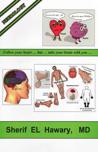
  • 1. Followyour heart ... but ... takeyour brain with you ... Giossitis + ve Babinski M~a/ol,lastlcantmla Sherif EL Hawary, glove and stocking hyposthesia MD
  • 3. Copyright © 2019, Sherif EL Hawary, MD Text copyright © 2019, Sherif EL Hawary, MD Figures, tables, illustrations and images copyright © 2019, Sherif EL Hawary, MD All rights reserved. No part ofthis book may be translated, reprinted, used or reproduced in any form or by any electronic, mechanical, or other means, now known or hereafter invented, including photocopying and recording, or in any information storage or retrieval system, without permission in writing from the author.
  • 4. INDEX CENTRAL NERVOUS SYSTEM---------------------------------- THE MOTOR SYSTEM--------------------------------------------- Differentiation between UMNL & LMNL ------------------------- THE SENSORY SYSTEM ------------------------------------------ AREAS OF THE CEREBRAL CORTEX----------------------- SPEECH DISORDERS ----------------------------------------------- THE CRANIAL NERYES ------------------------------------------- BELL'S PALSY ------------------------------------------------------- VERTIGO--------------------------------------------------------------- BULBAR PALSY ----------------------------------------------------- CEREBRO - VASCULAR DISEASE ------------------------------ STROKE----------------------------------------------------------------- TRANSIENT ISCHEMIC ATTACKS (TIAs) ------------------- HEMIPLEGIA ---------------------------------------------------------- BLOOD SUPPLY OF THE BRAIN------------------------------ NEUROGENIC BLADDER----------------------------------------- PARAPLEGIA ---------------------------------------------------------- SPECIAL FORMS OF PARAPLEGIA --------------------------- CAUDA EQUINA ----------------------------------------------------- SPONDYLOSIS-------------------------------------------------------- SCIATICA--------------------------------------------------------------- MOTOR NEURONE DISEASE------------------------------------ DD ofwasting ofthe small muscles ofthe hand ------------------- THE EXTRAPYRAMIDAL SYSTEM ---------------------------- THE CEREBELLUM------------------------------------------------- PERIPHERAL NEURITIS ------------------------------------------- MYASTHENIAS ------------------------------------------------------- MYOPATHIES --------------------------------------------------------- BRAIN TUMOURS--------------------------------------------------- EPILEPSY --------------------------------------------------------------- MULTIPLE SCLEROSIS -------------------------------------------- HEADACHE ------------------------------------------------------------ MIGRAINE -------------------------------------------------------------- SUBARACHNOID HEMORRHAGE ------------------------------ MENINGITIS----------------------------------------------------------- ENCEPHALITIS ------------------------------------------------------- COMA-------------------------------------------------------------------- 1 4 10 11 15 21 24 41 43 45 46 46 57 61 69 78 79 88 92 95 98 100 102 103 108 111 123 126 130 136 142 150 151 155 163 170 172
  • 5. A wise man once said ......... " Everyone has 2 cerebral hemispheres, right & left.... In the left: nothing is right........ and... In the right: nothing is left....."
  • 6. CENTRAL NERVOUS SYSTEM The CNS is formed of 2 parts: I. INTRACRANIAL PART 1. Two cerebral hemispheres. 2. Basal ganglia. 3. Brain stem. 4. Cerebellum. 1. Two cerebral hemispheres: • CONNECTION: They are connected to each other by the CORPUS CALLOSUM. • SURFACE: The surface ofeach hemisphere is divided into 4 LOBES: 1. Frontal 2. Parietal. 3. Temporal. 4. Occipital. • CEREBRAL LOBES: 1. Outer gray matter: o It is composed of: (cerebral cortex) NERVE CELLS. o It contains certain AREAS that control specific functions. 2. Inner white matter: (depth ofthe cerebral hemisphere) o It is composed of: NERVE FIBRES. o It CONDUCTS IMPULSES to & from the cerebral cortex. 2. Basal nuclei: - At the base ofeach cerebral hemisphere (At various levels deep in the white matter): • BASAL GANGLIA: Caudate, Putamen, Globuspallidus. • THALAMUS, SUBTHALAMUS, HYPOTHALAMUS. 3. Brain stem: "3 parts from above downwards" • MIDBRAIN: contains the motor nuclei of the cranial nerves 3 & 4. • PONS: contains the motor nuclei of the cranial nerves 5, 6, 7. • MEDULLA: contains the motor nuclei ofthe cranial nerves 9, 10, 11, 12. • The 1st, 2 nd & 8 th cranial nerves have no motor nuclei. • They are SENSORY NERVES concerned with special sensations. 4. Cerebellum: - It lies at the BACK & the BOTTOM ofthe cranium behind the brain stem, in the posterior cranial fossa. 1 l
  • 7. 2 2 II. SPINAL PART 1. Spinal Cord. 2. Cauda Equina. 1. Spinal Cord: o It lies in the spinal canal. o It ends at the lower border ofthe 1 st lumbar vertebra (Lt). Segments of the spinal cord: • Cervical segments: 8 segments. • Thoracic segments: 12 segments. • Lumbar segments: 5 segments. • Sacral segments: 5 segments. o CONUS MEDULLARIS: The lowermost 3 segments ofthe spinal cord (S3,4, 5). o EPICONUS: The 4 segments above the conus medullaris (L4, 5, S1, 2). Section in the spinal cord: - It contains: gray matter (cells) surrounded by white matter (fibres). - The GRAY MATTER: H - shaped in a transverse section & is formed of: • 2 anterior horns: MOTOR function. • 2 posterior horns: SENSORY function. - The WIDTE MATTER: contains nerve fibres arranged into tracts: 1. The important ascending tracts are: • Lateral & ventral spinothalamic: • Posterior column: • Spinocerebellar: 2. The important descending tracts are: for superficial sensations. for deep sensations. for cerebellar information. • The pyramidal tract (corticospinal tract). • The extrapyramidal tracts. • The cerebello-spinal tracts. 2. Cauda Eguina: " Collection of Lumbo - sacral roots " o It fills the lower part ofthe spinal canal. o It starts at the lower border ofthe 1 st lumbar vertebra (Lt).
  • 8. C2 cord segment C. nerve root Anterior S, cord segn1ent ~a..-"r' )..r-:~ ;;:.__ Spinous process T 1 vertebra Posterior Spinous process 1..1 vertebra Cavda equ;na 51nerve root - ----- THE SPlNAL CORD, LATERAL VIEW 3 3
  • 9. 4 4 THE MOTOR SYSTEM 1. The Pyramidal System (UMN) • ORIGIN: in the cerebral cortex (motor area" 4" & premotor area" 6"). • TERMINATION: at the AHCs of the different levels ofthe spinal cord. • CONTROL: it controls the opposite side ofthe body. • FUNCTIONS: o /NIT/ATION ofthe voluntary motor activity. o Inhibition ofthe deep r~fiexes. o Inhibition ofthe muscle tone. 2. The Extrapyramidal System • ORIGIN: from the basal ganglia. • TERMINATION: at the AHCs of the different levels ofthe spinal cord. • CONTROL: it controls the opposite side of the body. • FUNCTIONS: o REGULATJON ofthe voluntary motor activity. o Regulation ofthe emotional & associated movements. o Inhibition ofthe muscle tone. 3. The Cerebellar System • ORIGIN: from the cerebellum. • TERMINATION: at the AHCs ofthe different levels of the spinal cord. • CONTROL: it controls the same side of the body. • FUNCTIONS: o Co-ordination ofthe voluntmy motor activity initiated by the pyramidal system. o Maintenance ofequilibrium. 4. The Lower Motor Neurone (LMN) • ORIGIN: • TERMINATION: • COMPONENTS: • FUNCTIONS: in the AHCs of the different levels of the spinal cord. at the voluntary muscles. AHCs, PN, NMJ & Voluntary muscles. o Transmission ofthe motor impulsefrom the AHCs to the voluntary muscles.
  • 10. THE MOTOR The Pyramidal System "Controls opposite side" The Extra Pyramidal System "Controls opposite side" I LMN AHC SYSTEM The Cerebellar System "Controls same side" 5 5
  • 11. 6 6 MOTOR PATHWAY • ORIGIN: the order starts in the Cerebral cortex. • TERMINATION: on the Voluntar muscle which will respond by movement. From the origin to the termination, the impulse passes through 2 neurones: 1. Upper Motor Neurone ( UMN ): PYRAMIDAL TRACT. 2. Lower Motor Neurone ( LMN ): ARCS, Nerves, NMJ, Muscles. 1. UPPER MOTOR NEURONE (UMN) 1. MOTOR AREA " 4 " " CEREBRAL CORTEX " The voluntary motor impulse originates mainly in the large pyramidal cells (Betz cells) ofthe motor area" 4" & to a lesser extent in the cells ofthe premotor area" 6 ". 2. INTERNAL CAPSULE The axons of these cells (canying the impulse) descend in the depth of the Cerebral hemisphere in the Corona radiata to pass in the Internal capsule, and, continue their descent in the brain stem (midbrain, pons & medulla). 3. CORTICOBULBAR TRACT "BRAIN STEM" In the Brain Stem, some of the descending fibres separate to supply the motor nuclei of the cranial nerves of BOTH sides except the lower ½ ofthe facial nucleus and all the bypoglossal nucleus which are supplied only from the opposite pyramidal tract. These fibres are known as the corticobulbar tract as they do not reach the spinal cord. The pathway from the cerebral cortex down to the cranial nuclei in the brain stem is known as the " Corticobulbar Tract ". 4. CORT/CO SPINAL TRACT "SPINAL CORD" In the lower medulla: - 85 % of fibres cross (decussate) to descend in the white matter ofthe opposite side of the spinal cord, - 15 % ofthe fibres descend directly in the white matter ofthe same side of the spinal cord. The fibres ofthe pyramidal tract tenninate at different levels of the AHCs of the spinal cord (corticospinal tract). The pathway from the cerebral cm1ex down to the AHCs in the spinal cord is known as the " Corticospinal tract ".
  • 12. I Pathway of the voluntary motor impulse (UMN & LMN)I , - - - - - - Visual fibres Sensory fibres Internal capsule posterior hmb ~'c'--------- F bres for lower limb Internal capsule - - - Fibres to motor nuclei of Mid-brain other half or mid-brain Crus cerebri Fibres for lower imb ,,___ ____ 6th nerve nucleus Pons - - - - 7th nerve nucleus Corticospinal ----w, (pyramidal) tract Medulla - - -- 'Fibres to motor nuclei of other half of medulla V. muscle Spinal cord Antenor (direct) ______::::rr•........- cort,cospinal tract - --- Lateral {indirect) corticospinal tract -.. To anterior hom 7 7
  • 13. 8 8 2. LOWER MOTOR NEURONE I. AHCs (LMN) • They are: special type ofnerve cells situated in the anterior horns ofthe: "H-shaped gray matter o{the spinal cord". • They receive the voluntary motor impulse from the corticospinal pyramidal tract. • Their axons exit from the spinal cord as the anterior roots. 2. PERIPHERAL MOTOR NERVES • They carry the motor impulse from the AHCs to the voluntary muscles. 3. NEUROMUSCULAR JUNCTION. 4. VOLUNTARY MUSCLES. !Muscle Tonel 1. This is a SPONTANEOUS local axon stretch reflex. 2. It is spontaneous because: the length of any skeletal muscle is shorter than the distance between the origin and the insertion. So any muscle is always in a state of: Persistent spontaneous slight stretch. • This spontaneous persistent stretch will persistently activate the local axon reflex. • This will result in ersistent (maintained) contraction of the muscle (tone). 3. The Muscle tone receives inhibition from the Pyramidal & Extrapyramidal systems: a) UMNL results in loss of inhibition on the muscle tone, leading to: Increased muscle tone (spasticity) below the level ofthe lesion. b) LMNL results in inten-uption ofthe reflex arc, leading to: Decreased muscle tone (flaccidity) at the level ofthe lesion. lneep Reflex (Tendon Jerk)I 1. This is an INDUCED local axon stretch reflex. 2. It is induced by: sudden stretch of the muscle by tapping the tendon with a hammer. • This induced sudden stretch will suddenly & temporarily activate the local axon reflex. • This will result in sudden transient contraction ofthe muscle Uerk). 3. The deep reflex receives inhibition from the Pyramidal system: a) UMNL results in loss of inhibition on the deep reflex, leading to: Increased deep reflex (hyperreflexia) below the level ofthe lesion. b) LMNL results in inte1ruption ofthe reflex arc, leading to: Decreased deep reflex (hyporeflexia) at the level ofthe lesion.
  • 14. STRETCH REFLEX (Muscle tone & Deep reflex) Higher control "mainly inhibitory" Inhibitoryintemeuron Posterior root "~I lc1onusl - Sudden sustained stretch ofthe muscle tendon, results in: Rapid, Rhythmic, Regular contractions. 1. It indicates: Severe pyramidal lesion due to: loss of inhibition on the stretch reflex. 2. It is elicited in the: ankle, patella, wrist. 3. It stops when: the stretch is stopped. 9 9
  • 15. 10 How would you clinically differentiate between: UMNL & LMNL ?? UMNL LMNL 1. Muscle power Paralysis or weakness below the Paralysis or weakness at the level ofthe lesion. level of the lesion. 2. Muscle wasting No wasting, & if present it is late Early & marked wasting due and due to disuse atrophy. to loss ofmuscle tone. 3. Muscle tone Hypertonia (spasticity) below Hypotonia (flaccidity) at the level oftbe lesion. the level of the lesion. 4. Deep reflexes Hyperreflexia below the level. Hyporeflexia at the level. 5. Pathological May be present. Absent. deep reflexes 6. Clonus May be present. Absent. 7. Superficial Lost ifthe lesion is above the Lost if the lesion involves reflexes segmental supply of the reflex. the supply of the reflex. 8. Plantar reflex Positive, i.e. dorsiflexion of the Plantar flexion ofthe toes, or (Babinski) * big toe ± fanning of the other toes. no response. 9. Fasciculations Absent May be present in irritative lesion of the AHCs. · Joseph Babinski (1857-1932) was a French neLtrologist ofPolish origin, he described the plantar reflex. 10
  • 16. 11 THE SENSORY SYSTEM - There are 3 types of sensations in the body: I. SOMATIC SENSATIONS: * They are conducted to the CNS via the " somatic nerves." * They include: 1. Superficial sensations: • Pain. • Temperature. • Touch. 2. Deep sensations (proprioceptive): • Vibration sense. • Joint sense. • Muscle sense. • Nerve sense. 3. Cortical sensations: • Tactile localization. • Two points discrimjnation. • Stereognosis. • Graphosthesia. II. VISCERAL SENSATIONS: * They are conducted to the CNS via the "autonomic nerves." * They include all sensations coming from the internal viscera, e.g. heart & intestines. Ill. SPECIAL SENSATIONS: * They are conducted to the CNS via the " cranial nerves. " * They include: Smell, Vision, Hearing. 11
  • 17. 12 !somatic Sensations! - Allsomatic sensations, whether supe1ficial or deep pass through 3 ORDER NEURONES from receptors in the skin & deep structures to reach the cortical sensmy area ofthe opposite side. I. The cell of 1st order neurone is: always in the posterior root ganglion ofthe same side. 2. The cell of3rd order neurone is: always in the thalamus ofthe opposite side. 3. The cell of2nd order neurone: varies according to the type ofsensation. A) PATHWAY OF SUPERFICIAL SENSATIONS I. PathwaY- of Pain & Temperature: 12 1. The P' order neurone: • The 1st order neurone is the cell of the posterior root ganglion (PRG). • The afferent sensory nerve relays in the PRG. • The efferent sensory nerve enters the spinal cord in Lissauer's tract to relay in the cells ofthe Substantia Gelatinosa ofRolandi (SGR), at the posterior horn ofthe gray matter. 2. The 21/(1 order neurone: • The 2nd order neurone is the cell of SGR & its axon. • This axon crosses to the OPPOSITE side & ascends in the: Lateral Spinothalamic tract of the spinal cord, then in the: Lateral Lemniscus ofthe brain stem, to relay in the thalamus. 3. The 3 rd order neurone: • The 3rd order neurone starts in the cell ofthe thalamus. • Its axon ascends to pass through the internal capsule conducting the impulse to the cortical sensory area in the parietal lobe. II. Pathway of Touch: 1. Crude touch: has the same pathway as pain & temperature, BUT: In the 2nd order neurone: it ascends in the Ventral Spinothalamic tract of the OPPOSITE side ofthe spinal cord. 2. Fine touch: has the same pathway as deep sensations.
  • 18. IPathway of Superficial & Deep Sensationsl Arm area Face area Thigh area Parietal lobe {Cortical sensory area) (1,2,3) Thalamus (3rd order neurone) ' · ~ - - - - Medial lemnlscus Lower medulla Lateral lemniscus Spino-thalamic T - Pain & Temp. - Crude touch Spinal cord (Thoracic) Spinal cord (Lumbar) t PHC, SGR (2nd order neurone) Gracile & Cuneate nuclei Posterior column - Position & Vibration - Fine touch (Gracile & Cuneate tracts) 13 13
  • 19. 14 B) PATHWAY OF DEEP SENSATIONS 14 1. The I51 order neurone: • The 1st order neurone is the cell of the posterior root ganglion (PRG). • The afferent sensory nerve relays in the PRG. • The efferent sensory nerve enters the spinal cord & ascends in Gracile & Cuneate tracts within the posterior column ofthe SAME side (along with fibres carrying fine touch) to relay in Gracile & Cuneate nuclei in the medulla: - Gracile Tract: canies fibres from lower ½ ofbody & lies medially. - Cuneate Tract: canies fibres from upper ½ ofbody & lies laterally. 2. The 211d order neurone: • The 2nd order neurone is the cell of Gracile & Cuneate nuclei & its axon. • This axon crosses to the OPPOSITE side & ascends in the Medial Lemniscus through the brain stem, to relay in the thalamus. 3. The 3 rd order neurone: • The 3rd order neurone starts in the cell ofthe thalamus. • Its axon ascends to pass through the internal capsule conducting the in1pulse to the cortical sensory area in the parietal lobe. C) PATHWAY OF CORTICAL SENSATIONS • These are a mixture ofrefined superficial & deep sensations arriving to the thalamus via the Ist & 2nd order neurones & conducted from the thalamus to the cortical sensory area (1, 2, 3) in the parietal lobe.
  • 20. jAreas of the Cerebral Cortexl The Human Cerebral Cortex Primary motor area Premotor Primary somatosensory area 4 Supramarginal Gyn1s Prefrontal association I I f ,C: I -f~l I --- I 1 area (personality) ' 1 ~ 44 ~ ,, ...,_,...,._ , ,... 1 ' , I .,._,.__,,. Limbic .~~,,J; ~' association K' I Angular Gy111s Secondary visual area 17 Primary visual area I area ,"'-.., Secondary auditory area Pr,imary auditory area The surface of each cerebral hemisphere is divided into 4 LOBES: 1. Frontal. 2. Parietal. 3. Temporal. Each lobe consists of: MANY AREAS. Each area is responsible for a specificfunction. 4. Occipital. 15 15
  • 21. 16 ! FRONTAL LOBEi 1 . MOTOR AREA (4) • Function: - Initiation ofthe voluntary motor activity ofthe OPPOSITE halfofthe body. - In this area the body is represented upside down. • Lesion: - Irritative: 1. SITE: " Contralateral Motor Jacksonian Fits " Convulsions that involve the muscles of one side ofthe body. 2. ONSET: Focal onset, they start either in: • The thumb or angle ofthe mouth (if the lesion starts in the lower part). • The big toe ( ifthe lesion starts in the upper part). 3. SPREAD: (Special march) • e.g. Thumb ~ arm ~ shoulder ~ trunk ~ LL. 4. OFFSET: Followed by transient paralysis due to fatigue ofBetz cells: • Todd's paralysis. - Destructive: " Contralateral Motor Paralysis" 2. PREMOTOR AREA (6) • Function: - Partly supplies the pyramidal tract. - Partly supplies the extra-pyramidal tract. • Lesion: - Contralateral Hypertonia & Hyperreflexia. 16
  • 22. 3. AREA OF CONJUGATE EYE DEVIATION (8) • Function: - Voluntary conjugate eye deviation to the OPPOSITE side. • Lesion: - Irritative: Attacks of conjugate eye deviation to the OPPOSITE side ofthe lesion. - Destructive: Loss ofconjugate eye deviation to the OPPOSITE side ofthe lesion. 4. BROCA'S AREA (44) • Function: - It is the Motor center for speech. "In The Dominant Hemisphere Only'' - It is responsible for fonnulation ofspoken words. - It is present in the left cerebral cortex in right-handed persons & vice versa. • Lesion: "Motor Aphasia (Verbal Aphasia)" - The patient cannot express himself in spoken words. 5. EXNER'S AREA (45) "In The Dominant Hemisphere Only'' • Function: - It is the motor center for writing. - It is responsible for formulation ofwritten words. - It is present in the left cerebral cortex in right-handed persons, and vice versa. • Lesion: "Writing Aphasia (Agraphia)" - The patient cannot express himselfin written words. 6. PARACENTRAL LOBULE • Function: - Cortical inhibition (conh·ol) ofbladder voiding & bowel evacuation. • Lesion: - Incontinence ofurine & stools. 17 17
  • 23. 18 7. PREFRONTAL AREA • Function: - It is the higher center for: o Mentality (Memory & Intelligence). o Personality (Behaviour, Planning, Problem - solving). - It inhibits the primitive reflexes present in the newborn, e.g. grasp reflex. • Lesion: - Disturbances in: Mentality & Personality -+ DEMENTIA. - Reappearance of the primitive reflexes, e.g. grasp reflex. [PARIETAL LOBEi 18 1. CORTICAL SENSORY AREA ( 1, 2 , 3) • Function: - Perception of c01iical sensations from the OPPOSITE halfofthe body. - In this area the body is represented upside down. • Lesion: - Irritative: " Contralateral Sensory Jacksonian Fits" 1. SITE: Sensory fits (numbness or tingling) in one side ofthe body. 2. ONSET: Focal onset. 3. SPREAD: Special march. - Destructive: " Contralateral Cortical Sensory Loss " 2. ANGULAR G YRUS (3 9) "In The Dominant Hemisphere Only'' • Function: - It is responsible for Reading, (Recognition & Recall of letters & numbers). • Lesion: "Alexia (Visual Aphasia, Word Blindness)" - The patient is able to see the letters & numbers, BUT, He is not able to recognize (understand) them, He is not able to read. THEREFORE,
  • 24. 3. SUPRAMARGINAL GYRUS (37) "In The Dominant Hemisphere Only' • Function: - It is responsible for: Storage & Recall of: 1. IDEAS of speech. 2. IDEAS ofcomplex voluntary motor activity. • Lesion: 1. Jargon's Aphasia (Word salad). 2. Apraxia: inability to perform complex voluntary motor activity, in absence of: paralysis, sensory loss or incoordination. [TEMPORAL LOBEi 1. PRIMARY AUDITORY AREA (41, 42) • Function: - It is the center for HEARING. • Lesion: - Irritative: o Auditory Hallucinations. - Destructive: o Slight impairment of HEARING. o Never complete deafness because hearing is bilaterally represented. 2. SECONDARY AUDITORY AREA (22) "In The DominantHemisphere Only' • Function: - Recognition & Recall of sounds & heard words. • Lesion: "Auditory Agnosia" - The patient is able to hear the sounds, BUT, He is not able to recognize (understand) them. 19 19
  • 25. 20 3. LIMBIC SYSTEM (UNCUS) • Function: - It is the center for SMELL. • Lesion: - Irritative: o Olfactory Hallucinations. - Destructive: o Slight impairment of SMELL. o Never complete anosmia because smell is bilaterally represented. I OCCIPITAL LOBEi 1. PRIMARY VISUAL AREA ( 17) • Function: - It is the center for VISION. • Lesion: - Irritative: o Visual Hallucinations. - Destructive: o Contralateral Homonymous Hemianopia (bilateral halfblindness). o Never bilateral complete blindness because vision is bilaterally represented. 2. SECONDARY VISUAL AREA ( 18, 19) "In The Dominant Hemisphere Only'' • Function: - R ecognition & Recall of images. 20 • Lesion: " Visual Agnosia " The patient is able to see the images, BUT, He is not able to recognize (understand) them.
  • 26. SPEECH DISORDERS - Normal speech involves 2 stages: 1. I Formulationl whether Spoken or Written, .... This needs: A) Sensory System: 1. Visual areas: 17, 18, 19, 2. Auditory areas: 41, 42 B) Motor S stem: 1. Broca's area: 44. 2. Exner's area: 45. C) Associative S stem: - Supramarginal gyrus: 37. Areas 17, 41, 42 in BOTH hemispheres. and 39. and 22. Areas 18, 19, 39, 22, 44, 45, 37 in DOMINANT hemisphere ONLY. 2. [Articulation! This needs: A) UMN: Pyramidal tracts. B) LMN: • Cranial Nuclei (5, 7, 10, 12). • Nerves. • NMJ • Muscles. C) Cerebellum: for co-ordination ofthe muscles ofspeech. D) Extrapyramidal system: for making the speech expressive. 21 21
  • 27. 22 22 - Disorders of speech include: [APHASIA! Definition - Difficulty or inability ofthe fon1mlation of speech: o In the absence oflesions ofthe sense organs (e.g. Vision or Hearing). o In the absence ofmental defect. Types 1. Sensory Aphasia: " due to defect in comprehension " a) Visual: - Visual agnosia: (lesion in area 18, 19) The patient sees objects but does not recognize them. - Alexia: (lesion in area 39) The patient cannot read because he does not understand the letters & numbers (word blindness). b) Auditory: - Auditory agnosia (lesion in area 22) The patient hears sounds but does not understand them. 2. Motor Aphasia: a) Verbal aphasia: " due to defect in expression " (lesion in Broca's area) The patient cannot express himself in spoken words; (although he can understand visual & auditory stimuli). b) Agraphia: (lesion in Exner's area) The patient cannot express himself in written words (although he can understand letters & numbers). 3. Jargon's Aphasia (Word salad): "due to defect in association" - It is due to lesion in the supramarginal gyrus (area 37). - The patient can speak, BUT, the words are meaningless & not related. - This condition is usually associated with apraxia.
  • 28. JDYSARTHRIAI Definition - Difficulty in articulation of speech (with normal formulation of speech). Types 1. Slurred Speech: - Disturbance in the roduction of consonants es eciall _: o Labials (e.g. B, M). o Dentals (e.g. D, T). - Causes include: a) Bilateral pyramidal tract lesion (Cortico-bulbar lesion), as in : o Pseudobulbar palsy. b) LMNL ofthe cranial nerves concerned with speech, e.g.: o Nuclear: True bulbar palsy. o Nerve: o NMJ: o Muscle: 2. Staccato Speech: Facial nerve palsy. Myasthenia. Myopathy (Facio-scapulo-humeral myopathy). - The speech is explosive with separation ofthe syllables. - Causes include: Cerebellar lesions: o Hereditary ataxias. o DS. 3. Scanning Speech: (Slurred Staccato) - It occurs also in: Cerebellar lesions. 4. Monotonus Speech: - The s eech is ex ressionless & monotonus. - Causes include: Extrapyramidal lesions: o Parkinsonism. 23 23
  • 29. 24 24 THE CRANIAL NERVES It. OLFACTORY NERVE! • Function - Sense ofsmell. • Lesion 1. Anosmia: a) Bilateral anosmia: ENT causes, Congenital. b) Unilateral anosmia: Traumatic: Inflammatory: Neoplastic: " loss ofsense ofsmell" causes: e.g. atrophic rhinitis. causes: fracture cribrifonn plate ( in fracture base ofskull ). basal meningitis. meningioma ofthe olfactory groove. 2. Olfactory Hallucinations: "f alse perception ofbad smell " It occurs in irritative lesions ofthe limbic system in the temporal lobe. 1 2. OPTIC NERVE! • Pathway - From the retina, the nerve fibres converge to form the optic nerve. - The nasal fibres of the nerve (which carry the temporal field of vision), decussate at the optic chiasma to pass in the optic tract ofthe QJ2posite side. - The tern oral fibres ofthe nerve ( which carry the nasal field ofvision), pass in the optic tract of the same side. - The fibres relay in the lateral geniculate body, where new fibres alise & pass in the posterior limb ofthe internal capsule. - The fibres then pass in the optic radiation ( in the parietal & temporal lobes). - The fibres of the optic radiation finally terminate in area 17 of the occipital lobe (visual sensory area), where vision is perceived. - The neighbouring areas 18 and 19 (visual associative areas) are responsible for the recognition & recall ofimages. • Function - Sense ofvision.
  • 30. • Lesion differs according to the site in the pathway: 1. Lesion in the O tic Nerve: - Ipsilateral loss of vision. - Loss ofdirect & consensual. light reflex. 2. Lesion in the O tic Chiasma: - Bitemporal Hemianopia. 3. Lesion in the O tic Tract: - Contralateral Homonymous Hemianopia (bilateral half blindness). 4. Lesion in the O tic Radiation (lower fibres): - Upper quadrantic Contralateral Homonymous Hemianopia. - Preservation of the light reflex. 5. Lesion in the O tic Radiation (upper fibres): - Lower quadrantic Contralateral Homonymous Hemianopia. - Preservation of the light reflex. In complete lesion of the Optic Radiation, there will be: - Contralateral Homonymous Hemianopia (bilateral halfblindness). - Preservation ofthe light reflex. 6. Lesion in the Occi ital Lobe: - Contralateral Homonymous Hemianopia (bilateral halfblindness). - Preservation of the light reflex. - Preservation ofthe macular vis.ion (due to double blood supply). Vlsual field defects L A 1co 2 Visual L/fields~A - - - - Retina - - -- -- Optic nerve ,:,-- - - - - - Optic chiasma ' ' ' ' •~:-•,:-,,----- Optic tract ,.... " <lm)___ Lateral geniculate .. .. .... body ',:,:·'..~,, Optic radiation '. '.-;--- Lower fibres in :i -); temporal lobe ,:',',,,: ' Upper fibres in ,:::(:>' anterior parietal Jobe ,;:::~ , (., -.(;';: ,?"----..:;r--- All fibres in posterior ...__._.._ , ..-- parietal lobe ~ Occipital cortex 25 25
  • 31. 26 oc;1GHT REFLExj Definition - Exposure ofone eye to bright light results in : 1. Constriction ofthe same side (direct reflex). 2. Constriction ofthe opposite side (consensual reflex). Pathway - Impulses pass in the optic nerve, chiasma & then optic tract. - Fibres do not reach the lateral geniculate body, but pass to the Edinger-Westphal nuclei (EW nuclei) ofboth sides in the midbrain. - Fibres then pass from the EW nuclei ofboth sides to the oculomotor nerves. - Fibres finally terminate in the constrictor pupillae muscles ofboth eyes. Cranial nerve 3 EW nucleus Pathway oflight reflex 26
  • 32. ~- OCULOMOTOR NERVEI • Function - Extraocular movements. - Elevation of the upper eyelid. - Pupillary constriction. • Lesion A) External 0 1. Ptosis. 2. Divergent paralytic squint ( the eye looks out & down due to the unopposed action of the lateral rectus " Cr N 6 " & the superior oblique " Cr N 4 " ). 3. Diplopia. B) Internal Ophthalmoplegia: 1. lpsilateral mydriasis. 2. !psilateral loss oflight reflex: loss ofdirect light reflex on the affected eye. 1 4. TROCHLEAR NERVEI • Function - Extra.ocular eye movement: inwards & downwards (superior oblique muscle). • Lesion 1. Limitation ofeye movement on looking inwards & downwards. 2. Diplopia. 1 6. ABDUCENT NERVEI • Function - Extraocular eye movement: outwards (lateral rectus muscle). • Lesion 1. Limitation ofeye movement on looking outwards. 2. Diplopia. 27 27
  • 33. 28 28 WUPILLARY DISORDERS! A. Myosis: 1. Horner's syndrome ( due to sympathetic lesion ): • Ptosis. • Myosis. • Enophthalmos. • Anhydrosis. 2. Argyll-Robertson pupil ( due to neurosyphilis, encephalitis, DS, DM ): • Myotic, irregular, eccentric pupil. • Lost light reflex. It is due to a lesion in the MB destroying: • The sympathetic pupillodilator fibres. • The fibres ofthe light reflex. 3. Pontine hemorrhage. 4. Morphine poisoning. B. Mydriasis: 1. Oculomotor nerve lesion: • Dilated fixed pupil. } 2. Adie's pupil ( Heredo-familial ): • Dilated pupil. • Lost light reflex. • Lost ankle reflex. It is Heredo-familial: • In females. • Unilateral. Pin-point pupiJ
  • 34. I s . TRIGEMINAL NERVE! PATHWAY OF THE TRIGEMINAL NERVE MB Pons Med Trigeminal nerve Cortex Thalamus Mesenchephalic n. Spinal sensory n. (pain & temp) { - - ■ ■ ■ ■ ■ ■ • Motor (nucleus & Pons A v Main sensory n. (touch) 29 29
  • 35. 30 30 • Function 1- The Sensor Division: - It conducts sensations from the face ( except the angle of the mandible, supplied by C2 ), the anterior 2/3 s of the tongue & the buccal cavity. - Jt is formed of3 branches: ophthalmic, maxilla,y , and mandibular. - Pathway: a) Fibres carrying pain & temperature sensations relay in the spinal sensory nucleus in the medulla on the same side, where: • The upper face is represented in the lower part of the nucleus & vice versa. • The outer face is represented in the inner pait ofthe nucleus & vice versa. b) Fibres catTying dee sensations relay in the mesencephalic nucleus in the midbrain on the same side. c) Fibres carrying touch relay in the main sensory neucleus in the pons on the same side. - From these 3 nuclei, new fibres cross to the opposite side, then ascend with the medial & lateral lemnisci, to reach the thalamus. - From the thalamus, fibres canying cortical sensations ofthe face ascend to end in the cortical sensory area in the parietal lobe. 2- The Motor Division: - It arises from the motor nucleus in the pons. - It supplies the muscles ofmastication. • Lesion 1. Sensor affection: a) Peripheral lesion: - Loss ofsensations on the same side of the face (sparing the angle of the angle of the mandible). - Loss of general sensations over the anterior 2/3 s of the tongue, (in lesions ofthe mandibular division). b) Central lesion: (mainly affects the spinal sensoty nucleus in the medulla): - Ipsilateral dissociated sensory loss ofthe face, i.e. lost pain & temperature with preservation of touch & deep sensations. - The clinical picture varies according to the site of the lesion: • Lesion starting from above (lower pontine tumour): • Lesion starting from below (upper cervical lesions): • Lesion starting from periphery (Tabes dorsalis): • Lesion starting from midline (syringobulbia): affects the lower part of face. affects the upper part offace. affects the central part of face. affects the outer part of face.
  • 36. 2. Motor affection: a) Weakness ofthe muscles ofmastication on the same side ofthe lesion. b) Deviation ofthe jaw to the affected side due to the unopposed action ofthe pterygoid muscles ofthe healthy side. 3. Reflex affection: a) Ipsilateral loss ofthe corneal reflex (afferent 5, efferent 7). b) Ipsilateral loss ofthe palatal reflex (afferent 5, efferent 10). c) Appearance ofjaw reflex (afferent 5, efferent 5): in cases ofbilateral UMNL above the pons. ~D OF FACIAL PAIN[ 1. Trigeminal neuralgia. 2. Post-herpitic neuralgia: - The pain usually affects the o hthalmic division. - The pain is followed by herpetic skin eruption. 3. Sinusitis: - The pain is worse in the morning, with tenderness over the involved sinus. 4. Ocular (glaucoma): - The pain is behind the eye, with associated visual symptoms. 5. Dental: - The pain is around the mouth. 6. Cluster headache: - Refer to the chapter of "Migraine". 7. Costen's syndrome: (Temporo-mandibular joint dysfunction). 31 31
  • 37. 32 TRIGEMINAL NEURALGIA (Tic douloureux) EFINITION - Severe attacks ofpain along one or more ofthe sensory branches ofthe trigeminal nerve, usually the maxillar_ or mandibular. ETIOLOGY 32 - The exact cause is UNKNOWN, but there are some related diseases: o Nerve root compression (in the cerebello-pontine angle by): - Tumour. - Aneurysm. - Vascular anomaly: Abnormal vascular course ofthe SCA.* o DS & DM. CLINICAL PICTURE - The "Pain s ndrome" is dia nosed b the "Risto " alone: • Site & reference: - One or more branches of the TN (usually the maxilla or mandibular). - Pain is unilateral (rarely bilateral). • Character & duration: - Severe agonizing: stabbing, burning, electric shocks. - Several seconds to several minutes (rarely hours or even longer). - Recurrent: but the patient is completely free in between the attacks. * SCA: Superior cerebellar artery
  • 38. 33 • Precipitated by: - Movements ofthe jaw: e.g. laughing & mastication. • Relieved by: - Spontaneously, or by potent analgesics. • Associated symptoms: - Pain provokes briefmuscle spasm ofthe facial muscles (tic). TREATMENT - Medical: o Carbamazepine: 500 - 1000 mg/ day orally. o Analgesics. - Surgical: e.g. o Section ofthe affected sensory root. o MICROVASCULAR DECOMPRESSION. 33
  • 39. 34 1 7. FACIAL NERVE! • Function The facial nerve is a mixed nerve, containing: motor, sensory_ and autonomic fibres. The motor part: supplies the muscles ofexpression of the face as well as 4 other muscles, • Platysma. • Stapedius. • Posterior belly ofthe digastric muscle. • Stylohyoid. The sensory part: receives taste sensations from the anterior 2/3 s of the tongue. The autonomic part: supplies the lacrimal, the submandibular & sublingual saliva1y glands. • Anatomy 34 1. Anatomy of the motor part: " See figure " The motor nucleus of the facial nerve Lies in the pons, near the 6th nerve nucleus. It is controlled by 2 types ofsupranuclear fibres: a) Pyramidal Fibres: - The upper part of the nucleus is supplied from both pyramidal tracts. - The Lower part of the nucleus is supplied from the opposite pyramidal tract only. b) Extra-pyramidal Fibres: - For emotional & associated movements. From the nucleus, the motor fibres form a loop around the 6th nerve nucleus, then pass laterally to emerge at the lower part of the pons. The nerve runs laterally between the 6th and 8th cranial nerves, in the subarachnoid space of the cerebeUopontine angle to enter, through the internal auditory meatus (1AM), into the facial canal. In the facial canal, the motor part of the facial nerve becomes adherent to its senso1y and autonomic parts. It gives the nerve to Stapedius muscle. Finally, the facial nerve leaves the canal through the stylomastoid foramen SF, passes through the parotid gland to divide into its terminal branches to innervate the muscles of the face.
  • 40. 2. Anatomy of the sensory and autonomic arts: "See figure" - In the facial canal lies the geniculate ganglion. This ganglion gives 2 branches, a peripheral branch & a central branch. a. The ~ ~;....c.cc..~-'-"--'~"'-=..c..= runs laterally and divides into the greater superficial petrosal nerve and the chorda tympani. • The greater superficial petrosal GSP nerve passes forwards to relay in the sphenopalatine ganglion where a new set offibres gives autonomic supply to the lacrimal gland. • The chorda tympani leaves the facial nerve before the stylomastoid foramen, to supply the submaxillary and sublingual salivary glands and to carry taste sensations from the anterior 2/3s ofthe tongue. b. The central branch: passes centrally, joins the motor pait ofthe nerve, then enters the cranial cavity through the internal auditory meatus as the nervus intermedius. • This nervus intermedius then enters the brain stem to tenninate in the solitary nucleus in the medulla. • A new set offibres passes from the nucleus to the opposite side and runs upwards to terminate in the lower part of the cortical sensory area, where taste sensation from the anterior 2/3s of the tongue is perceived. 35 35
  • 41. 36 36 PATHWAY OF THE FACIAL NERVE MB Pons Submand & subling salivary glands
  • 42. • Lesions of The Facial Nerve The lesion may be: 1. UMNL: (Supra-nuclear lesions) • Lesions ofthe cortico-bulbar tract ( pyramidal tract ) at any point from: The motor area in the cortex ~ to the facial nucleus in the pons. 2. LMNL: (Nuclear & infra-nuclear lesions) • Lesions that affect the facial motor nucleus or the nerve itself. 1. UMNL: (SUPRA-NUCLEAR LESIONS) - Causes: 1. Vascular: 2. Traumatic: 3. Inflammatmy: 4. Neoplastic: 5. Demyelinating: 6. Degenerative: - Clinical/f!atures: Embolism, Thrombosis, Hemorrhage. Head injury. Post-encephalitic. Glioma. DS. MND. Features of UMN Facial paralysis: 37 Paralysis ofthe LOWER HALF ofthe face on the OPPOSITE SIDE ofthe lesion: • Dropping ofthe angle ofthe mouth. • Deviation of the mouth to the healthy side on showing the teeth. • Dribbling ofsaliva. • Flattening ofthe naso-labial fold. • Inability to blow the cheek. • Accumulation of food behind the cheek. 37
  • 43. 38 2. LMNL: {NUCLEAR & INFRA-NUCLEAR LESIONS) - Causes: 1. Pontine Lesions. } Nuclear Lesions 2. Cerebello-Pontine Angle 3. Facial Canal Lesions. 4. Extracranial Lesions. } Infra-nuclear Lesions - Clinical features: 1. Features according to the level of the lesion * (see later: ). 2. Features of LMN facial paralysis: Paralysis of: ALL MUSCLES ofthe face on the SAME SIDE oftbe lesion: • Dropping ofthe angle ofthe mouth. • Deviation ofthe mouth to the healthy side on showing the teeth. • Dribbling of saliva. • Flattening ofthe naso-labial fold. • Inability to blow the cheek. • Accumulation of food behind the cheek. PLUS • Inability to raise the eye brows. • Inability to close the eyes. • Absence ofwrinkles ofthe forehead on looking upwards. 38
  • 44. * ! LOCALISATION OF THE SITE OF LMNLI SITE OF LESION Pontine Lesions (Nuclear Lesions) 1. Paralysis of facial muscles. 2. No impaitment oftaste sensation. 3. No impairment of salivation or lacrimation. 4. May be other LMN cranial palsies on the same side or hemiplegia on the opposite side. Cerebello-Pontine Anf!le Lesion (ALL) CAUSES 1. Vascular: - Vetebro-basilar insufficiency. - Millard-Gubler syndrome. 2. Infective: - Encephalitis, Poliomyelitis. 3. Neoplastic: - Glioma. 4. Demyelinating: - DS. 1. Paralysis offacial muscles. 1. Infective: 2. l taste on anterior 2/3s oftongue. - Basal meningitis. 3. l salivary & lacrimal secretions. 4. Associated Cr. 5, 6 & 8 palsies on same side. 2· NeoplaStic: - Acoustic neuroma, Meningioma. 5. Ipsilateral cerebellar ataxia. Facial Canal Lesion (If) 1. Paralysis offacial muscles. 2. t taste on anterior 2/3s oftongue, and t salivation if chorda tympani is involved. 3. Diminished lacrimation if the greater superficial petrosal nerve is involved. 4. Hyperacusis ifthe nerve to Stapedius is involved. Extracranial Lesions (After its exit from the stylomastoid foramen) Paralysis offacial muscles only. 1. Traumatic: - Fracture base. 2. Infective: - Otitis media, Herpes zoster. 3. Neoplastic: - Facial neuroma. 4. Bell's palsy. 1. Neuropathy: Diabetes, Leprosy. 2. Myopathy: Facioscapulohumeral. 3. Myasthenia. 4. Invasion by a tumour: (parotid). 5. Injury to the nerve during surgery. 39 39
  • 45. 40 tf>ifferentiation Between UMNL & LMNL of the Facial Paralysis! UMNL LMNL 1. Affects the Pyramidal tract above the I. Affects the Facial Motor Nucleus, or, Facial Nucleus. the Nerve itself. 2. Paralysis of the muscles of LOWER HALF of 2. Paralysis of ALL MUSCLES ofthe face the face on the OPPOSITE SIDE of the lesion (upper & lower halves), on the SAME SIDE (supplied from the opposite pyramidal only). of the lesion). 3. Paralysis involves the voluntaiy movement, 3. Paralysis involves the voluntary, emotional BUT: spares the emotional & associated and associated movements. movements (supplied from the extra-pyramidal fibres ). 4. Paralysis is associated with Hypertonia 4. Paralysis is associated with Hypotonia and Hyperreflexia. and Hyporeflexia. 5. There is associated Hemiplegia on the SAME 6. If there is Hemiplegia, it is on the SIDE of the facial paralysis. OPPOSITE SIDE ofthe facial paralysis (crossed Hemiplegia). Lesion (Right) Lesion (Right) 40
  • 46. Bell's Palsy toEFINITIONI - It is an acute LMN paralysis ofthe face, due to a non-suppurative inflammation and oedema ofthe facial nerve in the FACIAL CANAL near the stylomastoid foramen. - It is usually unilateral, may be recurrent & sometimes runs in families. [ETIOLOGY] - UNKNOWN: 1. Air drafts: Many causes have been suggested: Exposure to air drafts may lead to inflammation, oedema, & compression ofthe nerve at the stylomastoid foramen. 2. Viral infection: It may be secondary to a neurotropic vims (Herpes zoster), Herpes simplex. 3. Autoimmune: There is evidence ofhigh levels ofimmunoglobulins in the patient's serum. !CLINICAL PICTURE! l. PAIN: • The entire course ofthe disease may be PAINLESS, OR, • The disease may also present with acute PAIN behind the ipsilateral ear. 2. PARALYSIS: "one or two days a(ter the pain " • Complete paralysis of the facial muscles on the affected side of LMN features, (features of FACIAL CANAL lesion). I COMPLICATIONSI I. CROCODILE TEARS: • Excessive tears: • Explanation: 2. CORNEAL ULCERS. flow from the affected eye during eating. the regenerating fibres to the submandibular salivary glands become misdirected and reach the lacrymal gland. 3. INCOMPLETE RECOVERY: • Tbi.s may result in residual facial asymmetry. 41 41
  • 47. 42 ITREATMENTI 1. REASSURANCE. 2. PHYSIOTHERAPY: a. Massage of the facial muscles. b. Infrared inadiation to the face. c. Galvanic stimulation to the facial muscles. 3. MEDICAL TREATMENT: a. Corticosteroids: Oral prednisolone (1 mg / Kg / day): to ! inflammation & oedema. b. Corneal protection: Eye ointment during sleep, sunglasses during daytime. 4. SURGICAL TREATMENT: a. For Facial nerve palsy: Decompression of the facial nerve or Facial nerve grafting. b. For Crocodile tears: Section of the regenerating fibres. C. For Residual asymmetry: Plastic surgery. IPROGNOSISI • Most patients ( 80% ) recover in few weeks. • The remaining ( 20%) need surgical interference. 42
  • 48. I s. cocHLEO-VESTIBULAR NERVEI It has 2 divisions: 1. Cochlear division: • Function: - Hearing. • Lesion: - Irritative: Tinnitus. - Destructive: Deafness. 2. Vestibular division: • Function: - Equilibrium. • Lesion: - Vertigo. - Nystagmus. - Incoordination in the same side ofthe body. Vertigo PEFINITIONI - It is the sense of rotation of the body in steady surroundings or the reverse i.e.: o The body may be felt to rotate or fall while the surroundings are steady, or o The surroundings themselves appear to rotate around the body. 43 It is aggravated by movements of the head and it persists in all positions: Sitting, S tanding or Supine. - It is usually associated with autonomic manifestations in the form of: pallor, sweating. Nausea, vomiting, - It sbould be differentiated from dizziness, where the unsteadiness is not associated with a sense ofrotation. 43
  • 49. 44 [ETIOLOGjj 1) Labvrintbioe: a) Physiological: b) Pathological: 2) Peri heral nerve: sea sickness, car sickness. labyrintbitis, Meniere's disease. - Cerebello-pontine angle lesion as acoustic neuroma. - Vestibular neuritis. 3) Brain stem: - Vertebra-basilar artery insufficiency. - Posterior inferior cerebellar artery occlusion: PICA. - Disseminated sclerosis: OS. - Encephalitis. 4) Cerebral: - Increased intracranjaJ tension. 5) Psychogenic: - It occurs in acute anxiety . It responds to tranquillisers. 6) Benign Positional Vertigo: - It is usually idiopathic or due to inner ear disease. - It is associated with nystagmus & is usually by changing the position such as turning in bed. - [t is usually mild, transient, lasting less than 30 seconds. ITREATMENjj 1) Treatment ofthe cause. 2) Cinnarizine (Stugeron) and Dramamine may be used. 44
  • 50. Bulbar Palsy loEFINITIONI - Paralysis of the muscles supplied by tbe cranial nerves arising from the Medulla: 9 , IO , 12. !CLINICAL PICTURE! 1. Dysarthria. 2. Dysphagia. 3. Hoarseness ofvoice. 4. Nasal regurgitation of food. I TYPESI I. True Bulbar Pals - Bilateral LMNL of the cranial nerves in the Medulla. - Common causes are: 1. Poliomyelitis. 2. MND. 3. Syringobulbia. II. Pseudo Bulbar Pals : - Bilateral UMNL ofthe cranial nerves in the Medulla. - Common causes are: 1. DS. 2. MND. 3. Brain stem tumours. True Bulbar Palsy Pseudo Bulbar Palsy 1. Palatal reflex Absent Exaggerated 2. Pharyngeal reflex Absent Exaf.!:2erated 3. Jaw reflex Absent Present 4. Quadriple~ia Absent Present 5. To11gue - Wasting - No wasting - Placidity - Spasticity - Fasciculations - No fasciculations 45 45
  • 51. 46 46 CEREBRO-VASCULAR DISEASE - Diseases ofthe BRAIN VESSELS include: 1. STROKE. 2. TRANSIENT ISCHEMIC ATTACKS ( TIAs ). STROKE "BRAIN ATTACK" • ACUTE INJURY to the brain due to a VASCULAR problem. • It produces neurological deficits: - Acute in onset. - Persistentfor more than 24 hours. ETIOLOGY 1. Ischemic stroke:1 " The most common cause " 85 % OCCLUSION ofa blood vessel to a part ofthe brain ~ Ischemia. • Thrombosis: • Embolism: Acute occlusion. Sudden occlusion. 2. Hemorrhagic stroke: "Less common cause " 15% RUPTURE of a blood vessel in a part of the brain ~ Bleeding. 1. Thrombosis: "clot in situ resulting in cerebral infarction" 1. Vessel wall diseases: ~- - Cerebral ATHEROSCLEROSIS: "THE MOST COMMON" - Vasculitis: PAN & SLE. 2. H'i/l_ercoaaulability_: e.g. - Polycythemia ( i RBCs). - Leukemia ( i WBCs ). - Thrombocytosis ( j Platelets ). 3. Slow circulation e.g. - Heart failure. - Systemic hypoperfusion, e.g. Dehydration & shock. 1 Lacunar infarct is the most common type ofstroke (Refer to Hypertension). Sickle cell disease can also cause ischemic stroke.
  • 52. 2. EmboliSm: "wandering clot resulting in cerebral infarction" 1. Heart: The source ofthe embolus may be: " The most common source " - MS with AF, Vegetations of SBE, Mural thrombus in AMI. 2. Distal vessels: - Arterial: Detached atheromatous plaque from carotid vessels. - Venous: DVT ~ detached thrombus ~ paradoxical embolism through VSD or ASD. 3. Other (unusual): " very rare " - Fat emboli, air emboli, parasitic emboli, malignant cell emboli. 3. Hemorrhage: Types of lntracranial Hemorrhage 1. lntracerebral: • The bleeding is in the brain substance & may leak into the ventricles. • This is very serious as the blood may compress vital centres. • The commonest artery causing intracerebral hemorrhage is the "lenticulo-striate branch ofthe middle cerebral artery" 2. Subarachnoid: Bleeding is in the subarachnoid space. 3. Subdural or extradural: Bleeding often forms a hematoma. Causes of lntracranial Hemorrha e 1. Hypertension: The commonest cause of: Intracerebral haemorrhage. 2. Rupture of intracranial aneurysm: The commonest cause of: SAH. 3. Head trauma: The commonest cause of: Subdural haematoma & SAH. 1 4. Hemorrhagic blood diseases: Purpura & Hemophilia. 5. Anticoagulants. 1 The commonest causes of SAH are: head trauma & ruptw-e ofan intracranial aneurysm 47 47
  • 53. 48 48 Blood clot stops Lhe now of blood to an area ofthe brain lschemic Stroke 85 % of STROKES are ischemic i ., )( Blood leaks into brain tissue Hemorrhagic Stroke Weakened/diseased blood vessels rupture. 15 °/o of STROKES are hemorrhagic
  • 54. 1. Non - modifiable: • Age: • Sex: • Type A personality: • Genetic factors: 2. Modifiable: a) High risk: "For Stroke & TIAs" old. male: female = 3: 1. nervous, intellectual. positive family history. • Hea1t diseases: especially valvular heart diseases & AF. • Hyperlipidemia: j total cholesterol, j LDL, t HDL. • HYPERTENSION: causes endothelial damage. • Diabetes mellitus. • Cigarette smoking. b) Less risk: • Obesity. • Diet: rich in saturated fat & cholesterol. • Physical inactivity. • Psychological stress. • Hyperuricemia. • Homocysteinemia. • Heavy alcohol intake. • CCPs. CLINICAL PICTURE - Neurological deficits commonly roduced de end on the affected brain area: 1. HEMIPLEGIA. 2. HEMIANESTHESIA. 3. Speechproblems: 4. Vision problems_: 5. Ataxia: 6. Cranial nerve paralysis. Aphasia or Dysarthria. especially field defects. Lack ofco-ordination or Lack of balance. 49 49
  • 55. 50 CLINICAL TYPES 1. Stroke in evolution: (Progressing stroke) An enlarging brain infarct with neurologic deficits that worsen over 24 to 48 h. 2. Completed stroke: (Non - progressing stroke) A stable brain infarct with neurologic deficits that do not worsen with time. INVESTIGATIONS ~-~------------------' I. BRAIN IMAGING a) CT scan • Image acquisition is faster with CT scanning than with MRI. • CT is more available i11 emergencies than MRI. • NonContrast CT (NCCT) scanning is the most commonly used form. • CT also includes: CT angiography (CTA) & CT perfusion (CTP) scanning. 1. lschemic stroke: • Infarctions may not appear until 24 hours or more after onset. • Small infarctions may not appear at all. 2. Hemmorrhagic stroke: •Bleeding appears immediately (even very small lesions). • Therefore: CT scan can help to rule out a hemorrhagic stroke. b) MRI - Recent Brain MRI protocols reliably diagnose both types of stroke. II. OTHER IMAGING a) CARDIAC IMAGING: CXR, ECG, Echocardiography. b) VASCULAR IMAGINMG: DOPPLER ultrasonic imaging. III. LABORATORY TESTS a) CBC. b) PT & APTT. C) Risk factors: e.g. blood glucose, lipid profile, UA, homocysteine. d) !Jypercoagulability: e.g. protein C, protein S, Antithrombin III. 50
  • 56. TREATMENT I■ GENERAL CA~E: "For: Hemiplegic patient or a Comatosed patient" " Stroke Unit " 1. Skin: • Frequent change of the patient's position (every 2 hours), & ofthe bed sheets. • Frequent wash of the back & pressure points by alcohol followed by powder. 2. Swallowin & nutrition: • Tube feeding & IV fluids. 3. Balance of fluids: • Controlled IV fluids. 4. Breathing: • Suction of secretions. • Oxygen inhalation. 5. Bladder: • Catheterisation & urinary antiseptics. 6. Bowels: • Daily enema. II. SYMPTO~ATIC: 1. EARLY: • Cerebral dehydrating agents (e.g. Mannitol): to l brain oedema. • Prophylaxis against STRESS ULCER: "Refer to Cardiology". 2. LATE: PHYSIOTHERAPY. 51 51
  • 57. 52 III. SPECIFIC: " According to the type " 52 A) ISCHEMIC STROKE: 1. Limitation of infarct size. "Time is brain" In case ofthrombotic stroke; there are 3 brain areas ofparticular concern: 1) A central portion ofthe ischemic area is permanently damaged. 2) An immediately surrounding area, the penumbra, may be salvaged with appropriate and rapid treatment. 3) A healthy area outside the penumbra. 1) Central area {permanent damage) 2) Penumbra {salvageable damage) For this type of stroke, there are J treatment options to limit the infarct size: 1) Medical revascularization: "Fibrinolytic therapy" • Drug: IV Recombinant tPA (tissue plasminogen activator). • Duration: Infused within 3 to 4.5 hours after the onset ofsymptoms. • Contraindications: Refer to "Cardiology" ... plus some details; - Bleeding { General (platelets< 100,000/mm2, coagulopathy), Local (ICH) }. - Severe Hypertension > 185 / 110 mmHg unless a drug such as [V Labetalol was successful in reducing BP below this range. - Severe Hypoglycemia or Hyperglycetnia: PG < 50 or > 400 mg/dL. - Rapidly improving symptoms suggestive ofTIA (relative contraindication). - Advanced age: older than 75 years (relative contraindication). 2) Mechanical revascularization (thrombectomy): "Endovascular therapy" • A recent alternative when fibrinolytic therapy is ineffective or contraindicated.
  • 58. Mechanical revascularization (thrombectomy) methods 1 Stent - retriever system Blood vessel ..._................................... ... (arteries) Blood clot •·······································•• (caught in stent and removed) Stent············································· Penumbra System This microcatheter probes & breaks up the red clot, aspirating it from the tube with a vacuum pump Mechanical Embolus Retrieval in Cerebral lschemia (MERCI) Corkscrew-shaped device that captures &engages clots 1 American Hea11 Association/American Stroke Association 2015 53 53
  • 59. 54 54 2. Blood Pressure Control. A) H ertension control in non-rt-PA candidates: 1) BP < 220 /120 mm H : "In absence of TOD" 1. BP should be just monitored, and, 2. Symptoms (e.g. ~ ICP, seizures) should be treated. 2) BP > 220 /120 mm H : l. Labetalol: is the initial drug ofchoice. Dose: 10 - 20 mg IV for 1-2 min, Dose may be repeated or doubled every 10 minutes to a maximum dose of 300 mg. 2. Nicardipine: is an alternative; given IV at an initial rate of 5 mg/hand titrated by increasing the infusion rate 2.5 mg/h every 5 minutes, to a maximum of 15 mg/h. 3. Nitroprusside: may be used in severe cases, (BP > 230 I 140 mmHg), at 0.5 mcg/kg/min IV. !Target BP: reduction in BP of 10 - 15%1 B) H ertension control in rt-PA candidates: 1) BP< 185 /110 mm H : l. BP should be just monitored. 2. Symptoms (e.g. ~ ICP, seizures) should be treated. 2) BP> 185 /110 mm H : 1 1. BP should be reduced and continuously monitored. 2. Same 3 drugs (Labetalol or Nicardipine or Nitroprusside) are given before and after rt-PA. ! Target BP: reduction in BP of 15 - 25%1 1 BP > 185 /110 mm Hg is a contraindication to rt-PA, so we should reduce BP below 185 / 110 mrnHg ifthe patient should receive 1t-PA
  • 60. 3. Neuroprotective agents. A) Rationale: Reducing the release ofexcitatory neurotransmitters by neurons in the ischemic penumbra may enhance the survival of these neurons. B) Limitations: Despite very promising results in animal studies, however, no single neuroprotective agent in ischemic stroke has as yet been supp01ied by human studies. Nevertheless, substantial research is under way evaluating different neuroprotective strategies. C) Medications: 1) Prevention of Early lschemic Injury: N-methyl-D-aspartate (NMDA) receptor antagonists: e.g. Dextrophan. (Side effects: Hallucinations & Agitation) 2) Prevention of Reperfusion Injury: Despite the good outcome generally associated with reopening a blood vessel, additional. brain injury may result when reperfusion occurs. When WBCs reenter a previously hypoperfused region via returning blood, they can occlude small vessels, producing additional ischemia. In addition, leukocytes release toxic products that can lead to free radical formation. Enlimomab: a Monoclonal antibody that can block an intercellular 4. Antiplatelets. adhesion molecule (ICAM) on the endothelium to prevent adhesion ofWBCs to the vessel wall. This will reduce the formation offree radicals, promote neuronal repair & may protect the brain from additional injury during reperfusion. • Aspirin (low dose): 75 - 300 mg I day (single dose). 75 mg twice daily. • Dipyridamole: • Ticlopidine: 250 mg twice daily. • Clapidagrel: 75 mg once daily. - Recent AHA/ASA1 guidelines recommend giving aspirin, 325 mg orally, within 24 - 48 hours ofischemic stroke onset. Jauch EC, Saver JL, Adams HP Jr, Bruno A, Connors JJ, Demaerschalk BM, et al. Guidelines for the Early Management ofPatients With Acute Ischemic Stroke: A Guideline for Healthcare Professionals From the American Heart Association/American Stroke Association. StTOke. 2013 Jan 31. 55 55
  • 61. 56 56 5. Anticoagulants. Indications 1. Stroke in evolution. 2. Recurrent TIAs. 3. Embolic hemiplegia: esp. in cardiac cases to prevent recunent embolization. Contraindications 1. Hem01Thagic infarction; it is excluded by using the CT scan. 2. Hemon-hagic blood diseases. 2. Hepatic & Renal failure. 3. Subacute bacterial endocarditis (SBE). 6. Treatment of the cause. • Thrombotic stroke: Pro er control of risk factors of atherosclerosis; e.g. DM. • Embolic stroke: Treatment ofthe source ofemboli; e.g. MS with AF. B) HEMORRHAGIC STROKE: 1. Blood Pressure Control. - Refer to: Hypertensive emergencies in Cardiology. - No controlled studies have defined optimum BP levels for patients with acute hemorrhagic stroke, but greatly elevated BP is thought to lead to rebleeding and hematoma expansion. Intensive BP reduction early in the treatment ofpatients with intracerebral hemorrhage appears to ~ the absolute growth of hematomas. 2. Intracranial Pressure Control. - ti ICT may result from hematoma itselfor the smTounding edema. - ti the head ofthe bed to 30°; this improves jugular venous flow and 4- ICT. - Cerebral dehydrating agents (e.g. Mannitol): to t brain oedema. 3. Antifibrinolytic drugs. - Tranexamic acid (TXA) and epsilon-aminocaproic acid (EACA). - They delay clot lysis & thus prevent rebleeding, They may ti cerebral ischemia. 4. Surgery. However: - Evacuation ofthe hematoma provided it is not intraventricular.
  • 62. TRANSIENT ISCHEMIC ATTACKS (TIAs) DEFINITION "Mini - Stroke" "Sudden attacks of temporary cerebral ischemia too short to cause infarction" - DURATION: few min. or hours (maximum 24 hours) followed by complete recovery. - VALUE: A warning for an approaching ischemic stroke. - If the attack lasts over 24 hours & then the patient recovers, it is called: RIND; "Reversible Jschemic Neurological Deficit" ETIOLOGY - Temporary reduction or cessation of cerebral blood flow in a specific neurovascular distribution can be due to an acute thromboembolic event or to low flow through a partially occluded vessel. 1. Most commonly: • Ulcerated atherosclerotic plaques: • Thrombi: 2. Less commonly: in the carotid or vertebral arteries. in a diseased heart, e.g. AF. • Vasculitis (e.g. SLE & PAN), & Hyperviscosity (e.g. Polycythemia). Internal carotid artery Plaque Common carotid artery f 57 57
  • 63. 58 58 CLINICAL PICTURE 1. CAROTID TIAs. 2. VERTEBRO-BASILAR TIAs. PROGNOSIS (see later) (see later) TIA is a warning for an approaching ischemic stroke. Ifthe time period of blood supply impairment lasts more than a few minutes, the nerve cells of that area ofthe brain die and cause permanent neurologic deficit. One third ofpeople who had a TIA will develop a stroke. ·sk ofdeveloping stroke is stratified according to the A B C D2 score A Age > 60 years - 1 point. B BP > 140 / 90 mmHg - 1 point. C Clinical features: 0 Unilateral weakness 0 Speech disturbance without weakness D Duration ofthe attack: o 2:: 60 minutes o 10 to 59 minutes D Diabetes = 1 point. !Interpretation ofscore: Score I Risk ratiol • Score 1 - 3 (low). • Score 4 - 5 (moderate). • Score 2:: 6 (high). = 2 points. = 1 point. = 2 points. = 1 point. - Important question: "Self - assessment" "What is the DD of TIA" ??
  • 64. INVESTIGATIONS I. BRAIN IMAGING - CT scan: to rule out intracranial hem01Thage. II. OTHER IMAGING 1. CARDIAC IMAGING: CXR, ECG, Echo (TTE, TEE). 2. VASCULAR IMAGINMG: • DOPPLER ultrasonic imaging. • MRA. • CTA. III. LABORATORY TESTS 1. CBC. 2. PT & APTT. 3. Risk factors: e.g. blood glucose, lipid profile, uric acid, homocysteine. 4. Hypercoagulability: e.g. protein C, protein S, Antithrombin III. TREATMENT A) Medical: 1. Antiplatelets: "For Non - Cardioe1nbolic TIAs" • Aspi,in (low dose): • Dipyridamole: • Ticlopidine: • Clopidog,el: 75 - 300 mg I day (single dose). 75 mg twice daily. 250 mg twice daily. 75 mg once daily. 2. Anticoa lants: "For Cardioembolic TIAs" • Warfarin (target INR, 2-3). 3. Treatment ofrisk factors. e.g. proper control of HTN. 59 59
  • 65. 60 60 B) Surgical: 1. ENDARTRECTOMY: in carotid artery stenosis ofmore than 70 %. Technique: An incision is made to open the artery, the plaques are removed, and the artery is closed. Value: This preventive sw·gery clears carotid arteries of fatty deposits (atherosclerotic plaques) before another TIA or stroke occurs. Carotid artery 2. Surgical BYPASS: 1 Carotid endarterectom,y may prelent a stroke if you have a severely narrowed caroftd airtery~ - Bypass between the ECA & MCA is generally not beneficial. C) Carotid angioplasty & Stent: Technique: • Angioplasty: Balloon to dilate the stenotic artery. • Stent: Intra-luminal Stent to maintain patency ofthe dilated artery. Value: • It maintains patency in the carotid circulation. • It may offer benefits over surgical procedures.
  • 66. HEMIPLEGIA DEFINITION Paralysis of one side ofthe body. ETIOLOGY Pyramidal tract lesion at any point from its origin in the cerebral cortex, down to the 5th cervical segment ofthe spinal cord. I. VASCULAR: {STROKE) The most common - Causes ofStroke: Ischemic (Thrombosis, Embolism), Hemorrhagic. II. INFECTIVE: Encephalitis, Meningitis, Brain abscess. III. NEOPLASTIC: Glioma, Meningioma. IV. DEMYELINATING: Disseminated Sclerosis (DS), DEM. V. TRAUMATIC: Cerebral laceration, Subdural haematoma. VI. CONGENITAL: Cerebral palsy. VII. HYSTERICAL: Absence of organic pyramidal lesion. 61 61
  • 67. 62 62 ETIOLOGY "GENERAL & SPECIFIC" C 1. GENERAL CLINICAL :> ------------------------ Onset & Course - Acute onset & variable course: - Gradual onset & progressive course: - Remittent & Relapsing course: Symptoms & Signs " Varv according to the lesion " Vascular, Infective, Traumatic lesions. Neoplastic lesions. Demyelinating lesions (DS). " Vary according to the onset " I) Acute lesions: the hemiplegia passes through 2 stages: I. Stage of flaccidity: this is due to neuronal shock. II. Stage of spasticity; this is the stage ofestablished hemiplegia. 2) Gradual lesions: the hemiplegia passes directly to the stage of spasticity. I. Stage of Flaccid Paralysis: (Shock Stage) I. On the paralysed side there are: • Muscle tone: Completely lost. (flaccidity) • Deep reflexes: Absent. • Plantar reflex: Absent. (no Babinski sign) !DURATION: 2- 6 weeksl 2. If the onset is associated with coma, the paralysed side is determined by: • Muscle tone asymet,y. • Deep reflex asymmetry. • Facial asymetry. } Lateralizing signs 3. Finally, RECOVERY from the shock stage occurs: • Muscle tone: Reappearance, then gradual increase. • Deep reflexes: Reappearance, then gradual increase. • Plantar reflex: Positive Babinski sign. ! This means that the Stage of spasticity starts!
  • 68. II. Stage of Spastic Paralysis: (Stage of established hemiplegia) 1. Paralysis ofone side ofthe body: "Paralysis shows a pyramidal distribution" - Affects the progravity (weak muscles) more than the antigravity (strong muscles). In UL: are weaker than the flexors InLL: - Affects the distal more than the proximal muscles. The hand is weaker than the shoulde1 The foot is weaker than the hip.......... 2. Hypertonia (spasticitv) ofthe paralysed side ofclasp-knife type: - Affects antigravity (stronger muscle tone) more than progravity (weaker muscle tone). 11 UL: are more s astic than the extensors InLL: are more s astic than the flexors 3. Exaggerated deep reflexes: • Deep reflexes in both UL & LL are exaggerated 011 the paralysed side. • Pathological deep reflexes (normally absent) may appear. • Clonus may be present. 4. Lost superficial reflexes. 5. Extensor Plantar reflex. (Positive Babinski sign) 6. Gait: (Circumduction) Ifthe patient can walk, his gait is circumduction due to: spasticity ofthe extensors ofLL. 63 63
  • 69. 64 64 2. SPECIFIC CLINICAL PICTURE It varies according to: A. SITE OF LESION B. CAUSE OF LESION ~- ACCORDING TO THE SITE OF THE LESION! The lesion causing hemiplegia may occur at 3 main levels: " LEVELiNG " I. Spinal cord II. Brain stem Motor area ~ Ill. Cerebral hemiplegia II. Brain stem hemiplegia Midbrain level - - Pons level ---- • Medulla level - - Decussation - -~- Spinal cord ---+ • • III. Cerebral Cerebral hemisphere pyramidal T pyramidal T I. Spinal hemiplegia
  • 70. I. SPINAL HEMIPLEGIA (Brown-Sequard Syndrome) • The lesion is on one side ofthe cord & exists between Cl & C5 segments. • It is caused by: stab wound, disc prolapse, DS or tumour. a) At the Level of the Lesion: 1. Ipsilateral localised LMNL ofthe muscles supplied by the affected segments. 2. Ipsilateral loss ofall sensations in the area supplied by the posterior roots of the affected segments. b) Below the Level of the Lesion: 1. Ipsilateral hemiplegia (UMNL). 2. Ipsilateral deep sensory loss. 3. Contralateral superficial sensory loss for pain & temperature. 4. Touch diminishes on both sides. II. BRAIN STEM HEMIPLEGIA (Crossed Hemiplegia) 1. Hemiplegia on the OPPOSITE SIDE ofthe lesion. 2. Cranial nerve paralysis of LMN nature on the SAME SIDE ofthe lesion. [1. Mid-Brain Lesion[ a) Weber's syndrome: 1. Hemiplegia on the opposite side ofthe lesion. 2. 3rd cranial nerve paralysis on same side of lesion. ' b) Benedict s syndrome: 1. Hemiplegia & Hemiataxia on the opposite side ofthe lesion. 2. 3 rd cranial nerve paralysis on the same side oflesion. 65 65
  • 71. 66 66 12. Pontine Lesion! a) Millard Gubler Syndrome: 1. Hemiplegia on the opposite side ofthe lesion. 2. 6th & ih cranial nerve paralysis on the same side oflesion. b) Foville Syndrome: 1. Hemiplegia on the opposite side ofthe lesion. 2. Loss ofconjugate deviation ofthe eyes to the same side ofthe lesion due to lesion in the medial longitudinal bundle (MLB). 13. Medullary Lesion! a) Avellis Syndrome: 1. Hemiplegia on the opposite side ofthe lesion. 2. 9th & 10th cranial nerve paralysis on the same side ofthe lesion. b) Jackson's Syndrome: 1. Hemiplegia on the opposite side ofthe lesion. 2. 10th & 1th cranial nerve paralysis on the same side ofthe lesion. III. CEREBRAL HEMIPLEGIA 1. Hemiplegia plus UMNL 7th & li11 cranial nerves on the opposite side ofthe lesion. 2. Absence ofany cranial nerve paralysis on the same side of the lesion. 1. Cortical: "characterised by one or more ofthe following" 1. Coma: 2. Convulsions: 4. Contralateral cortical senso1y loss: ifthe lesion is extensive. ifthe lesion is i1Titative. ifthe lesion involves the parietal lobe. 4. Homonymous hemianopia: ifthe lesion involves the occipital lobe. 5. Aphasia and agraphia: ifthe lesion is in the dominant hemisphere. 6. The paralysis usually involves one limb (monoplegia) especially in vascular lesions.
  • 72. 67 2. Subcortical : - It is similar to cortical hemiplegia except that the paralysis is more extensive. 3. Capsular: "characterised by the following" 1. Complete hemiplegia associated with UMNL 7 th & 12 th cranial nerves: "All are on the opposite side of the lesion". 2. Hemihyposthesia on the opposite side of the lesion. 3. Hemianopia may occur: (affection offibres ofthe optic radiation in the capsule). 4. No convulsions, No aphasia, No coma. le. ACCORDING TO THE CAUSE OF THE LEs10NI - The most common cause ofhemiplegia is: VASCULAR "STROKE" ' Therefore the DD between: thrombosis, embolism & hemorrhage is important. Feature Thrombosis Embolism Hemorrhage 1.Age Old age Any age Commonly old age 2.0nset Rapid (taking hours) Sudden (taking sec.) Dramatic or apoplectic 3.Prodromata TIAs TIAs Absent 4. Vomiting Absent Absent Common 5.Consciousness Usually preserved Usually preserved DCL up to deep coma 6.Convulsions May occur May occur Frequent 7.Pupils Normal and equal Nonnal and equal Dilated and ineactive 8.Fever Absent Absent Present 9.Bloodpressure May be high Normal Usually high 10. Heart May be CAD Usually valvular lesion LV hypertrophy 11. CSF Clear Clear Bloody, j tension 12. CTsca11, MRI D I A G N 0 s T I C 67
  • 73. 68 68 Hysterical hemiplegia • Usually: young neurotic ~ . • The cause: psychological & there is no organic lesion. • Incidence: usually occurs in the presence ofpeople. • It is associated with: anxiety, palpitaion & hyperventilation. • CT & MRJ: n01mal. CAUSES OF TRANSIENT HEMIPLEGIA 1. Transient ischemic attacks (TIAs). 2. Todd's paralysis 3. Demyelinating disease 4. Hemiplegic migraine. 5. Hysterical. Case (post-epileptic). (DS). A 65 yo man with no previous history of neurological events presents with an abrupt onset of: right-sided hemiparesis (arm> leg) & hemisensory loss. He is also noted to have a motor aphasia. He is right-handed. The onset was not associated with fever or trauma, but was associated with vomiting & DCL. 1. What is the most likely diagnosis ?? 2. What is the most important investigation??
  • 74. BLOOD SUPPLY OF THE BRAIN The blood reaches the brain through two Systems ofblood vessels: I. THE CAROTID SYSTEM. II. THE VERTEBROBASILAR SYSTEM. 69 69
  • 75. 70 70 I. frHE CAROTID SYSTEM! INTERNAL CAROTID ARTERY <ANATOMY) Each internal carotid artery enters the cranial cavity through the: Carotid foramen and Canal to the Cavernous sinus where it lies lateral to the optic Chiasma. The artery in the sinus gives off 3 SMALL BRANCHES: • The ophthalmic a11ery. • The anterior choroidal artery. • The posterior communicating artery. The artery finally divides into its 2 MAIN TERMINAL BRANCHES: The middle cerebral arte and the anterior cerebral artery. INTERNAL CAROTID ARTERY <OCCLUSION ) • Etiology: • Age: • Sex: • Clinical Picture: most commonly atherosclerosis ofthe artery. most commonly the 4th -6th decades. most commonly affecting the males. I. Recurrent Carotid TIAs: "may preceed complete occlusion" 1. Transient 2. Transient 3. Transient 4. Transient 5. Transient sudden Headache. Convulsions. Ipsilateral Blindness. Contralateral Hemiparesis & Hemihyposthesia. Aphasia in lesions ofthe DOMINANT HEMISPHERE. "These manifestations indicate insufficiency ofthe Internal Carotid Artery, (Carotid TlAs) which may terminate in complete occlusion ofthe artery. " II. STROKE (Complete Carotid Occlusion): "may be preceeded by TIAs" 1. Ipsilateral Blindness. 2. Contralateral Hemiplegia & Hemihyposthesia. 3. Aphasia in lesions ofthe DOMINANT HEMISPHERE. 4. Contralateral Homonymous Hemianopia.
  • 76. 1. THE MIDDLE CEREBRAL ARTERY It runs on the lateral surface ofthe cerebral hemisphere. It supplies the lateral as ect of the anterior 3/Ss of the cerebral ofhemis here. It gives the following branches: 1. CAPSULAR BRANCH 2. CORTICAL BRANCHES. 1. CAPSULAR BRANCH (Lenticulo-striate artery): - It supplies all the dorsal halfof the IC. CANATOMY::> CAPSULAR BRANCH (Lenticulo-striate artery): OCCLUSION - It is the most commonly occluded artery resulting in: I. Contralateral complete hemiplegia affecting the UL & LL to the same extent. 2. Contralateral UMNL of the facial & hypoglossal nerves. 3. Contralateral hernihyposthesia. 4. Contralateral hemianopia may occur. 5. No coma, No convulsions, No aphasia. 2. CORTICAL BRANCHES: ~ Frontal branch • Motor area of/ace & UL • Motor speech area (44) • Motor writing area (45) Parietal branch • Sensory area of UL • Angular gyrus (39) • Supramarginal gyrus (37) • Upper.fibres ofoptic radiation Temporal branch • Auditory areas • Lowerfibres ofoptic radiation CORTICAL BRANCHES: C OCCLUSION ::> Frontal branch Contralateral Faciobrachial monoplegia Motor aphasia (in lesions ofDomin Hemi) Agraphia (in lesions ofDomin Hemi) Parietal branch Cortical senso,y Iass in UL Alexia (in lesions ofDomin Hemi) Apraxia ofboth sides (in lesions ofDomin Hemi) Contralateral Lower quadrantic Homo Hemianopia Tern oral branch Auditory agnosia (in lesions ofDomin Hemi) Contralateral Upper quadrantic Homo Hemianopia MAIN ARTERY OCCLUSION! 1. Coma & may be Convulsions at the onset ( because the lesion is extensive). 2. Contralateral hemiplegia affecting UL more than LL. 3. Contralateral bemihyposthesia with more cortical sensory loss in UL. 4. Contralateral homonymous hemianopia. 5. APHASIA (Motor aphasia, Agraphia, Alexia, Auditory agnosia) and APRAXIA (in lesions ofDomin Hemi). 71 71
  • 77. 72 72 2. THE ANTERIOR CEREBRAL ARTERY It runs on the medial surface of the cerebral hemisphere. It supplies the medial as ect of the anterior 3/Ss of the cerebral of hemis here. It gives the following branches: 1. CAPSULAR BRANCH 2. CORTICAL BRANCHES. 1. CAPSULAR BRANCH (Heubner's artery): - It supplies the ventral half of tbe anterior limb ofthe IC. C ANATOMY:> CAPSULAR BRANCH (Heubner's artery): - Contralateral Facio-brachia! monoplegia. (proximal more than distal) OCCLUSION 2. CORTICAL BRANCHES: CORTICAL BRANCHES: ~ Frontal branch Prefrontal area • Mentality • Personality • Inhibition a/primitive reflexes Paracentral branch • Motor area of LL • Sensory area of LL • Paracentral lobule Callosal branch • Corpus callosum C OCCLUSION :> Frontal branch Prefrontal area Mentality changes Personality changes Positive grasp reflex Paracentral branch Contralateral Monoplegia in LL Contralateral Cortical sensory loss in LL Incontinence ofurine (jn bilateral lesions) Callosal branch Apraxia ofthe same side ofthe domin hemi IMAIN ARTERY OCCLUSION! I. Contralateral hemiplegia affecting LL more than UL. 2. Contralateral cortical sensory loss in LL. 3. Mentality & Personality changes & Positive grasp reflex. 4. Incontinence ofurine (in bilateral lesions). 5. Apraxia ofthe same side ofthe domin hemi.
  • 78. jINTERNAL CAPSULE[ - It is a broad band of white fibres lying in the depth ofthe cerebral hemisphere. - It is formed of: I. The anterior limb: placed between the caudate nucleus and the lentiform nucleus. 2. Thegenu. 3. The posterior limb: placed between the thalamus and the lentiform nucleus . Assoc ~ Sens A V Blood Supply of the Internal Capsule: 1. The dorsal halfof the anterior limb, knee, and posterior limb ofthe internal capsule is supplied by the Capsular branch ofthe middle cerebral artery (Lenticulo striate arter . 2. The ventral half ofthe anterior limb is supplied by the capsular branches of the anterior cerebral artery (Heuhner's arter . 3. The ventral half of the knee and posterior limb is supplied by the capsular branches of the posterior cerebral artery (Thalamo-qeniculate artery). Fibres passing through the Internal Capsule: l. Pyramidal fibres descend in the genu, adjacent part ofAL and anterior I/2 of PL. The.fibres supplying the arm are.followed by those supplying the head, trunk, and lastly the LL. 2. Sensory fibres from the thalamus ascend in the PL. 3. Auditory and optic radiations pass in the posterior part of PL. 4. Associative fibres pass in the anterior part ofAL. 73 73
  • 79. 74 II. S,ERTEBRO-BASILAR SYSTEM CANATOMY :) 74 - Each VERTEBRAL ARTERY passes upwards through the vertebral foramina to enter the cranial cavity through the foramen magnum and runs upwards on each side of the medulla. - Both ve1tebral arteries meet at the lower border of the pons to fo1 m one midline single artery: the BASILAR ARTERY. - The basilar artery runs upwards on the ventral surface of the pons. • It gives: Small branches known as the: paramedian arteries to the brain stem. • It then divides: into its two terminal branches the: posterior cerebral arteries. - Each POSTERIOR CEREBRAL ARTERY supplies the following ~ : • The whole occipital lobe. } • The posterior part ofthe temporal lobe. posterior 2/5s of the cerebral hemisphere - Each POSTERIOR CEREBRAL ARTERY gives the following branches: 1. Posterior Communicating artery. 2. Capsular branch supplying the ventral half ofthe posterior limb of the internal capsule, the thalamus and the geniculate bodies (Thalamo-geniculate artery). 3. Cortical branches to the posterior 2/5s ofthe cerebral hemisphere. - In its cow-se, the VERTEBRO - BASILAR system gives the following branches: - Two spinal arteries which unite to form the anterior spinal artery. - Three cerebellar arteries on each side: Superior, Middle & Inferior. - FORMATION: 1. Anteriorly: 2. Posteriorly: CIRCLE OF WILLIS by the union of both anterior cerebral a1teries through the anterior communicating a1tery. by the union ofeach posterior cerebral artery with the ICA of the same side th.rough the posterior communicating aitery. Therefore, in the circle ofWillis: - The two carotid arteries communicate with each other, and with the vertebra-basilar system. - SITE: - RELATIONS It is present in the subarachnoid space at the base of the brain. • Anteriorly: • In the middle: • Posteriorly: to the olfactory tract to the cavernous sinus to the midbrain & & & optic chiasma. optic chiasma. oculomotor nerve.
  • 80. CIRCLE OF * middle / cerebral a With *= Carotid System Without *= Vertebro - Basilar System WILLIS posterior ommunicating a deep penetrating branches of PCA ~ superior --- cerebellar a basilar a - short circumferential basilar vessels anterior inferior cerebellar a posterior inferior cerebellar a anterior spinal a 75 75
  • 81. 76 II. [vERTEBRO-BASILAR SYSTEM! 76 VERTEBRO-BASILAR ARTERY MAIN VESSEL OCCLUSION a) Recurrent Vertebro-Basilar TIAs: "may preceed complete occlusion" 1. Syncope. 2. Diplopia. 3. Ophthalmoplegia. 4. Vertigo or tinnitus. 5. Bulbar symptoms (dysphagia, dysa1thria, nasal regurgitation, and hoarseness of voice). 6. Hemiparesis, hemianaesthesia. 7. Ataxia. "These manifestations indicate insufficiency ofthe Vertebra-Basilar Arte1y, (Vertebra-Basilar TJAs) which may terminate in complete occlusion ofthe artery. " b) STROKE (Complete occlusion): "may be preceeded by TIAs" - Deep coma. } - Complete quadriplegia with decerebrate rigidity. _ Bulbar paralysis. Usually fatal - Hypoventilation _. acute respiratory failure. L..___ __ _ ____, !POSTERIOR INFERIOR CEREBELLAR ARTERY! (PICA) - It arises as a branch of the vertebral aitery & terminates to supply the inferior part ofcerebellum. - It runs upwards on the lateral aspect of the medulla &supplies its upper lateral part which contains: 1. Some of the reticular formation nuclei. 2. Part of the vestibular nucleus. 3. Sympathetic fibres to the eye and face. 4. The nuclei of 9th , I0th & 11 th cranial nerves. 5. Spinal sensmy nucleus of the 5th nerve for pain & temp sens of the same side of the face. 6. Spinal lemniscus canying pain & temp sens from the opposite side of the body. [POSTERIOR INFERIOR CEREBELLAR ARTERYj (PICA) 1. Acute onset associated with syncope, hiccup, vomiting, vertigo and pain over the face. 2. Ipsilateral cerebellar ataxia (nystagmus, dysarthria, incoordination). 3. Ipsilateral Homer's syndrome. 4. Ipsilateral palato-pharyngeo-laryngeal paralysis & weakness ofstemomastoid & trapezius. 5. Ipsilateral loss ofpain and temperature sensations over the face. 6. Contralateral loss ofpain and temperature sensations over the body.
  • 82. BRAIN STEM BRANCHES OCCLUSION "Syndromes of Brain Stem Hemiplegia: Crossed Hemiplegia" POSTERIOR CEREBRAL ARTERY OCCLUSION 1. CAPSULAR BRANCH (Thalamo-geniculate artery): - It supplies: the ventral halfof the posterior limb ofthe internal capsule, the thalamus and the geniculate bodies. CAPSULAR BRANCH (Thalamo-geniculate artery): COCCLUSION:> A) Thalamic Syndrome: 1. Thalamic pain: severe burning pain on the contralateral side. 2. Contralateral hemianaesthesia. 3. Extrapyramidal manifestations: due to ischemia ofthe basal ganglia. BJ LL monoplegia. 2. CORTICAL BRANCH: - It supplies: the posterior 2/5s ofthe cerebral hemisphere. CORTICAL BRANCH: COCCLUSION :> l. Contralateral homonymous hemianopia + preserved light reflex + preserved macular vision. 2. Visual agnosia (in lesions ofDomin Hemi). jMAIN ARTERY OCCLUSIONj 1. Thalamic Syndrome. 2. Contralateral homonymous hemianopia + preserved light reflex + preserved macular vision. 3. Visual agnosia (in Lesions ofDomin Hemi). 4. LL monoplegia. 77 77
  • 83. 78 NEUROGENIC BLADDER I. LMNL ( Lesions at the level ofthe reflex arc) 1. Sensory Atonic Bladder (Lesion in the afferent sensory fibres) • Absence ofthe sense offullness ofthe bladder. • Retention ofurine with overflow. 2. Motor Atonic Bladder (1esion in the efferent motor fibres) • Preservation of the sense offullness ofthe bladder. • Retention of urine due to inability to evacuate the bladder voluntarily. • Catheterisation is needed. 3. Autonomic Bladder ~esion in spinal center S2,3,4 or in both afferent & efferent fibres) • Involuntary & Incomplete evacuation of the bladder . • Evacuation ofthe bladder occurs by its myogenic contraction. II. UMNL ( Lesions above the level ofthe reflex arc) 1. Acute: • Retention of urine with overflow. 2. Gradual: • Partial lesion: Precipitancy of rnicturition. • Complete lesion: Automatic Bladder: - Complete evacuation ofthe bladder. - Evacuation ofthe bladder occurs by the spinal reflex arc. For any disturbance in bladder function to occur, the lesion should be bilateral. I. I UMNLI 1. Acute: - Retention with overflow. 2. Gradual: a. Partial: Precipitancy. b. Complete: Automatic bladder. II. ILMNLI 1. Sensory atonic bladder. CS) 2. Motor atonic bladder. • 3. Autonomic bladder. ~ 78
  • 84. PARAPLEGIA DEFINITION Paralysis or Paraparesis ofboth lower limbs. • SPASTIC PARAPLEGIA: due to UMNL (any lesion in the Pyramidal pathway). (any lesion from the AHCs till the muscles). • FLACCID PARAPLEGIA: due to LMNL SPASTIC PARAPLEGIA EFINITION • Paralysis or paresis ofboth LLs due to BILATERAL PYRAMIDAL tract lesion. I. SPINAL PARAPLEGIA The most common typ_e paraplegia with a level 1. Focal: 2. Systemic 3. Disseminated } paraplegia without a level II. CEREBRAL PARAPLEGIA The least common ty]2_e A. Causes in the Parasagittal Region: (area of cortical presentation ofLLs) 1. Traumatic e.g. Depressed fracture ofskull, Subdural hematoma. 2. Vascular e.g. Superior Sagittal Sinus thrombosis. 3. Inflammato1y e.g. Encephalitis, Meningio-encephalitis. 4. Neoplastic e.g. Parasagittal meningioma. 5. Degenerative e.g. Cerebral palsy (Little's disease). B. Causes in the Brain Stem: - Syringobulbia. - Midline brain stem tumours. 79 79
  • 85. 80 80 CAUSES OF SPASTIC PARAPLEGIA 1. FOCAL PARAPLEGIA: due to: A. Compression: Acute, Subacute, Chronic It. Acutel (extramedullary) • TB ofthe spine: Pott's disease. } • Fracture or fracture-dislocation ofthe vertebrae. } • Disc prolapse. 12. Subacut~ (extramedullary) or • TB ofthe spine: Pott's disease. } • Tumours ofthe spine. } I Inflammatory I Traumatic (intramedullary) I Inflammatory I Neoplastic Tumours that com ress the s inal cord ma arise from: 1. Extramedullary: • Vertebrae: ( Outside the spinal cord: Extradural: e.g. Vertebrae, Meninges, Roots ) lry osteosarcoma, or 2ry deposits. • Meninges: Dural e.g. menmg10ma. • Roots: Intradural e.g. neurofibroma. 2. Intramedullary: ( Inside the spinal cord parenchyma itself) • Glioma or Ependymoma. p. Chronic! (intramedullary) or (extramedullary) • Glioma or Ependymoma. • Syringomyelia. • Spondylosis. B. nflammatot:Yi: • Transverse myelitis. C. Vascular: • Anterior spinal artery occlusion. } } } Neoplastic Cavitary Degenerative
  • 86. 81 2. SYSTEMIC PARAPLEGIA: due to: • Systemic disease affects 1or more systems selectively & is usually bilateral &symmetrical. • Systemic disease affects the pyramidal tracts, either alone or with other tracts - paraplegia. A. J-Ieridofamiliaj: 1. Hereditary spastic paraplegia. 2. Hereditary ataxias, e.g. Friedreich's ataxia or Marie's ataxia. B. Secondar_y: 1. Pellagra! lateral sclerosis. 2. Subacute combined degeneration (SCD). C. Idiopathic: Motor neurone disease (MND). 3. DISSEMINATED PARAPLEGI~: dueto: • A disseminated disease is a multi ocal disease o the same nature: 1. p isseminated Sclerosis (DS): " Refer to DS ". 2. isseminated Encephalo-myelitis (DEM). DIAGNOSIS OF SPASTIC PARAPLEGIA I. CLINICAL DIAGNOSIS Criteria to diagnose Spastic Paraplegia 1. Paralysis or paresis ofboth LLs. 2. Hypertonia (clasp knife spasticity). 3. Hyper-reflexia (and may be clonus). 4. Positive Babinski sign. 5. Sphincteric troubles, especially precipitancy o{micturition. 81
  • 87. 82 82 II. ANATOMICAL DIAGNOSIS ~ 00 Is it: Systemic, Disseminated or Focal paraplegia ?? • Systemic: Paraplegia without a level - Onset: Gradual Course: Progressive. - Affection: Symmetrical affection. • Disseminated: Paraplegia without a level - Onset: Acute Course: Remissions & exacerbations. - Affection: Multiple nervous system affection (dissemination). • Focal: Paraplegia with a level - Below the level: UMN manifestations & Sensory loss. - At the level: LMN manifestations & Radicular manifestations. ~ Clinical Picture of Focal Paraplegia: 1. Vertebral manifestations: " in vertebral causes only " - Localised pain & tenderness. - Localised swelling & deformity. 2. Radicular manifestations: "in extra-medulla1y causes only" a) Posterior root affection: - Early: Girdle pain: back pain referred to the distribution of the affected root. - Late: Sensory loss: in the dem1atome supplied by the affected root. b) Anterior root affection: - Localised LMN weakness in the muscles supplied by the affected root. 3. Cord manifestations: a- Sensor affection: '1¢xtra-medulla,y lesion~ Sensory level: below which all types ofsensations are diminished. Sacral affection: early loss ofsensations in the saddle area ( S3,4,5). Vntra-medulla,y lesion~ Jacket sensory loss: ofdissociated nature (lost pain & temp. & preserved touch & deep). Sacral spare: preserved sensations in the saddle area ( S3,4,s ). b- Motor affection: - LMNL ( with fasciculations): at the level ofthe lesion due to affection ofthe AHCs. - UMNL: below the level ofthe lesion due to affection of the descending pyramidal tracts. c- S hincteric affection: Early in Intra-medul!aty & Late in Extra-medullaty lesions - Acute lesions: Retention with overflow. - Gradual lesions: Precipitancy (partial lesion), then: Automatic bladder (complete lesion).
  • 88. ~ Is it: Extra-medullar or Intra-medullary paraplegia?? - Refer to the table. Ill. DIAGNOSIS OF THE STAGE 1. Stage of flaccid paralysis (shock stage) - It occurs in acute lesions & lasts from 2-6 weeks. - It is characterized by: o Paralysis ofboth LLs. o Hypotonia & Hyporeflexia. o No plantar response. o Retention ofurine with overflow. 2. Stage of spastic paralysis (established paraplegia) - It occurs in: a) Recovery of acute lesions, or b) Gradual lesions from the start. - It passes into 2 stages: A. Paraplegia in Extension: - It occurs in INCOMPLETE injury ofthe spinal cord, where there is: affection ofthe pyramidal & sparing ofthe extra-pyramidal tracts. - It is characterized by: • Hypertonia (spasticity): affects the extensors more than the flexors. • Deep reflexes: exaggerated & may be CLONUS. • PRECIPITANCY ofmicturition. B. Paraplegia in Flexion: - It occurs in COMPLETE transection ofthe spinal cord, where there is: affection ofboth pyramidal & extra-pyramidal tracts. - It is characterized by: • Hypertonia (spasticity): affects the flexors more than the extensors. • Deep reflexes: less exaggerated & NO CLONUS. • AUTOMATIC BLADDER. • Positive mass reflex: Any stimulus below the level of the lesion, results in: withdrawal ofthe legs, sweating below the level, micturition, defecation, & ejaculation. 83 83
  • 89. 84 84 Paraplegia in extension Paraplegia in flexion 1. Cause Pyramidal lesion Pyramidal & extrapyramidal 2. Hyypertonia More in extensors More in flexors 3. Position ofLLs Extended Flexed 4. Deep reflexes Exaggerated Less exaggerated 5. Clonus Present Absent 6. Mass reflex Absent May be present 7. Bladder Precipitancy Automatic bladder IV. ETIOLOGICAL DIAGNOSIS - Differential Diagnosis of the causes. NVESTIGATIONS ~ CSF Formation & circulation: - Normally the CSF is a clear colourless fluid, formed in the cerebral ventricles. - It leaves the ventricles through the lateral and medial foramina ofthe fourth ventricle to the subarachnoid space covering the brain & the spinal cord. - It is then finally absorbed into the blood to internal jugular veins. Functions: - Shock absorption: - Nutrition: - Waste disposal: it acts as a cushion for the CNS and protects it from traumatic injury. it contains sugars & other elements that are used by the CNS cells. it removes waste products produced by metabolism of CNS cells. Absorption ofthe CSF from the subarachnoid space into the blood stream takes place through the arachnoid villi. Normally, when the CSF pressure is greater than the venous pressure, CSF will flow into the blood stream. However, the arachnoid villi act as "one way valves". Ifthe CSF pressure is less than the venous pressure, the arachnoid villi will NOT let blood pass into the ventricular system.
  • 90. 1) CSFpressure: It is measured by tapping the CSF in the lumbar region, using a needle connected to a spinal manometer. - Normally it is 120 - 140 mmH2O. - In extra-medullary compression: it is markedly diminished. - In intra-medullary compression: it is moderately diminished. 2) Queckenstedt's test: "UNRELIABLE TEST" - Bilateral jugular vein compression n01mally l the flow ofthe CSF from the SAS to the blood & leads to rapid rise ofCSF pressure in the manometer. Release ofthe compression leads to rapid drop ofpressure to normal. - In Extra-rnedullary Compression: (complete dynamic block) There is no change of CSF pressure, due to complete obstruction ofthe SAS. - In Intra-rnedullary compression: (partial dynamic block) There is a slower & lesser rise ofpressure, due to incomplete obstrnction ofthe SAS. 3J CSF contents: • Proteins: Normal CSF contents 20 - 40 mg I I00 ml. • Cells: 0 - 5 I HPF., mainly lymphocytes. • Chlorides: 720 - 750 mg / 100ml. • Sugar: 50 - 80 mg / 100 ml. ression: There is marked increase in proteins leading to: " Froin's Syndrome" • Spontaneous coagulation. • Xanthochromia (yellowish discoloration). • Cyto-albuminous dissociation (i proteins BUT near-nonnal cell count). - In Intra-medullary comression: - There is no significant change. ~ CT scan & MRI - Accurate in localization ofthe lesion. 85 85
  • 91. 86 ~ Plain X-Ray of the Spine " in ve1iebral causes only" - Destruction of the vertebrae: Pott's disease, Trauma or Tumours. - Narrowing of intervertebral spaces ± osteophytes: Spondylosis & acute disc prolapse. ~ Myelograeh¥ A radio-opaque substance is injected into the SAS and then X-ray pictures are taken: • Extra-medullary compression: complete block with short tails (saddle shape). • Intra-medulla1y compression: i width ofthe cord with long tails (fusifo1m shape). Extra- Intra- medullary medullary HISTORY Onset Painful Painless Sphincteric affection Late or Absent Early CLINICAL PICTURE Sensory level: Jacket sensoQ!_ loss: below which ill_types of of dissociated nature (lost sensations are diminished. pain & temp. & preserved Sensory touch & deep). Sacral afjj!ction: Sacral spare: early loss ofsensations in the preserved sensations in the saddle area ( S3,4,S ). saddle area ( S3,4,S ). Sphincteric affection Late or Absent Early INVESTIGATIONS Marked ! ofpressure. Moderate ! ofpressure. CSF Complete dynamic block. Partial dynamic block. Froin's syndrome. No Froin's syndrome. CT & MRI Diagnostic Diagnostic Plain X-Ray Possible vertebral lesion Normal Myelography Saddle-shaped block Fusifom1-shaped block 86
  • 92. 87 TREATMENT I. GENERAL CARE 1. Care of the skin: • Frequent change of: the patient's position (every 2 hours), and of the bed sheets. • Frequent wash of: skin ofthe back & pressure points by alcohol followed by powder. 2. Care of nutrition & fluid balance: • Tube feeding & IV fluids. 3. Care of the bladder: • Catheterisation & urinary antiseptics. • Parasympatbomimetics: in case ofretention. 4. Care of the bowel: • Daily enema. III. PHYSIOTHERAPY. IV. SPECIFIC: " treatment ofthe cause " - e.g. Anti-tuberculous drugs in case ofPott's disease. 87
  • 93. 88 88 SPECIAL FORMS OF PARAPLEGIA TRANSVERSE MYELITIS It is an ACUTE INFLAMMATION affecting the gray & white matter in one or more adjacent spinal cord segments; commonly the thoracic segments. ETIOLOGY 1. IDIOPATHIC. 2. INFECTION: • Viral: HS, HZ, CMV, EBV, • Bacterial: TB, Syphilis, 3. IMMUNOLOGIC: • Following vaccination. • Associated with vasculitis: 4. IATROGENIC & TOXIC: • Post lumbar. • IV heroin. Influenza, Diphtheria. e.g. SLE. 5. DEMYELINATING: DS & DEM. Shock stage: (2 - 6 weeks) Coxsakie B or Echo virus. • Paraplegia: Flaccid paraplegia + sensory level below which all types ofsensations are lost. • Sphincteric: Retention ofurine with overflow. Recovery stage: • Paraplegia: Spastic paraplegia + sensory level below which all types ofsensations are lost. • Sphincteric: Precipitancy of micturition.
  • 94. DIFFERENTIAL DIAGNOSIS • Acute cord compression. • Anterior spinal artery occlusion. • Guillain-Barre syndrome. INVESTIGATIONS CSF examination: o Increased both cells and proteins. o In DS there is high immunoglobulin level. MRI: o To exclude acute cord compression. TREATMENT 1. Nonspecific symptomatic ttt. 2. CORTICOSTEROIDS: • Prednisone ( I mg / Kg / day, orally): m: Idiopathic cases, SLE, DS. • ACTH ( IM): m: DS. ANTERIOR SPINAL ARTERY OCCLUSION ETIOLOGY • Thrombosis. • E1nbolism. CLINICAL PICTURE Similar to Transverse Myelitis but there is sparing ofthe deep sensations because the posterior columns ofthe spinal cord are supplied by the posterior spinal artery. 89 89
  • 95. 90 SYRINGOMYELIA DEFINITION It is a chronic disorder characterised by: 1. Lesion: Long CAVITIES smrnunded by GLIOSIS. 2. Site: In the CENTRAL pru1 ofthe gray matter of the spinal cord: • Most commonly: in the lower cervical & upper thoracic segments. • Less commonly: in the lumbar segments OR may extend up to the medulla (Syringobulbia). ETIOLOGY l . Congenital. 2. Idiopathic. CLINICAL PICTURE • Age: Commonly between 15 & 35 years. • Onset and course: Gradual onset & slowly progressive course. I. Cervical Syringomyelia: 1. Motor manifestations: • Early: In UL: Localised LMN weakness+ fasciculations due to encroachment on AHCs. • Late: In LL: Spastic paraplegia (UMN) due to encroachment on the ramidal tracts. 2. Sensory manifestations: • Jacket sensory loss of dissociated nature in the area of skin supplied by the affected segments with sacral spare. 3. Autonomic manifestations: • Morvan's syndrome. • Homer's syndrome. 4. Associated skeletal anomalies pes cavus, & spina bifida. II. Lumbar Syringomyelia: Picture of: epiconus lesion (see later). 90
  • 96. 91 INVESTIGATIONS - MRI. - Myelography. TREATMENT 1. X-ray in-adiation ofthe affected region ofthe cord. 2. Physiotherapy & symptomatic treatment. SYRINGOBULBIA • It is a lesion in the MEDULLA similar to that in syringomyelia. • The lesion may start in the medulla OR may be an upward extension ofa cervical syringomyelia. • It involves: the spinal nucleus of 5th cranial nerve Bulbar nuclei & Vestibular nucleus. CLINICAL PICTURE • PAlN IN THE FACE followed by loss ofsensation ofdissociated nature. • BULBAR SYMPTOMS with lost palatal and pharyngeal reflexes + wasting of the tongue. • VERTIGO. 91
  • 97. 92 92 CAUDA EQUINA ANATOMY - Dw-ing intra-uterine life, the rate ofgrowth ofthe vertebral column is faster than the rate of growth of the spinal cord. - So, normally the spinal cord ends at the junction of L1 & L2vertebrae. - From tbjs level downwards, the spinal canal is not empty; it is occupied by the collection of lumbo-sacral roots, known as the " Cauda Equina ." EFINITIONS Cauda Equina Conus MedulJaris Epiconus = collection of Jumbo-sacral roots ion the lower part of the spinal canal. the lowermost 3 segments of the spinal cord: S3,4,s. the 4 segments ofthe spinal cord above the conus medullaris: L4.5 S1.2. CAUDA EQUINA LESION ETIOLOGY Fracture or fracture-dislocation of the lumbar vertebra. - Disc prolapse. } Traumatic - TB ofthe spine: Pott's disease. } Inflammatory - Neoplastic diseases ofthe spine: }- Neoplastic • Primary: e.g. osteosarcoma. • Secondary ( metastatic ) from a primary carcinoma. - Lumbar spondylosis. }- Degenerative