5.4 Caerina
•
1 gostou•724 visualizações

Artículo que describe el eaislamiento, purificación y aplicación de la Caerina, péptido natural antimicrobiano de la piel de la Rana Litoria
![VOL. 79, 2005 AMPHIBIAN PEPTIDE INHIBITION OF HIV INFECTION 11599
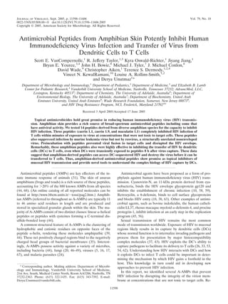
TABLE 1. Amphibian antimicrobial peptides
Peptide Species of origin Sequence
Caerin 1.1 Litoria caerulea GLLSVLGSVAKHVLPHVVPVIAEHL-NH2
Caerin 1.9 Litoria chloris GLFGVLGSIAKHVLPHVVPVIAEKL-NH2
Caerin 4.1 Litoria caerulea GLWQKIKSAAGDLASGIVEGIKS-NH2
Dahlein 5.6 Litoria dahlii GLLASLGKVFGGYLAEKLKPK
Dermaseptin Phyllomedusa sauvagii ALWKTMLKKLGTMALHAGKAALGAAADTISQGTQ
Esculentin-1ARb Rana areolata GLFPKFNKKKVKTGIFDIIKTVGKEAGMDVLRTGIDVIGCKIKGEC
Esculentin-2P Rana pipiens GFSSIFRGVAKFASKGLGKDLARLGVNLVACKISKQC
Maculatin 1.1 Litoria genimaculata GLFGVLAKVAAHVVPAIAEHF-NH2
Magainin II Xenopus laevis GIGKFLHSAKKFGKAFVGEIMNS
MRP Rana tagoi AIGSILGALAKGLPTLISWIKNR-NH2
Palustrin-3AR Rana areolata GIFPKIIGKGIVNGIKSLAKGVGMKVFKAGLNNIGNTGCNNRDEC
Ranatuerin-6 Rana catesbeiana FISAIASMLGKFL-NH2
Ranatuerin-2P Rana pipiens GLMDTVKNVAKNLAGHMLDKLKCKITGC
RCCP Rana catesbeiana Natural mixture of peptides
Uperin 3.6 Uperoleia mjobergii GVIDAAKKWNVLKNLF-NH2
markably, these peptides also inhibit the transfer of HIV from crobial peptides at various time points and concentrations. Infection of T cells
DCs to T cells, even when DCs are transiently exposed to was analyzed through GFP expression after 3 days with a FACSCalibur four-
color cytometer (BD Biosciences) and CELLQuest software (BD Biosciences).
peptides hours after capture of virus. These findings suggest Aliquots of cells were removed at different time points post-peptide treatment
that A-AMPs could be utilized as an approach for HIV pro- and incubated with propidium iodide (PI; 25 g/ml, Sigma). Cells were analyzed
phylaxis and importantly reveal that HIV is not fully protected by flow cytometry for PI exclusion as an indicator of viability. Infection and PI
after DC capture. data were normalized against phosphate-buffered saline (PBS) treatment con-
trols set at 100% infectivity or viability, respectively.
T-cell proliferation assay. The effect of A-AMPs on T-cell activation and
MATERIALS AND METHODS proliferation was measured by labeling the T cells with carboxy-fluorescein di-
Amphibian peptides. The sequence of the peptides tested and species of origin acetate succinimidyl ester (CFSE; Molecular Probes). Purified cells were washed
can be found in Table 1. Caerin 1.1, caerin 1.9, caerin 4.1, dahlein 5.6, maculatin and resuspended in PBS. CFSE was added at a final concentration of 1.65 M
1.1, and uperin 3.6 were synthesized by Chiron Mimotopes, Clayton, Victoria, and incubated at 37°C for 3 min. Labeling was quenched by addition of 50% fetal
Australia, using L-amino acids and standard 9-fluorenylmethoxycarbonyl chem- calf serum in PBS, followed by three washes with RPMI-supplemented medium.
istry. Dermaseptin and magainin II were purchased from Sigma (St. Louis, MO). All CFSE labeling and cell culture were performed in the dark. Cells were
Esculentin-2P and ranatuerin-2P were synthesized by Sigma Genosys (Houston, activated by plate bound anti-CD3 and anti-CD28 as previously described (41).
TX). Esculentin-1ARb, melittin-related peptide (MRP), and palustrin-3ARa The proliferation index was calculated as the sum of the total cells in all gener-
were isolated from skin secretions of Rana areolata and Rana tagoi by J. M. ations divided by the estimated number of original parent cells (determined by
Conlon (1, 19). Ranatuerin-6 was synthesized using standard 9-fluorenylme- dividing the final number of cells in each division by the squared generation
thoxycarbonyl chemistry. number).
Cell culture and primary cell isolation. Peripheral blood mononuclear cells Reovirus and MLV infection assays. Caerin 1.9 was added to samples of T1L
were isolated from adult blood using a Ficoll-Hypaque gradient. Monocytes and virions or ISVPs at a final concentration of 2.5 M, 5 M, or 7.5 M and
CD4 T cells were isolated from peripheral blood mononuclear cells as previ- incubated at 37°C for 45 min. Virus was incubated with a related but inactive
ously described (36, 40). DCs were generated by culturing CD14 monocytes in peptide, caerin 4.1, at a final concentration of 5 M or buffer only (PBS) as
RPMI complete medium (10% fetal bovine serum, 2 mM L-glutamine, 100 U/ml controls. Virus was adsorbed to confluent HeLa cell monolayers at a multiplicity
of penicillin G, and 100 g/ml of streptomycin) supplemented with interleukin-4 of infection (MOI) of 2 PFU per cell at 4°C for 1 h. Following two washes with
(50 g/ml; R&D Systems) and granulocyte-macrophage colony-stimulating fac- PBS, fresh medium was added, and cells were incubated at 37°C for 24 h. Cells
tor (50 g/ml; R&D Systems) for 5 days and subsequently matured by addition and medium were subjected to three cycles of freezing and thawing, and virus
of lipopolysaccharide (100 ng/ml; Sigma) for 1 to 2 days. Mature DC production titer was determined by plaque assay (21). Assays were performed in triplicate,
was assessed by staining cells with antibodies to CD14, CD83, CD86, and and the data presented are representative of two experiments. Caerin 1.9 and
HLA-DR (all from BD Biosciences). Hut 78 T cells expressing CCR5 (Hut/ caerin 4.1 were added to MLV (30,000 infectious units [IFU]) and incubated at
CCR5) were prepared and maintained as previously described (40, 65). 293T, 37°C for 45 min. Virus was added to 3 104 Mus dunni cells and cultured for 3
Mus dunni, and HeLa cells were maintained in Dulbecco’s modified Eagle days before cells were fixed and analyzed for infection by GFP expression by flow
medium. Murine L cells used to titer reovirus were maintained in Joklik minimal cytometry.
essential medium supplemented to contain 5% fetal bovine serum and 100 mg/ml DC-mediated infection assays. Monocyte-derived DCs were pulsed with rep-
of penicillin G, 100 g/ml of streptomycin/ml, and 250 ng/ml of amphotericin B. lication-competent HIV-R5 at an MOI of 2. Virus-cell mixtures were centrifuged
Virus production. Vesicular stomatitis virus glycoprotein (VSV-G)- at 2,000 rpm for 1 h and cultured for an additional 2 h to allow DCs to efficiently
pseudotyped replication-incompetent HIV particles (HDV-VSV-G), or replica- capture virus. DCs were washed three times with complete RPMI medium to
tion-competent virus expressing either the HIV envelope BaL that uses CCR5 as remove non-cell-associated virus. Peptide was added to cells at a final concen-
a coreceptor (HIV-R5) or LAI that uses CXCR4 as coreceptor (HIV-X4) were tration of 1 to 32 M and incubated for 45 min. DCs were washed three times
generated as previously described (60). All these viruses also contain enhanced with complete RPMI medium and incubated with 3 104 Hut/CCR5 cells for 3
green fluorescent protein (EGFP) (Clontech) in place of the nef gene. Reovirus days. Cells were harvested, fixed with 1% paraformaldehyde, and analyzed for
strain type 1 Lang (T1L) is a Dermody laboratory stock. Reovirus titers were expression of GFP by flow cytometry.
determined by plaque assay with L-cell monolayers (21). Infectious subvirion Virus-cell fusion assay. HIV fusion assays were performed essentially as pre-
particles (ISVPs) were prepared by treatment of purified T1L virions with N - viously described (13). Briefly, viruses carrying a -lactamase reporter protein
p-tosyl-L-lysine chloromethyl ketone-treated bovine chymotrypsin (Sigma) as fused to the amino terminus of the virion protein Vpr (BlaM-Vpr), were treated
previously described (2). Murine leukemia virus (MLV) pseudotyped with with caerin 1.9 (1 to 10 M) for 1 h. These viruses plus peptide mixtures were
VSV-G was produced by transfecting 293T cells with pCIG-B (7), pBABE- added to Hut/CCR5 cells at 37°C for 2 h to allow virus-cell fusion. CCF2/AM (20
EGFP, and pHCMV-G. M; Aurora Biosciences Corp.) was added, and the cultures were incubated for
HIV infection and cell viability assay. Virus was cultured with Hut or primary 14 h at room temperature. Cells were pelleted and resuspended in PBS, and the
CD4 T cells activated as previously described (41) in the presence of antimi- fluorescence was measured at 447 and 520 nm with a microplate fluorometer](data:image/gif;base64,R0lGODlhAQABAIAAAAAAAP///yH5BAEAAAAALAAAAAABAAEAAAIBRAA7)
Recomendados
Recomendados
Mais conteúdo relacionado
Mais procurados
Mais procurados (20)
Destaque
Destaque (20)
Semelhante a 5.4 Caerina
Semelhante a 5.4 Caerina (20)
Mais de Alfonso Enrique Islas Rodríguez
Mais de Alfonso Enrique Islas Rodríguez (20)
Último
Último (20)
5.4 Caerina
- 1. JOURNAL OF VIROLOGY, Sept. 2005, p. 11598–11606 Vol. 79, No. 18 0022-538X/05/$08.00 0 doi:10.1128/JVI.79.18.11598–11606.2005 Copyright © 2005, American Society for Microbiology. All Rights Reserved. Antimicrobial Peptides from Amphibian Skin Potently Inhibit Human Immunodeficiency Virus Infection and Transfer of Virus from Dendritic Cells to T Cells Scott E. VanCompernolle,1 R. Jeffery Taylor,1,5 Kyra Oswald-Richter,1 Jiyang Jiang,1 Bryan E. Youree,3,4 John H. Bowie,6 Michael J. Tyler,7 J. Michael Conlon,8 David Wade,9 Christopher Aiken,1 Terence S. Dermody,1,2,4 Vineet N. KewalRamani,10 Louise A. Rollins-Smith,1,2 and Derya Unutmaz1* Department of Microbiology and Immunology,1 Department of Pediatrics,2 Department of Medicine,3 and Elizabeth B. Lamb Center for Pediatric Research,4 Vanderbilt University School of Medicine, Nashville, Tennessee 37232; AdvancMed, LLC, Lexington, Kentucky 405135; Department of Chemistry, The University of Adelaide, Australia6; Department of Environmental Biology, The University of Adelaide, Australia7; Department of Biochemistry, United Arab Emirates University, United Arab Emirates8; Wade Research Foundation, Somerset, New Jersey 088739; and HIV Drug Resistance Program, NCI, Frederick, Maryland 2170210 Received 3 April 2005/Accepted 17 June 2005 Topical antimicrobicides hold great promise in reducing human immunodeficiency virus (HIV) transmis- sion. Amphibian skin provides a rich source of broad-spectrum antimicrobial peptides including some that have antiviral activity. We tested 14 peptides derived from diverse amphibian species for the capacity to inhibit HIV infection. Three peptides (caerin 1.1, caerin 1.9, and maculatin 1.1) completely inhibited HIV infection of T cells within minutes of exposure to virus at concentrations that were not toxic to target cells. These peptides also suppressed infection by murine leukemia virus but not by reovirus, a structurally unrelated nonenveloped virus. Preincubation with peptides prevented viral fusion to target cells and disrupted the HIV envelope. Remarkably, these amphibian peptides also were highly effective in inhibiting the transfer of HIV by dendritic cells (DCs) to T cells, even when DCs were transiently exposed to peptides 8 h after virus capture. These data suggest that amphibian-derived peptides can access DC-sequestered HIV and destroy the virus before it can be transferred to T cells. Thus, amphibian-derived antimicrobial peptides show promise as topical inhibitors of mucosal HIV transmission and provide novel tools to understand the complex biology of HIV capture by DCs. Antimicrobial peptides (AMPs) are key effectors of the in- Antimicrobial agents have been proposed as a form of pro- nate immune response of animals (31). The skin of anuran phylaxis against human immunodeficiency virus (HIV) trans- amphibians (frogs and toads) is a rich source of these peptides, mission. Cyanovirin-N, an 11-kDa protein derived from cya- accounting for 20% of the 880 known AMPs from all species nobacteria, binds the HIV envelope glycoprotein gp120 and (44, 68). (An online catalog of all reported molecules can be inhibits the establishment of chronic infection (10, 58, 59). found at http://www.bbcm.units.it/ tossi/pag2.htm.) Amphib- Retrocyclin, a -defensin, binds to cell surface glycoproteins ian AMPs (referred to throughout as A-AMPs) are typically 11 and blocks HIV entry (18, 38, 63). Other examples of antimi- to 46 amino acid residues in length and are produced and crobial agents, such as bovine indolicidin, the human catheli- stored in specialized granular glands within the skin. The ma- cidin LL37, rhesus macaque myeloid -defensin-4, and procine jority of A-AMPs consist of two distinct classes: linear -helical protegrin-1, inhibit infection at an early step in the replication peptides or peptides with cysteines forming a C-terminal dis- program (45, 56). ulfide-bonded loop (44). Sexual transmission of HIV remains the most common A common structural feature of A-AMPs is the clustering of mode of transmission worldwide. Exposure of HIV to mucosal hydrophobic and cationic residues on opposite faces of the regions likely results in its capture by dendritic cells (DCs) peptide -helix, rendering these molecules amphipathic (39, whose normal function is to internalize invading pathogens and 44). These net positively charged regions bind to the negatively process them for presentation by major histocompatibility charged head groups of bacterial membranes (35). Interest- complex molecules (37, 43). HIV exploits the DC’s ability to ingly, A-AMPs possess activity against a variety of microbes, capture pathogens to facilitate its delivery to T cells (26, 33, 53, including bacteria (44), fungi (20, 46–49), viruses (5, 16, 17, 54, 62). Understanding how HIV interacts with DCs and how 67), and malaria parasites (24). it exploits DCs to infect T cells could be important in deter- mining the mechanism by which HIV gains a foothold in the host. This knowledge in turn could aid in developing new * Corresponding author. Mailing address: Department of Microbi- approaches to prevent HIV infection. ology and Immunology, Vanderbilt University School of Medicine, 21st Ave. South, Medical Center North, Room AA5206, Nashville, TN In this report, we identified several A-AMPs that prevent 37232-2363. Phone: (615) 322-1435. Fax: (615) 343-7392. E-mail: HIV infection by disrupting the integrity of the virion mem- Derya.Unutmaz@vanderbilt.edu. brane at concentrations that are not toxic to target cells. Re- 11598
- 2. VOL. 79, 2005 AMPHIBIAN PEPTIDE INHIBITION OF HIV INFECTION 11599 TABLE 1. Amphibian antimicrobial peptides Peptide Species of origin Sequence Caerin 1.1 Litoria caerulea GLLSVLGSVAKHVLPHVVPVIAEHL-NH2 Caerin 1.9 Litoria chloris GLFGVLGSIAKHVLPHVVPVIAEKL-NH2 Caerin 4.1 Litoria caerulea GLWQKIKSAAGDLASGIVEGIKS-NH2 Dahlein 5.6 Litoria dahlii GLLASLGKVFGGYLAEKLKPK Dermaseptin Phyllomedusa sauvagii ALWKTMLKKLGTMALHAGKAALGAAADTISQGTQ Esculentin-1ARb Rana areolata GLFPKFNKKKVKTGIFDIIKTVGKEAGMDVLRTGIDVIGCKIKGEC Esculentin-2P Rana pipiens GFSSIFRGVAKFASKGLGKDLARLGVNLVACKISKQC Maculatin 1.1 Litoria genimaculata GLFGVLAKVAAHVVPAIAEHF-NH2 Magainin II Xenopus laevis GIGKFLHSAKKFGKAFVGEIMNS MRP Rana tagoi AIGSILGALAKGLPTLISWIKNR-NH2 Palustrin-3AR Rana areolata GIFPKIIGKGIVNGIKSLAKGVGMKVFKAGLNNIGNTGCNNRDEC Ranatuerin-6 Rana catesbeiana FISAIASMLGKFL-NH2 Ranatuerin-2P Rana pipiens GLMDTVKNVAKNLAGHMLDKLKCKITGC RCCP Rana catesbeiana Natural mixture of peptides Uperin 3.6 Uperoleia mjobergii GVIDAAKKWNVLKNLF-NH2 markably, these peptides also inhibit the transfer of HIV from crobial peptides at various time points and concentrations. Infection of T cells DCs to T cells, even when DCs are transiently exposed to was analyzed through GFP expression after 3 days with a FACSCalibur four- color cytometer (BD Biosciences) and CELLQuest software (BD Biosciences). peptides hours after capture of virus. These findings suggest Aliquots of cells were removed at different time points post-peptide treatment that A-AMPs could be utilized as an approach for HIV pro- and incubated with propidium iodide (PI; 25 g/ml, Sigma). Cells were analyzed phylaxis and importantly reveal that HIV is not fully protected by flow cytometry for PI exclusion as an indicator of viability. Infection and PI after DC capture. data were normalized against phosphate-buffered saline (PBS) treatment con- trols set at 100% infectivity or viability, respectively. T-cell proliferation assay. The effect of A-AMPs on T-cell activation and MATERIALS AND METHODS proliferation was measured by labeling the T cells with carboxy-fluorescein di- Amphibian peptides. The sequence of the peptides tested and species of origin acetate succinimidyl ester (CFSE; Molecular Probes). Purified cells were washed can be found in Table 1. Caerin 1.1, caerin 1.9, caerin 4.1, dahlein 5.6, maculatin and resuspended in PBS. CFSE was added at a final concentration of 1.65 M 1.1, and uperin 3.6 were synthesized by Chiron Mimotopes, Clayton, Victoria, and incubated at 37°C for 3 min. Labeling was quenched by addition of 50% fetal Australia, using L-amino acids and standard 9-fluorenylmethoxycarbonyl chem- calf serum in PBS, followed by three washes with RPMI-supplemented medium. istry. Dermaseptin and magainin II were purchased from Sigma (St. Louis, MO). All CFSE labeling and cell culture were performed in the dark. Cells were Esculentin-2P and ranatuerin-2P were synthesized by Sigma Genosys (Houston, activated by plate bound anti-CD3 and anti-CD28 as previously described (41). TX). Esculentin-1ARb, melittin-related peptide (MRP), and palustrin-3ARa The proliferation index was calculated as the sum of the total cells in all gener- were isolated from skin secretions of Rana areolata and Rana tagoi by J. M. ations divided by the estimated number of original parent cells (determined by Conlon (1, 19). Ranatuerin-6 was synthesized using standard 9-fluorenylme- dividing the final number of cells in each division by the squared generation thoxycarbonyl chemistry. number). Cell culture and primary cell isolation. Peripheral blood mononuclear cells Reovirus and MLV infection assays. Caerin 1.9 was added to samples of T1L were isolated from adult blood using a Ficoll-Hypaque gradient. Monocytes and virions or ISVPs at a final concentration of 2.5 M, 5 M, or 7.5 M and CD4 T cells were isolated from peripheral blood mononuclear cells as previ- incubated at 37°C for 45 min. Virus was incubated with a related but inactive ously described (36, 40). DCs were generated by culturing CD14 monocytes in peptide, caerin 4.1, at a final concentration of 5 M or buffer only (PBS) as RPMI complete medium (10% fetal bovine serum, 2 mM L-glutamine, 100 U/ml controls. Virus was adsorbed to confluent HeLa cell monolayers at a multiplicity of penicillin G, and 100 g/ml of streptomycin) supplemented with interleukin-4 of infection (MOI) of 2 PFU per cell at 4°C for 1 h. Following two washes with (50 g/ml; R&D Systems) and granulocyte-macrophage colony-stimulating fac- PBS, fresh medium was added, and cells were incubated at 37°C for 24 h. Cells tor (50 g/ml; R&D Systems) for 5 days and subsequently matured by addition and medium were subjected to three cycles of freezing and thawing, and virus of lipopolysaccharide (100 ng/ml; Sigma) for 1 to 2 days. Mature DC production titer was determined by plaque assay (21). Assays were performed in triplicate, was assessed by staining cells with antibodies to CD14, CD83, CD86, and and the data presented are representative of two experiments. Caerin 1.9 and HLA-DR (all from BD Biosciences). Hut 78 T cells expressing CCR5 (Hut/ caerin 4.1 were added to MLV (30,000 infectious units [IFU]) and incubated at CCR5) were prepared and maintained as previously described (40, 65). 293T, 37°C for 45 min. Virus was added to 3 104 Mus dunni cells and cultured for 3 Mus dunni, and HeLa cells were maintained in Dulbecco’s modified Eagle days before cells were fixed and analyzed for infection by GFP expression by flow medium. Murine L cells used to titer reovirus were maintained in Joklik minimal cytometry. essential medium supplemented to contain 5% fetal bovine serum and 100 mg/ml DC-mediated infection assays. Monocyte-derived DCs were pulsed with rep- of penicillin G, 100 g/ml of streptomycin/ml, and 250 ng/ml of amphotericin B. lication-competent HIV-R5 at an MOI of 2. Virus-cell mixtures were centrifuged Virus production. Vesicular stomatitis virus glycoprotein (VSV-G)- at 2,000 rpm for 1 h and cultured for an additional 2 h to allow DCs to efficiently pseudotyped replication-incompetent HIV particles (HDV-VSV-G), or replica- capture virus. DCs were washed three times with complete RPMI medium to tion-competent virus expressing either the HIV envelope BaL that uses CCR5 as remove non-cell-associated virus. Peptide was added to cells at a final concen- a coreceptor (HIV-R5) or LAI that uses CXCR4 as coreceptor (HIV-X4) were tration of 1 to 32 M and incubated for 45 min. DCs were washed three times generated as previously described (60). All these viruses also contain enhanced with complete RPMI medium and incubated with 3 104 Hut/CCR5 cells for 3 green fluorescent protein (EGFP) (Clontech) in place of the nef gene. Reovirus days. Cells were harvested, fixed with 1% paraformaldehyde, and analyzed for strain type 1 Lang (T1L) is a Dermody laboratory stock. Reovirus titers were expression of GFP by flow cytometry. determined by plaque assay with L-cell monolayers (21). Infectious subvirion Virus-cell fusion assay. HIV fusion assays were performed essentially as pre- particles (ISVPs) were prepared by treatment of purified T1L virions with N - viously described (13). Briefly, viruses carrying a -lactamase reporter protein p-tosyl-L-lysine chloromethyl ketone-treated bovine chymotrypsin (Sigma) as fused to the amino terminus of the virion protein Vpr (BlaM-Vpr), were treated previously described (2). Murine leukemia virus (MLV) pseudotyped with with caerin 1.9 (1 to 10 M) for 1 h. These viruses plus peptide mixtures were VSV-G was produced by transfecting 293T cells with pCIG-B (7), pBABE- added to Hut/CCR5 cells at 37°C for 2 h to allow virus-cell fusion. CCF2/AM (20 EGFP, and pHCMV-G. M; Aurora Biosciences Corp.) was added, and the cultures were incubated for HIV infection and cell viability assay. Virus was cultured with Hut or primary 14 h at room temperature. Cells were pelleted and resuspended in PBS, and the CD4 T cells activated as previously described (41) in the presence of antimi- fluorescence was measured at 447 and 520 nm with a microplate fluorometer
- 3. 11600 VANCOMPERNOLLE ET AL. J. VIROL. after excitation at 409 nm. Uncleaved CCF2 fluoresces green, due to fluores- cence resonance energy transfer between the coumarin and fluorescein groups; however, cleavage by BlaM results in the dissociation of these fluorophores, and the emission spectrum shifts to blue. Thus, the ratio of blue to green cellular fluorescence is proportional to the overall extent of virus-cell fusion. Fluores- cence ratios were calculated after subtraction of the average background fluo- rescence of control cultures containing no virus (blue values) and wells contain- ing PBS (green values). HIV p24 release assay. HIV-VSV-G was incubated with either peptides or PBS at a concentration range of 1 to 32 M for 30 min in complete RPMI medium. The medium was then assayed for the presence of viral core protein p24 by enzyme-linked immunosorbent assay (ELISA) as previously described (40). Plates were analyzed by microplate reader (Molecular Devices) at 405 nm ab- sorbance. Total p24 was calculated using linear regression analysis from stan- dards included on each plate. RESULTS Amphibian-derived AMPs inhibit HIV infection of T cells. Because AMPs have activity against some viruses, including frog virus 3, channel catfish virus, and herpes simplex virus 1 infection (5, 17, 67), we investigated whether A-AMPs can inhibit infection by HIV. AMPs from diverse amphibian spe- cies (Table 1) were tested for their ability to block HIV infec- tion of T cells (Fig. 1A). Prior to fixation, a portion of the cells were removed and stained with PI to determine cell viability. We identified three peptides (caerin 1.1, caerin 1.9, and mac- ulatin 1.1) that inhibited HIV infection without adversely af- fecting T-cell viability. Other peptides, including esculentin- 1ARb, esculentin-2P, magainin II, MRP, palustrin-3AR, and ranatuerin-2P, were not effective in inhibiting HIV infection of T cells at nontoxic concentrations (Fig. 1A). Dermaseptin par- tially inhibited HIV infection; however, higher doses were toxic to cells (Fig. 1A and data not shown). To determine the MIC, we incubated HIV and T cells with increasing doses of the effective peptides for 3 days. All three peptides were effective FIG. 1. Amphibian peptides inhibit HIV infection of T cells. over a relatively narrow concentration range, with caerin 1.9 (A) Fourteen purified peptides and one natural peptide mixture (RCCP) derived from the skin of anuran amphibians were incubated at having the lowest IC50 (calculated as the concentration of 10 g/ml (RCCP), 1 M (ranatuerin-2P, esculentin-1ARb, palustrin- peptide necessary to inhibit 50% of PBS-treated HIV infection 2AR, and esculentin-2P), 5 M (caerin 1.9 and magainin II) or 10 M of T cells) of 1.2 M, caerin 1.1 having an IC50 of 7.8 M, and (all others) with 3 104 T cells and HIV-R5 at an MOI of 1 per cell maculatin 1.1 having an IC50 of 11.3 M (Fig. 1B). Because of at 37°C for 3 days. Cells were harvested and analyzed for GFP expres- the higher concentration of maculatin 1.1 required for inhibi- sion. The data are presented as the mean of three replicate samples from one representative experiment of two independent experiments. tion, we focused on caerin 1.1 and caerin 1.9 for the remainder Error bars indicate standard deviations. Data are presented as percent of the study. A related noninhibitory peptide, caerin 4.1, was inhibition normalized to infection in the presence of PBS. (B) T cells used as a control in all experiments. (3 104) were incubated with HIV-R5 at an MOI of 1 and increasing It has been proposed that amphibians secrete these peptides concentrations of caerin 1.1, caerin 1.9, maculatin 1.1, caerin 4.1, or PBS at 37°C for 3 days. Cells were harvested and analyzed for GFP to combat topical microbial pathogens (68). Because endopep- expression. The data are presented as the mean of three replicate tidases rapidly degrade them, the peptides are at a high con- samples from one representative experiment of six independent exper- centration for only a short time (52). Thus, they must exert iments. Error bars indicate standard deviations. IC50s were calculated their cytopathic effects faster than the generational doubling by regression analysis. time of the pathogen. We therefore tested the kinetics of A-AMP-mediated inhibition of HIV infection. Both caerin 1.9 and caerin 1.1 inhibited infection by 99% and 90%, respec- there was no effect on T-cell proliferation (Fig. 2C). The pep- tively, after only 5 to 10 min of exposure to HIV (Fig. 2A). tides were equally effective at blocking HIV infection of either Furthermore, these short incubation periods also increased the Hut/CCR5 or primary CD4 T cells (Fig. 2B). These results tolerance of T cells to much higher concentrations of peptide show that A-AMPs potently and rapidly inhibit HIV infection while retaining the effective IC50 of the peptides to block HIV at significantly lower concentrations than those required to (Fig. 2B). In addition, to exclude any adverse effects of the affect the viability of the T cells. peptides on T-cell activation and proliferation, we measured Amphibian AMPs can inhibit other enveloped viruses. To cell division upon activation using CFSE labeling (Fig. 2B, determine whether the effects of A-AMPs are limited to a inserts). These experiments demonstrate that even at twofold- particular HIV strain or envelope coreceptor usage, we prein- higher concentrations that completely inhibit HIV infection, cubated caerin 1.9 with HIV-LAI (CXCR4-tropic virus), HIV
- 4. VOL. 79, 2005 AMPHIBIAN PEPTIDE INHIBITION OF HIV INFECTION 11601 pseudotyped with VSV-G (coreceptor independent), HIV pseudotyped with the MLV amphotropic envelope glycopro- teins, or MLV pseudotyped with VSV-G. T cells (or Mus dunni cells for the MLV infections) were then added and analyzed for infection at day 3. Caerin 1.9 potently inhibited infection of cells by HIV with different envelopes, as well as VSV-G pseudotyped MLV (Fig. 3A). These results suggest that A- AMPs inhibit both HIV and MLV infection independently of envelope glycoproteins. We next asked whether the A-AMPs could inhibit infection of a nonenveloped virus. For this experiments we used mam- malian reoviruses, which are nonenveloped viruses that con- tain a segmented, double-stranded RNA genome. After en- gagement of cell-surface receptors including junctional adhesion molecule-A (4) and sialic acid (14, 23, 28, 42), reo- virus virions enter cells by receptor-mediated endocytosis and are uncoated to produce ISVPs (9, 50, 55). ISVPs penetrate endosomal membranes, thereby delivering transcriptionally ac- tive core particles into the cytoplasm (8, 30, 34, 57). In sharp contrast to the results gathered using retroviruses HIV and MLV, the peptides did not inhibit reovirus infection (Fig. 3B). Amphibian AMPs directly inactivate HIV by disrupting vi- ral envelope integrity. To define the mechanism by which A- AMPs inhibit HIV, we determined whether caerin 1.9 directly affects the integrity of the virus or interacts with the target cell to prevent infection. Caerin 1.9 was added to T cells at various intervals extending to 48 h and then washed away prior to virus exposure. If the peptide interfered with cellular components required for infection, fewer GFP-positive cells would be ex- pected following this pretreatment. Instead, there was no dif- FIG. 2. Peptide inhibition occurs rapidly with no effect on cell vi- ability. (A) Caerin 1.1 (20 M), caerin 1.9 (5 M), caerin 4.1 (20 M), or PBS was incubated with 3 104 IFU of HIV-R5. At the times shown, virus-peptide solutions were diluted to a concentration that resulted in minimal activity based the results shown in Fig. 1B (four- fold) in complete RPMI medium with 3 104 T cells and incubated at 37°C for 3 days. Cells were harvested, fixed, and analyzed for GFP expression by flow cytometry. Data were normalized to infection fol- lowing PBS treatment and are presented as the mean of three replicate samples from one representative experiment of three independent experiments, with error bars indicating standard deviation. (B) Pep- tides were incubated for 5 min at increasing concentrations (1 to 48 M) with 5 104 activated primary CD4 T cells (circles) or Hut/ CCR5 cells (squares) infected with HIV-R5 at an MOI of 1, diluted fourfold with complete RPMI medium, and incubated at 37°C for 3 days. Cells were harvested and analyzed for GFP expression (closed circles and squares). Data are normalized to infection following PBS treatment and are presented as the mean of three replicate samples from one representative experiment of three independent experiments with error bars indicating standard deviation. At 2 h postinfection, 2 104 T cells were removed from the culture, stained with PI, and ana- lyzed for viability by flow cytometry (open circles and squares). (C) Peptides were incubated for 5 min at increasing concentrations (1 to 50 M) with 5 104 CFSE-labeled primary CD4 T cells. Cells were then diluted fourfold with complete RPMI medium and activated by T-cell receptor engagement as previously described (41). The acti- vated cells were cultured for 4 days, and CFSE fluorescence intensity was measured by flow cytometry. Representative histograms (panel B, insets) from each peptide treatment are shown (40 M caerin 1.1, 10 M caerin 1.9, and 40 M caerin 4.1), which were identical to those of the cells not treated with peptides.
- 5. 11602 VANCOMPERNOLLE ET AL. J. VIROL. FIG. 3. Peptide activity against HIV with different envelopes and diverse viral species. (A) Caerin 1.9 (5 M) and caerin 4.1 (5 M) were incubated with 3 104 T cells and HIV-R5, HIV-X4, HDV-VSV-G, or FIG. 4. Caerin 1.1 and caerin 1.9 disrupt the HIV envelope. HDV pseudotyped with the MLV amphotropic envelope at an MOI of (A) HIV-R5 carrying a -lactamase reporter protein was incubated 1 at 37°C for 3 days. Caerin 1.9 (10 M) and caerin 4.1 (10 M) were with 5 M caerin 1.9 (lane 3) or PBS (lane 1) for 1 h prior to added to MLV (30,000 IFU) and incubated at 37°C for 45 min. Virus inoculation of Hut/CCR5 cells (105) by centrifugation at 600 g for was then added to 3 104 Mus dunni cells and cultured at 37°C for 3 1.5 h at 35°C. These cells or uninoculated cells (lane 4) were incubated days. Following incubation, cells were harvested and analyzed for GFP at 37°C for 2 h to allow virus-cell fusion. CCF2/AM (20 M) was expression by flow cytometry. Data are normalized to infection follow- added, the cultures were incubated at room temperature for 14 h, and ing PBS treatment and are presented as the mean of three replicate fluorescence was measured at 447 and 520 nm by using a microplate samples from one representative experiment of three independent fluorometer after excitation at 409 nm. Fluorescence ratios were cal- experiments, with error bars indicating standard deviation. (B) Reovi- culated after subtraction of the average background fluorescence and rus T1L virions or ISVPs were incubated with caerin 1.9, caerin 4.1, or are presented as relative fusion. Caerin 1.9 (5 M) was added during PBS (mock) at the concentrations shown at 37°C for 45 min. Virus was the CCF2/AM incubation to control for peptide effects on dye loading adsorbed to confluent HeLa cell monolayers at an MOI of 2 PFU per (lane 2). Duplicate determinations were performed for each virus cell at 4°C for 1 h. Following two washes with PBS, fresh medium was dilution. (B) HIV-VSV-G was incubated with peptides or PBS at the added, and cells were incubated at 37°C for 24 h. Cells and medium concentrations shown for 30 min in complete RPMI. Medium was then were subjected to three cycles of freezing and thawing, and virus titer diluted 1:1,000 in assay buffer without detergent and analyzed for the was determined by plaque assay. The results are presented as the presence of viral core protein p24 by ELISA. Total p24 was calculated means of two independent experiments. Error bars represent standard from dilutions of replicate samples using linear regression analysis deviations. from p24 standards included on each plate. Data are presented as the mean of two replicate samples and are representative of two experi- ments. Error bars indicate standard deviation. ference in infection of the T cells regardless of the caerin 1.9 pretreatment interval (data not shown). To determine the stage of HIV life cycle that is inhibited by of HIV with caerin 1.9 inhibited viral fusion to target cells (Fig. these A-AMPs, we first analyzed the effect of delayed addition 4A), suggesting that these A-AMPs block infection prior to of peptide to T cells after infection with HIV. The reverse viral entry of the target cells. transcription of HIV after infection of cells was assessed by the The inhibition of HIV entry by caerin 1.9 may have been presence of strong-stop, minus-strand viral DNA (R/U5 DNA) caused by peptide attack of the virion membrane prior to cell by quantitative real-time PCR. Addition of caerin 1.9 to cells binding and fusion. To determine whether A-AMPs directly after initiation of reverse transcription (4 to 8 h after infection) inactivate virions by disrupting the envelope, virus was prein- did not have any inhibitory effect (data not shown). These data cubated with increasing concentrations of A-AMPs or controls suggest that caerin 1.9 inhibits HIV infection prior to the as described above and the release of HIV core protein p24 initiation of reverse transcription. was measured by an ELISA. In this assay, p24 would only be We therefore asked whether these A-AMPs prevent the detected if the viral envelope was disrupted by A-AMPs. In- entry of HIV by blocking its fusion to target cells. For this deed, treatment of virions with A-AMPs released p24 into experiment, we employed a recently developed reporter assay culture supernatant (Fig. 4B), indicating disruption of the viral to quantify HIV particle entry (13). In this assay, pretreatment membrane.
- 6. VOL. 79, 2005 AMPHIBIAN PEPTIDE INHIBITION OF HIV INFECTION 11603 Amphibian AMPs inhibit DC-mediated trans infection of T cells. DCs resident at mucosal sites are thought to be among the first cells that interact with HIV upon exposure to the virus and possibly pass the virus on to T cells (26). An ideal topical antimicrobial would interfere with this “capture” process and inactivate HIV particles that have already been captured by DCs before they are transmitted to T cells. We therefore tested whether A-AMPs inhibit DC-mediated infection of T cells after the virus has been captured by DCs. For this experiment, DCs were first incubated with HIV, washed to remove cell-free virus, and subsequently incubated with different concentrations of peptide for 45 min. DCs were again washed to remove the peptide and incubated with T cells for 3 days to allow trans infection. Treatment of HIV-adsorbed DCs with A-AMPs in- hibited the subsequent infection of T cells with similar IC50 values (caerin 1.1 to 12.6 M and caerin 1.9 to 1.6 M) (Fig. 5A) found for inhibition of direct T-cell infection (Fig. 1B). The A-AMPs also inhibited the trans infection of T cells with X4-tropic viruses captured by DCs (data not shown). We then asked how long after capture of HIV by DCs A-AMPs were able to inhibit virus transmission to T cells. To answer this question, DCs were first incubated with HIV and washed to remove unbound virus. The A-AMPs were then added after different time intervals, washed to remove the peptide, and cultured with T cells for 3 days. Exposure of DCs to A-AMPs as late as 8 h post-HIV capture resulted in 95% inhibition of trans infection (Fig. 5B). In these experiments, the FIG. 5. Amphibian antimicrobial peptides inhibit DC-mediated viability of the DCs was not affected, as assessed by PI staining, trans infection of T cells. (A) DCs (4 104) were pulsed with HIV-R5 even at threefold-higher peptide concentrations (data not at an MOI of 2. Virus-cell mixtures were centrifuged for 1 h at 2,000 shown). rpm and incubated at 37°C for 2 h to allow DCs to efficiently capture the virus. DCs were washed three times with complete RPMI medium The inhibition of DC-mediated transfer of virus by A-AMPs to remove unbound virus. Peptide was then added to the DCs at the was remarkable, since free peptide was washed away before concentrations shown and incubated at 37°C for 45 min. DCs were addition of the target T cells. These findings suggested that the washed and incubated with 3 104 T cells at 37°C for 3 days. Cells peptides somehow access HIV captured by DCs either on the were harvested, fixed with 1% paraformaldehyde, and analyzed for cell surface or intracellularly. It is also conceivable that some GFP expression. Data are normalized to infection following PBS treat- ment and are presented as the mean of three replicate samples from peptide sticks to the DC surface and interacts with the virus as one representative experiment of three independent experiments, with it is being passed on to T cells. To test this possibility, we first error bars indicating standard deviation. (B) DCs (4 104) were incubated the DCs with high doses of peptides (8 M caerin incubated with HIV-R5 at an MOI of 1. At the times shown postin- 1.9, 16 M caerin 1.1, 16 M caerin 4.1, or PBS) for 30 min, oculation, 30 M caerin 1.1, 4 M caerin 1.9, and 30 M caerin 4.1 were added to cultures and incubated at 37°C for 45 min. DCs were subsequently washed the cells several times to remove free washed three times in complete RPMI medium, incubated with 2 peptide, and added virus to DCs. After 2 h of incubation, the 104 T cells at 37°C for 3 days, and analyzed for GFP expression. Data DC-plus-virus cultures were extensively washed, and target T are normalized as above and are representative of four independent cells were added. We did not observe any inhibition of the experiments. infection of T cells (data not shown). We concluded that pre- treatment of the DCs with A-AMPs, prior to addition of the virus, does not alter the capacity of DCs to capture HIV virions and trans infect T cells. DCs are thought to “capture” HIV virions at the mucosal surfaces and transfer the captured virus to its primary targets, CD4 T cells (26). This capture process is generally thought to DISCUSSION occur via a protein highly expressed on the surface of mucosal Our findings demonstrate that A-AMPs are highly effective DCs, DC-SIGN (DC-specific ICAM grabbing nonintegrin) in inhibiting HIV infection by disrupting the viral membrane (22, 26, 27). After binding to DC-SIGN or as-yet-unidentified and preventing the entry of the virus into target cells. Remark- molecules on DCs, HIV is internalized into late-endosomal- ably, the A-AMPs also were capable of inhibiting the transfer stage compartments near the surface, where the virus remains of HIV by DCs to T cells even when DCs were transiently in an infectious state for up to 5 days (26, 32). However, it is exposed to peptides 8 h after virus capture. These data suggest not clear whether these internalized virions are indeed infec- that A-AMPs can access DC-sequestered HIV and destroy the tious or whether the few viral particles that remain on the cell virus before it can be transferred to T cells. This finding has surface are preferentially transmitted to T cells. Our finding broader implications for understanding how HIV is captured that the virus captured by DCs is effectively neutralized by and transmitted to T cells. A-AMPs suggests that either the virions are destroyed at the
- 7. 11604 VANCOMPERNOLLE ET AL. J. VIROL. DC cell surface or the peptides gain access to the same com- HIV. An exciting prospect will be the utilization of A-AMPs as partment within DCs in which internalized virus resides. tools to dissect these unresolved questions about the mecha- The prevailing mechanistic model of A-AMP activity, called nism of HIV capture and presentation by DCs. the “carpet” mechanism, is that positively charged regions of Our findings demonstrate that the A-AMPs inhibit HIV the -helical peptides bind to negatively charged lipids in the infection by disrupting the virion envelope. Importantly, this membrane (35, 51, 66). The peptides integrate into the outer disruption occurs at much lower peptide concentrations than membrane, causing an increase in surface area that strains the that which is toxic to cells. This observation could be explained bilayer. This alteration leads to transient pore formation, by the small surface area of the virion relative to that of the transport of outer lipids to the inner leaflet, and finally collapse cell. If we assume that the peptides function by carpeting the of the membrane into small AMP-coated vesicles. An alterna- outer leaflet of the membrane, we would expect an increase in tive model of activity called the “barrel-stave” mechanism pro- surface area that strains the lipid bilayer (35, 51). The extreme poses that -helical peptides bundle on the surface of the rate of curvature in the viral membrane compared to the cell membrane and assume an orientation to allow the hydropho- membrane could cause the viral bilayer to succumb to stresses bic surfaces to interact with the lipid core, while the hydro- induced by peptide binding on the outer leaflet at lower con- philic surfaces orient inward (51). This arrangement creates an centrations. Another plausible explanation is that differences aqueous channel. Death of the microbe would then result from in the lipid content of the HIV envelope could increase the either loss of polarization (64), leaking of cellular contents binding rate or affinity of the amphibian peptides. Although (66), disturbance of membrane function from lipid redistribu- HIV acquires its envelope by budding through cellular mem- tion (35), or activation of hydrolases that destroy the cell wall branes, the lipid constituency of the viral envelope differs from (6). In both of these models, A-AMPs are likely to be bound, that of a normal human cell. HIV envelopes enrich in Brij either to the outer leaflet of the membrane or to internal 98-insoluble lipid rafts, which contain elevated levels of cho- regions of the bilayer. Based on these models, a likely mech- lesterol, sphingomyelin, and glycophospholipids (11, 12). The anistic action of peptides would be to disrupt virions while they presence of cholesterol and sphingomyelin contributes to are exposed at the cell surface of the DCs. However, if most tighter packing of the membrane lipids, resulting in a stabilized infectious HIV is internalized by DCs, then the destruction of bilayer. Transient disruptions of the glycerophospholipid re- only cell surface-bound virus would be hard to reconcile with gions of the envelope by peptides that adhere to the viral this mode of action. Indeed, it has been shown that transient surface could be preferentially stabilized by the high choles- trypsin treatment of DCs that have captured HIV does not terol content of the envelope, similar to fusion pore stabiliza- prevent transmission of virus to T cells, suggesting that DCs tion. protect the virus from cleavage at the cell surface (32). Fur- The rapid kinetics and the large dose range of peptides that thermore, the DC-captured HIV is also protected from neu- inhibit virus infectivity without producing cell toxicity, coupled tralizing anti-HIV antibodies (25). with the capacity to inhibit DC-captured HIV, render these A particularly appealing scenario would be that the virus peptides ideal candidates as topical microbicides for prevent- cycles from a vesicular compartment to the surface of the DC, ing HIV transmission. It is conceivable that certain A-AMPs where it is transiently exposed to peptides. This mechanism might elicit a specific immune response, which would limit the would explain how A-AMPs could inhibit trans infection even long-term use of A-AMPs. However, peptides in the absence after 8 h of capture, since the internalized virus would be of an adjuvant are typically not immunogenic. For example, exposed to peptide during the cycling period. This model could studies with the HIV fusion inhibitor peptide T20 (enfuvirtide) also provide a mechanism for viral transmission from the sur- suggest that even upon repeated systemic application this pep- face of the DC to CD4 T cells at the immunological synapse tide did not elicit functionally relevant antibody response (29). (36). In support of this model, we observed that A-AMP ac- Subtle modifications in the amino acid sequence of A-AMPs tivity in the DC capture assays reached maximal inhibition may improve their inhibitory characteristics, decreasing IC50s after 30 min (compared to 10 min for virus alone) (Fig. 2A and while further increasing 50% lethal doses. A veritable treasure data not shown). This slower rate of neutralization could be trove of unexplored A-AMPs (61) might hold even greater due to time required for viral particles to cycle to the cell promise as antiviral compounds against a wide array of impor- surface and thus be exposed to destruction by the peptides. A tant human pathogens. The remarkable capacity of A-AMPs to radical alternative explanation for the destruction of DC-cap- gain access to HIV sequestered within DCs also suggests that tured HIV by A-AMPs could be that only the virions that these peptides serve as highly effective probes and may help to remain on the cell surface of the DCs are infectious and there- gain insight into the complex and intricate biology of DC- fore their disruption by A-AMPs is sufficient to neutralize trans mediated HIV capture and trans infection of T cells. infection by DCs to T cells. ACKNOWLEDGMENTS Although the above possibilities are consistent with the mode of action of the peptides, they do not rule out the inhi- This work was supported by Public Health Service awards R01 bition of trans infection of HIV by A-AMPs by accessing the AI049131 (D.U.), R01 AI47506 (C.A.), and R01 AI32539 (T.S.D.); the Elizabeth B. Lamb Center for Pediatric Research (B.E.Y. and T.S.D.); internal DC compartments to seek and destroy viral particles. and National Science Foundation awards DEB-0213851 and Because the interaction of A-AMPs with biomembranes is IBN-0131184 (L.A.R.-S). dependent on many variables, such as lipid constituency, REFERENCES charge, and the presence of proteins that might affect stability 1. Ali, M. F., K. R. Lips, F. C. Knoop, B. Fritzsch, C. Miller, and J. M. Conlon. (3, 15, 66, 68), it is conceivable that they may uniquely be 2002. Antimicrobial peptides and protease inhibitors in the skin secretions of internalized by DCs to gain access to the same compartment as the crawfish frog, Rana areolata. Biochim. Biophys. Acta 1601:55–63.
- 8. VOL. 79, 2005 AMPHIBIAN PEPTIDE INHIBITION OF HIV INFECTION 11605 2. Baer, G. S., and T. S. Dermody. 1997. Mutations in reovirus outer-capsid human immunodeficiency virus type 1 promotes cell-mediated transmission protein 3 selected during persistent infections of L cells confer resistance to of virus resistant to broadly neutralizing antibodies. J. Virol. 78:11980–11987. protease inhibitor E64. J. Virol. 71:4921–4928. 26. Geijtenbeek, T. B., D. S. Kwon, R. Torensma, S. J. van Vliet, G. C. van 3. Balla, M. S., J. H. Bowie, and F. Separovic. 2004. Solid-state NMR study of Duijnhoven, J. Middel, I. L. Cornelissen, H. S. Nottet, V. N. KewalRamani, antimicrobial peptides from Australian frogs in phospholipid membranes. D. R. Littman, C. G. Figdor, and Y. van Kooyk. 2000. DC-SIGN, a dendritic Eur. Biophys. J. 33:109–116. cell-specific HIV-1-binding protein that enhances trans-infection of T cells. 4. Barton, E. S., J. C. Forrest, J. L. Connolly, J. D. Chappell, Y. Liu, F. Schnell, Cell 100:587–597. A. Nusrat, C. A. Parkos, and T. S. Dermody. 2001. Junction adhesion mol- 27. Geijtenbeek, T. B., R. Torensma, S. J. van Vliet, G. C. van Duijnhoven, G. J. ecule is a receptor for reovirus. Cell 104:441–451. Adema, Y. van Kooyk, and C. G. Figdor. 2000. Identification of DC-SIGN, a 5. Belaid, A., M. Aouni, R. Khelifa, A. Trabelsi, M. Jemmali, and K. Hani. novel dendritic cell-specific ICAM-3 receptor that supports primary immune 2002. In vitro antiviral activity of dermaseptins against herpes simplex virus responses. Cell 100:575–585. type 1. J. Med. Virol. 66:229–234. 28. Gentsch, J. R., and A. F. Pacitti. 1985. Effect of neuraminidase treatment of 6. Bierbaum, G., and H. G. Sahl. 1985. Induction of autolysis of staphylococci cells and effect of soluble glycoproteins on type 3 reovirus attachment to by the basic peptide antibiotics Pep 5 and nisin and their influence on the murine L cells. J. Virol. 56:356–364. activity of autolytic enzymes. Arch. Microbiol. 141:249–254. 29. Greenberg, M., N. Cammack, M. Salgo, and L. Smiley. 2004. HIV fusion and 7. Bock, M., K. N. Bishop, G. Towers, and J. P. Stoye. 2000. Use of a transient its inhibition in antiretroviral therapy. Rev. Med. Virol. 14:321–337. assay for studying the genetic determinants of Fv1 restriction. J. Virol. 30. Hooper, J. W., and B. N. Fields. 1996. Role of the 1 protein in reovirus 74:7422–7430. stability and capacity to cause chromium release from host cells. J. Virol. 8. Borsa, J., B. D. Morash, M. D. Sargent, T. P. Copps, P. A. Lievaart, and J. G. 70:459–467. Szekely. 1979. Two modes of entry of reovirus particles into L cells. J. Gen. 31. Kimbrell, D. A., and B. Beutler. 2001. The evolution and genetics of innate Virol. 45:161–170. immunity. Nat. Rev. Genet. 2:256–267. 9. Borsa, J., M. D. Sargent, P. A. Lievaart, and T. P. Copps. 1981. Reovirus: 32. Kwon, D. S., G. Gregorio, N. Bitton, W. A. Hendrickson, and D. R. Littman. evidence for a second step in the intracellular uncoating and transcriptase 2002. DC-SIGN-mediated internalization of HIV is required for trans-en- activation process. Virology 111:191–200. hancement of T cell infection. Immunity 16:135–144. 10. Boyd, M., K. Gustafson, J. McMahon, R. Shoemaker, B. O’Keefe, T. Mori, 33. Lee, B., G. Leslie, E. Soilleux, U. O’Doherty, S. Baik, E. Levroney, K. R. Gulakowski, L. Wu, M. Rivera, C. Laurencot, M. Currens, J. Cardellina Flummerfelt, W. Swiggard, N. Coleman, M. Malim, and R. W. Doms. 2001. II, R. Buckheit, Jr., P. Nara, L. Pannell, R. Sowder II, and L. Henderson. cis expression of DC-SIGN allows for more efficient entry of human and 1997. Discovery of cyanovirin-N, a novel human immunodeficiency virus- simian immunodeficiency viruses via CD4 and a coreceptor. J. Virol. 75: inactivating protein that binds viral surface envelope glycoprotein gp120: 12028–12038. potential applications to microbicide development. Antimicrob. Agents Che- 34. Lucia-Jandris, P., J. W. Hooper, and B. N. Fields. 1993. Reovirus M2 gene mother. 41:1521–1530. is associated with chromium release from mouse L cells. J. Virol. 67:5339– 11. Brown, D. A., and E. London. 2000. Structure and function of sphingolipid- 5345. and cholesterol-rich membrane rafts. J. Biol. Chem. 275:17221–17224. 35. Matsuzaki, K. 1999. Why and how are peptide-lipid interactions utilized for 12. Campbell, S., K. Gaus, R. Bittman, W. Jessup, S. Crowe, and J. Mak. 2004. self-defense? Magainins and tachyplesins as archetypes. Biochim. Biophys. The raft-promoting property of virion-associated cholesterol, but not the Acta 1462:1–10. presence of virion-associated Brij 98 rafts, is a determinant of human im- 36. McDonald, D., L. Wu, S. M. Bohks, V. N. KewalRamani, D. Unutmaz, and munodeficiency virus type 1 infectivity. J. Virol. 78:10556–10565. T. J. Hope. 2003. Recruitment of HIV and its receptors to dendritic cell-T 13. Cavrois, M., C. De Noronha, and W. C. Greene. 2002. A sensitive and specific cell junctions. Science 300:1295–1297. enzyme-based assay detecting HIV-1 virion fusion in primary T lymphocytes. 37. McGreal, E. P., L. Martinez-Pomares, and S. Gordon. 2004. Divergent roles Nat. Biotechnol. 20:1151–1154. for C-type lectins expressed by cells of the innate immune system. Mol. 14. Chappell, J. D., J. L. Duong, B. W. Wright, and T. S. Dermody. 2000. Immunol. 41:1109–1121. Identification of carbohydrate-binding domains in the attachment proteins of 38. Munk, C., G. Wei, O. O. Yang, A. J. Waring, W. Wang, T. Hong, R. I. Lehrer, type 1 and type 3 reoviruses. J. Virol. 74:8472–8479. N. R. Landau, and A. M. Cole. 2003. The theta-defensin, retrocyclin, inhibits 15. Chia, C. S., J. Torres, M. A. Cooper, I. T. Arkin, and J. H. Bowie. 2002. The HIV-1 entry. AIDS Res. Hum. Retrovir. 19:875–881. orientation of the antibiotic peptide maculatin 1.1 in DMPG and DMPC 39. Nicolas, P., and A. Mor. 1995. Peptides as weapons against microorganisms lipid bilayers. Support for a pore-forming mechanism. FEBS Lett. 512:47–51. in the chemical defense system of vertebrates. Annu. Rev. Microbiol. 49: 16. Chinchar, V. G., L. Bryan, U. Silphadaung, E. Noga, D. Wade, and L. 277–304. Rollins-Smith. 2004. Inactivation of viruses infecting ectothermic animals by amphibian and piscine antimicrobial peptides. Virology 323:268–275. 40. Oswald-Richter, K., S. M. Grill, M. Leelawong, and D. Unutmaz. 2004. HIV 17. Chinchar, V. G., J. Wang, G. Murti, C. Carey, and L. Rollins-Smith. 2001. infection of primary human T cells is determined by tunable thresholds of T Inactivation of frog virus 3 and channel catfish virus by esculentin-2P and cell activation. Eur. J. Immunol. 34:1705–1714. ranatuerin-2P, two antimicrobial peptides isolated from frog skin. Virology 41. Oswald-Richter, K., S. M. Grill, N. Shariat, M. Leelawong, M. S. Sundrud, 288:351–357. D. W. Haas, and D. Unutmaz. 2004. HIV infection of naturally occurring and 18. Cole, A. M., T. Hong, L. M. Boo, T. Nguyen, C. Zhao, G. Bristol, J. A. Zack, genetically reprogrammed human regulatory T-cells. PLoS Biol. 2:E198. A. J. Waring, O. O. Yang, and R. I. Lehrer. 2002. Retrocyclin: a primate 42. Pacitti, A., and J. R. Gentsch. 1987. Inhibition of reovirus type 3 binding to peptide that protects cells from infection by T- and M-tropic strains of host cells by sialylated glycoproteins is mediated through the viral attach- HIV-1. Proc. Natl. Acad. Sci. USA 99:1813–1818. ment protein. J. Virol. 61:1407–1415. 19. Conlon, J. M., A. Sonnevend, M. Patel, V. Camasamudram, N. Nowotny, E. 43. Reis e Sousa, C. 2004. Toll-like receptors and dendritic cells: for whom the Zilahi, S. Iwamuro, P. F. Nielsen, and T. Pal. 2003. A melittin-related bug tolls. Semin. Immunol. 16:27–34. peptide from the skin of the Japanese frog, Rana tagoi, with antimicrobial 44. Rinaldi, A. C. 2002. Antimicrobial peptides from amphibian skin: an expand- and cytolytic properties. Biochem. Biophys. Res. Commun. 306:496–500. ing scenario. Curr. Opin. Chem. Biol. 6:799–804. 20. Conlon, J. M., A. Sonnevend, M. Patel, C. Davidson, P. F. Nielsen, T. Pal, 45. Robinson, W. E., Jr., B. McDougall, D. Tran, and M. E. Selsted. 1998. and L. A. Rollins-Smith. 2003. Isolation of peptides of the brevinin-1 family Anti-HIV-1 activity of indolicidin, an antimicrobial peptide from neutro- with potent candidacidal activity from the skin secretions of the frog Rana phils. J. Leukoc. Biol. 63:94–100. boylii. J. Pept. Res. 62:207–213. 46. Rollins-Smith, L. A., C. Carey, J. M. Conlon, L. K. Reinert, J. K. Doersam, 21. Coombs, K. M., S. C. Mak, and L. D. Petrycky-Cox. 1994. Studies of the T. Bergman, J. Silberring, H. Lankinen, and D. Wade. 2003. Activities of major reovirus core protein sigma 2: reversion of the assembly-defective temporin family peptides against the chytrid fungus (Batrachochytrium den- mutant tsC447 is an intragenic process and involves back mutation of Asp- drobatidis) associated with global amphibian declines. Antimicrob Agents 383 to Asn. J. Virol. 68:177–186. Chemother. 47:1157–1160. 22. Curtis, B. M., S. Scharnowske, and A. J. Watson. 1992. Sequence and 47. Rollins-Smith, L. A., C. Carey, J. Longcore, J. K. Doersam, A. Boutte, J. E. expression of a membrane-associated C-type lectin that exhibits CD4-inde- Bruzgal, and J. M. Conlon. 2002. Activity of antimicrobial skin peptides from pendent binding of human immunodeficiency virus envelope glycoprotein ranid frogs against Batrachochytrium dendrobatidis, the chytrid fungus as- gp120. Proc. Natl. Acad. Sci. USA 89:8356–8360. sociated with global amphibian declines. Dev. Comp. Immunol. 26:471–479. 23. Dermody, T. S., M. L. Nibert, R. Bassel-Duby, and B. N. Fields. 1990. A 1 48. Rollins-Smith, L. A., J. K. Doersam, J. E. Longcore, S. K. Taylor, J. C. region important for hemagglutination by serotype 3 reovirus strains. J. Vi- Shamblin, C. Carey, and M. A. Zasloff. 2002. Antimicrobial peptide defenses rol. 64:5173–5176. against pathogens associated with global amphibian declines. Dev. Comp. 24. Efron, L., A. Dagan, L. Gaidukov, H. Ginsburg, and A. Mor. 2002. Direct Immunol. 26:63–72. interaction of dermaseptin S4 aminoheptanoyl derivative with intraerythro- 49. Rollins-Smith, L. A., L. K. Reinert, V. Miera, and J. M. Conlon. 2002. cytic malaria parasite leading to increased specific antiparasitic activity in Antimicrobial peptide defenses of the Tarahumara frog, Rana tarahumarae. culture. J. Biol. Chem. 277:24067–24072. Biochem. Biophys. Res. Commun. 297:361–367. 25. Ganesh, L., K. Leung, K. Lore, R. Levin, A. Panet, O. Schwartz, R. A. Koup, 50. Rubin, D. H., D. B. Weiner, C. Dworkin, M. I. Greene, G. G. Maul, and W. V. and G. J. Nabel. 2004. Infection of specific dendritic cells by CCR5-tropic Williams. 1992. Receptor utilization by reovirus type 3: distinct binding sites
- 9. 11606 VANCOMPERNOLLE ET AL. J. VIROL. on thymoma and fibroblast cell lines result in differential compartmentaliza- transmission of SHIV89.6P in macaques. AIDS Res. Hum. Retrovir. 19:535– tion of virions. Microb. Pathog. 12:351–365. 541. 51. Shai, Y. 1999. Mechanism of the binding, insertion and destabilization of 60. Unutmaz, D., V. N. KewalRamani, S. Marmon, and D. R. Littman. 1999. phospholipid bilayer membranes by alpha-helical antimicrobial and cell non- Cytokine signals are sufficient for HIV-1 infection of resting human T lym- selective membrane-lytic peptides. Biochim. Biophys. Acta 1462:55–70. phocytes. J. Exp. Med. 189:1735–1746. 52. Steinborner, S. T., R. J. Waugh, J. H. Bowie, J. C. Wallace, M. J. Tyler, and 61. Vanhoye, D., F. Bruston, P. Nicolas, and M. Amiche. 2003. Antimicrobial S. L. Ramsay. 1997. New caerin antibacterial peptides from the skin glands peptides from hylid and ranin frogs originated from a 150-million-year-old of the Australian tree frog Litoria xanthomera. J. Pept. Sci. 3:181–185. ancestral precursor with a conserved signal peptide but a hypermutable 53. Steinman, R. M. 2000. DC-SIGN: a guide to some mysteries of dendritic antimicrobial domain. Eur. J. Biochem. 270:2068–2081. cells. Cell 100:491–494. 62. van Kooyk, Y., A. Engering, A. N. Lekkerkerker, I. S. Ludwig, and T. B. 54. Steinman, R. M., A. Granelli-Piperno, M. Pope, C. Trumpfheller, R. Igna- Geijtenbeek. 2004. Pathogens use carbohydrates to escape immunity induced tius, G. Arrode, P. Racz, and K. Tenner-Racz. 2003. The interaction of by dendritic cells. Curr. Opin. Immunol. 16:488–493. immunodeficiency viruses with dendritic cells. Curr. Top. Microbiol. Immu- 63. Wang, W., A. M. Cole, T. Hong, A. J. Waring, and R. I. Lehrer. 2003. Retrocyclin, an antiretroviral theta-defensin, is a lectin. J. Immunol. 170: nol. 276:1–30. 4708–4716. 55. Sturzenbecker, L. J., M. L. Nibert, D. B. Furlong, and B. N. Fields. 1987. 64. Westerhoff, H. V., D. Juretic, R. W. Hendler, and M. Zasloff. 1989. Magainins Intracellular digestion of reovirus particles requires a low pH and is an and the disruption of membrane-linked free-energy transduction. Proc. Natl. essential step in the viral infectious cycle. J. Virol. 61:2351–2361. Acad. Sci. USA 86:6597–6601. 56. Tanabe, H., A. J. Ouellette, M. J. Cocco, and W. E. Robinson, Jr. 2004. 65. Wu, L., T. D. Martin, R. Vazeux, D. Unutmaz, and V. N. KewalRamani. 2002. Differential effects on human immunodeficiency virus type 1 replication by Functional evaluation of DC-SIGN monoclonal antibodies reveals DC- alpha-defensins with comparable bactericidal activities. J. Virol. 78:11622– SIGN interactions with ICAM-3 do not promote human immunodeficiency 11631. virus type 1 transmission. J. Virol. 76:5905–5914. 57. Tosteson, M. T., M. L. Nibert, and B. N. Fields. 1993. Ion channels induced 66. Yang, L., T. M. Weiss, R. I. Lehrer, and H. W. Huang. 2000. Crystallization in lipid bilayers by subvirion particles of the nonenveloped mammalian of antimicrobial pores in membranes: magainin and protegrin. Biophys. J. reoviruses. Proc. Natl. Acad. Sci. USA 90:10549–10552. 79:2002–2009. 58. Tsai, C. C., P. Emau, Y. Jiang, M. B. Agy, R. J. Shattock, A. Schmidt, W. R. 67. Yasin, B., M. Pang, J. S. Turner, Y. Cho, N. N. Dinh, A. J. Waring, R. I. Morton, K. R. Gustafson, and M. R. Boyd. 2004. Cyanovirin-N inhibits AIDS Lehrer, and E. A. Wagar. 2000. Evaluation of the inactivation of infectious virus infections in vaginal transmission models. AIDS Res. Hum. Retrovir. Herpes simplex virus by host-defense peptides. Eur. J. Clin. Microbiol. In- 20:11–18. fect. Dis. 19:187–194. 59. Tsai, C. C., P. Emau, Y. Jiang, B. Tian, W. R. Morton, K. R. Gustafson, and 68. Zasloff, M. 2002. Antimicrobial peptides of multicellular organisms. Nature M. R. Boyd. 2003. Cyanovirin-N gel as a topical microbicide prevents rectal 415:389–395.