blood flow measurement
•Transferir como PPTX, PDF•
70 gostaram•45,214 visualizações

This ppt provides basic idea about different blood flow measurement systems. It is helpful in biomedical instrumentation and nursing field.

Recomendados
Mais conteúdo relacionado
Mais procurados
Mais procurados (20)
Semelhante a blood flow measurement
Semelhante a blood flow measurement (20)
Último
Último (20)
blood flow measurement
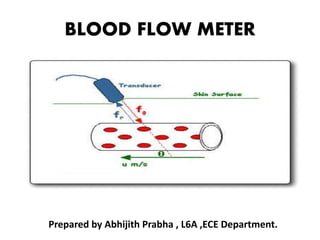
- 1. BLOOD FLOW METER Prepared by Abhijith Prabha , L6A ,ECE Department.
- 2. INTRODUCTION • Blood flow is the one of the important physiological parameter and the most difficult to measure accurately. • The average velocities of blood flow vary over a wide range depending on diameter of blood vessel. • There are many techniques for measuring the blood flow and velocity. • They are categorized into 1.invasive(surgical). 2.non invasive(through the skin).
- 3. Need for blood flowmeter • Inspection for block in blood flow. • Testing artificial blood vessels during organ transplantation. • During Fistula creation in dialysis.
- 4. Typical values of blood flow Type of blood vessels Total cross-section area Blood velocity in cm/s Aorta 3–5 cm2 40 cm/s Capillaries 4500–6000 cm2 0.03 cm/s Vena cava inferior and superior 14 cm2 15 cm/s
- 5. Introduction • Most widely used blood flow meters are: Electromagnetic blood flow meter Ultrasonic blood flow meters NMR blood flow meter Laser Doppler blood flow meter
- 7. ELECTROMAGNETIC BLOOD FLOWMETER • Measures instantaneous pulsatile flow of blood • Works based on the principle of electromagnetic induction • The voltage induced in a conductor moving in a magnetic field is proportional to the velocity of the conductor • The conductive blood is the moving conductor
- 8. Principle of EM blood flow meter.
- 9. ELECTROMAGNETIC BLOOD FLOWMETER • A permanent magnet or electromagnet positioned around the blood vessel generates a magnetic field perpendicular to the direction of the flow of the blood. • Voltage induced in the moving blood column is measured with stationary electrodes located on opposite sides of the blood vessel and perpendicular to the direction of the magnetic field. • This method requires that the blood vessel be exposed so that the flow head or the measuring probe can be put across it.
- 10. • The Induced emf • Where • B = magnetic flux density, T • L = length between electrodes, m • u = instantaneous velocity of blood, m/s
- 11. Fig: Typical flow transducer • The electromagnetic flow-transducer is a tube of non-magnetic material to ensure that the magnetic flux does not bypass the flowing liquid and go into the walls of the tube.
- 12. Types of EM Flowmeters • basically, all modern flowmeters consist of a generator of AC, a probe assembly , a series of capacitance coupled amplifiers, a demodulator, a DC amplifier and a suitable recording device. • Basing shape of the energizing current waveform for the electromagnet 2 types of EM Flowmeters are : A. Sine wave flometer. B. Square wave flowmeter.
- 13. Sine wave Flowmeters • Probe magnet is energized with a sine wave and the induced voltage will also be sinusoidal. • Since the flow of blood acts as a secondary terminal of a transformer w.r.t probe magnet, an additional artifact voltage induced is called transformer voltage. • This voltage is 900 out of phase with the original signal corresponding to flow of blood . • A method for eliminating transformer voltage by using a gated amplifier(amplify signal only during flow induced voltage is maximum). • This type of instrument is known as ‘gated sine wave flowmeter’ ,
- 14. Square wave flowmeter • Probe magnet is energized with a square wave and induced voltage is also square wave. • It is easier to control magnitude and wave shape of energizing current. • Separation of transformer voltage is easy . • For the measurement action square wave is amplitude modulated by variation in blood flow.
- 15. Block diagram of square wave flowmeter
- 16. Block diagram Transducer • consist of an electromagnet, a pair of electrodes. • Electrodes may be in contact with either flowing blood or outer surface of the blood vessel Preamplifier • The induced voltage pick up by the electrodes, is given to a low noise differential amplifier through a capacitive coupling • Must have a very high CMRR and input impedance
- 17. Block diagram Gating circuit • It helps to remove spurious voltages generated during magnet current reversal • The gating action is controlled by the circuit which provides an excitation current to the electromagnet Band pass filter • It is an active RC band pass amplifier , which selectively pass through it the amplified square wave signal • Peak response is kept for 400Hz
- 18. Block diagram Detector • A phase sensitive detector is used to recover the signal • It also helps in the rejection of interfering voltages at frequencies below the carrier frequency Low pass filter and output stage • Demodulated signal is given to an RC LPF , which provides a uniform frequency response and a linear phase shift • Followed by an integrator provide output corresponding to the mean flow
- 19. Block diagram Magnet current drive • The square wave input to the power amplifier stage which supplies current to the electromagnet is fed from a free running multivibrator Zero flow reference line • it establish the signal corresponding to zero-flow before measurement.
- 21. Ultrasonic blood flowmeter • An ultrasonic flow meter is a type of flow meter that measures the velocity of a fluid with ultrasound to calculate volume flow. • Using ultrasonic transducers, the flow meter can measure the average velocity along the path of an emitted beam of ultrasound. • By averaging the difference in measured transit time between the pulses of ultrasound propagating into and against the direction of the flow or by measuring the frequency shift from the Doppler effect.
- 22. Fig : ultra sonic flow meter
- 23. (a)Doppler-shift flow-velocity meters • It is an non invasive method. • It is based on the analysis of echo signals from erythrocytes(RBCs) in blood. • The incident ultrasound is scattered by the blood cells and scattered wave is received by the second receiver. • The frequency shift of the scattered wave gives idea about velocity of scatterers. • The Doppler frequency shift is a measure of size and direction of the flow velocity.
- 24. Doppler-shift flow-velocity meters 5 MHz exciter Rx Tx R F amplifierdetector LPF Audio amplifier Zero crossing detector LPF Blood vessel Fig : block diagram output
- 25. (b)Range gated pulsed Doppler flow meter • Baker(1970) stated that recording blood flow using Doppler shift are sometimes misleading and inaccurate. • These difficulties can be overcome , if the ultrasonic source is pulsed. • If the returning signal is range gated ,then the diameter and velocity of blood stream can measure together.
- 26. Receiver limiter Phase detector LPF Sample and amplifier Power amp Pulse amp Sample pulse gen. LPF Freq division Master Oscillator (4.5-5.5 MHz) Rx element Tx element Audible Doppler output Fig : block diagram of pulsed doppler flow meter.
- 27. (c) Flow measurement by Doppler Imaging • Doppler ultrasound is not only used for measurement of the absolute value of blood velocity and volume, but it also helps to visualize blood flow. • The probe this imaging equipment is mechanically coupled to position resolver, which gives electrical output. • Imaging is done by moving the probe through the skin, and developing a 3D image using a computer. • Thus, it is possible to construct anterior-posterior , lateral and cross-sectional scans of blood vessel.
- 28. Flow measurement by Doppler Imaging Figs : Flow measurement by Doppler Imaging
- 29. Flow measurement by Doppler Imaging • Using this recording of mean blood flow, peak flow, reverse peak flow etc. of cardiac cycle are possible. • This method is also helpful for taking measurements from brain , which is difficult to access.
- 31. NMR Blood Flowmeter • Non invasive method by Nuclear magnetic resonance principle. • For flow measurement , behaviour of the 2 Hydrogen atoms of water is studied.(Since blood is approximately 83% water). • Due to magnetic moment of the H atom , the nucleus behaves as a micro miniature magnet which can affected by externally applied magnetic field. • the hydrogen nuclei orient themselves to produce a net parallel alignment to a steady magnetic field.
- 32. NMR Blood Flowmeter • Orientation of H atoms will be change according to the status of blood flow. • This orientation will results in the magnetization of H atoms. • Therefore, magnitude of magnetization can be related to either velocity of blood or flow rate. • A crossed coil configuration is used to detect the level of magnetization in body. • The voltage induced in coil(NMR signal) is proportional to velocity and area of vessel carrying blood. • NMR flow meters are limited in their application to the measurement in limbs.
- 33. Laser Doppler blood flowmeter
- 34. Fig : schematic diagram of laser doppler blood flow meter
- 35. THE END