
Recomendados
Mais conteúdo relacionado
Semelhante a chapter6-themuscularsystem-151207133246-lva1-app6891.pdf
Semelhante a chapter6-themuscularsystem-151207133246-lva1-app6891.pdf (20)
Mais de VeenaMoondra
Mais de VeenaMoondra (20)
Último
Último (20)
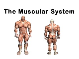
chapter6-themuscularsystem-151207133246-lva1-app6891.pdf
- 2. Did you know that ? - more than 50% of body weight is muscle ! - And muscle is made up of proteins and water
- 4. The Muscular System • Muscles are responsible for all movement of the body • There are three basic types of muscle – Skeletal – Cardiac – Smooth
- 5. Info About Muscles • Only body tissue able to contract • create movement by flexing and extending joints • Body energy converters (many muscle cells contain many mitochondria)
- 6. 3 Types of Muscles
- 7. Three types of muscle Skeletal Cardiac Smooth
- 8. Classification of Muscle Skeletal- found in limbs Cardiac- found in heart Smooth- Found in viscera Striated, multi- nucleated Striated, 1 nucleus Not striated, 1 nucleus voluntary involuntary involuntary
- 9. Characteristics of Muscle • Skeletal and smooth muscle are elongated • Muscle cell = muscle fiber • Contraction of a muscle is due to movement of microfilaments (protein fibers) • All muscles share some terminology – Prefixes myo and mys refer to muscle – Prefix sarco refers to flesh
- 10. Shapes of Muscles • Triangular- shoulder, neck • Spindle- arms, legs • Flat- diaphragm, forehead • Circular- mouth, anus
- 12. Skeletal Muscle • Most are attached by tendons to bones • Cells have more than one nucleus (multinucleated) • Striated- have stripes, banding • Voluntary- subject to conscious control • Tendons are mostly made of collagen fibers • Found in the limbs • Produce movement, maintain posture, generate heat, stabilize joints
- 13. Structure of skeletal muscle • Each cell (fibre) is long and cylindrical • Muscle fibres are multi-nucleated • Typically 50-60mm in diameter, and up to 10cm long • The contractile elements of skeletal muscle cells are myofibrils
- 14. Skeletal muscle - Summary • Voluntary movement of skeletal parts • Spans joints and attached to skeleton • Multi-nucleated, striated, cylindrical fibres
- 15. Smooth Muscle • No striations • Spindle shaped • Single nucleus • Involuntary- no conscious control • Found mainly in the walls of hollow organs
- 16. Smooth muscle • Lines walls of viscera • Found in longitudinal or circular arrangement • Alternate contraction of circular & longitudinal muscle in the intestine leads to peristalsis
- 17. Smooth Muscle
- 18. Structure of smooth muscle • Spindle shaped uni-nucleated cells • Striations not observed • Actin and myosin filaments are present( protein fibers)
- 19. Smooth muscle - Summary • Found in walls of hollow internal organs • Involuntary movement of internal organs • Elongated, spindle shaped fibre with single nucleus
- 20. Cardiac Muscle • Striations • Branching cells • Involuntary • Found only in the heart • Usually has a single nucleus, but can have more than one
- 21. Cardiac muscle • Main muscle of heart • Pumping mass of heart • Critical in humans • Heart muscle cells behave as one unit • Heart always contracts to it’s full extent
- 22. Structure of cardiac muscle • Cardiac muscle cells (fibres) are short, branched and interconnected • Cells are striated & usually have 1 nucleus • Adjacent cardiac cells are joined via electrical synapses (gap junctions) • These gap junctions appear as dark lines and are called
- 23. Cardiac muscle - Summary • Found in the heart • Involuntary rhythmic contraction • Branched, striated fibre with single nucleus and intercalated discs
- 24. Muscle Control Type of muscle Nervous control Type of control Example Skeletal Skeletal Controlled by CNS Voluntary Lifting a glass Cardiac Regulated by ANS Involuntary Heart beating Smooth Controlled by ANS Involuntary Peristalsis
- 25. Types of Responses • Twitch- – A single brief contraction – Not a normal muscle function • Tetanus – One contraction immediately followed by another – Muscle never completely returns to a relaxed state – Effects are compounded
- 26. Where Does the Energy Come From? • Energy is stored in the muscles in the form of ATP • ATP comes from the breakdown of glucose during Cellular Respiration • This all happens in the Mitochondria of the cell • When a muscle is fatigued (tired) it is unable to contract because of lack of Oxygen
- 27. Fast Twitch and Slow Twitch Fibers Fast Twitch vs Slow Twitch
- 28. Exercise and Muscles • Isotonic- muscles shorten and movement occurs ( most normal exercise) • Isometric- tension in muscles increases, no movement occurs (pushing one hand against the other)
- 29. How are Muscles Attached to Bone? • Origin- attachment to immovable bone • Insertion- attachment to a movable bone • Muscles are always attached to at least 2 points • Movement is attained due to a muscle moving an attached bone
- 31. Muscle Attachments • The origin is on the clavicle and sternum. • The insertion is on the skull. • When the muscle contracts it will shorten the distance between the origin and insertion. • The head will move when this muscle contracts.
- 32. Flexion Types of Musculo-Skeletal Movement
- 33. Extension
- 34. Hyperextension
- 36. Rotation
- 37. More Types of Movement…… • Inversion- turn sole of foot medially • Eversion- turn sole of foot laterally • Pronation- palm facing down • Supination- palm facing up • Opposition- thumb touches tips of fingers on the same hand
- 38. The Skeletal Muscles There are about 650 muscles in the human body. They enable us to move, maintain posture and generate heat. In this section we will only study a sample of the major muscles.
- 40. Sternocleidomastoideus • Sometimes called the sternocleitomastoid. • It is the same neck muscle shown on the previous slide. • This muscle has two origins. – The first origin is on the sternum manubrium. – The second origin is on the clavicle. • The insertion is on the mastoid process of the skull. • Contraction of both sternocleidomastoideus muscles will flex the head. If just one of the muscles contracts, the head will rotate.
- 42. Masseter • The masseter is one of major chewing muscles. • The origin of the masseter is on the zygomatic arch. • The insertion is on the mandible. • Contraction of the masseter will elevate the jaw.
- 43. Temporalis Elevate & Retract Mandible
- 44. Temporalis • The temporalis is another chewing muscle. – Note how it attaches on the side of skull. • It also elevates the mandible. • You do not need to know the insertions and origins for this muscle
- 45. Trapezius Extend Head, Adduct, Elevate or Depress Scapula
- 46. Trapezius • The trapezius is a large muscle in the upper back. • It attaches to the skull, shoulder and vertebrae of the back. • When this muscle contracts it will cause the head to extend. • It will also move the scapula. • The direction the scapula moves depends on which part of the trapezius contracts. • The trapezius may elevate or depress the scapula.
- 47. Latissimus Dorsi Extend, Adduct & Rotate Arm Medially
- 48. Latissimus Dorsi • The latissimus dorsi is a large muscle in the back. – It is often referred to as a lat. • It has origins on the vertebrae, ilium ribs and scapula. • The insertion is on the humerus. – When it contracts it moves the humerus. • It can extend, adduct and rotate the arm medially. • This is the main muscle used in movement such as pounding a nail with a hammer.
- 49. Deltoid Abduct, Flex & Extend Arm
- 50. Deltoid • The deltoid covers the shoulder and has the shape of a delta. • It has origins on the scapula and clavicle. – The deltoid inserts on the deltoid tuberosity of the humerus. • Contraction of the deltoid will adduct the arm. • If only the anterior fibers of the muscle contract it will flex the arm. • Contraction of the posterior fibers will extend the arm.
- 51. Pectoralis Major Flexes, adducts & rotates arm medially
- 52. Pectoralis Major • The pectoralis major is a large muscle in the pectoral region of the body. • It has origins on the clavicle and sternum. – The insertion is on the greater tubercle of the humerus. • Contraction of the pectoralis major will flex the arm. • It will also adduct and rotate the arm medially. • The pectoralis major is used in movements such a climbing, throwing and doing pushups.
- 53. Biceps Brachii Flexes Elbow Joint
- 54. Biceps Brachii • The biceps brachii is located on the anterior side of the upper arm. • It is often just called the biceps. – There is a biceps femoris in the leg we will study shortly. • The biceps has two origins. One origin is on the corocoid process and the other on the Glenoid cavity of the scapula. • The “bi” in biceps refers to the two origins. – It inserts on the radial tuberosity. • Contraction of the biceps will cause flexing at the elbow joint.
- 55. Triceps Brachii Extend Elbow Joint
- 56. Triceps Brachii • The triceps is on the back of the upper arm. • It has three origins. • Two origins are on the back of the humerus and one on the scapula. • The triceps inserts on the olecranon. • Movement of the triceps will extend the elbow joint.
- 58. Rectus Abdominus • Rectus abdominus is a long muscle in the abdomen. • The muscle originates on the pubis. • It inserts on the xiphoid process of the sternum and also on cartilage of the ribs. • When rectus abdominus contracts it will flex the abdomen.
- 60. External Oblique • Another muscle in the abdomen is the external oblique. • It has muscle fibers that run in an oblique direction across the abdomen. • Contraction of the external oblique will compress the abdomen.
- 62. External Intercostals • There are two groups of muscles that run between the ribs. • The first are the external intercostals. • They will elevate the ribs.
- 64. Internal Intercostals • The internal intercostals are also located between the ribs. • They will depress the ribs.
- 66. Diaphragm • This is an inferior view of the diaphragm. • This muscle separates the abdominal cavity from the thoracic cavity. • When it contracts it will cause inspiration.
- 67. Forearm Muscles
- 68. Forearm Muscles • Flexor carpi—Flexes wrist • Extensor carpi—Extends wrist • Flexor digitorum—Flexes fingers • Extensor digitorum—Extends fingers • Pronator—Pronates • Supinator—Supinates
- 69. Gluteus Maximus Extends & Rotates Thigh Laterally
- 70. Gluteus Maximus • The large muscle on the posterior side of the body at the top of each leg is the gluteus maximus. • The gluteus maximus originates on the ilium, sacrum and coccyx. • It inserts on the gluteal tuberosity of the femur. • This muscle will extend and rotate the thigh laterally.
- 71. Rectus Femoris Flexes Thigh, Extends Lower Leg
- 72. Rectus Femoris • Rectus femoris is located on the anterior side of the thigh. • It originates on the ilium. • The insertion is on the patella and the tibial tuberosity. • When rectus femoris contracts it will flex the thigh and extend the lower leg.
- 73. Gracilis Adducts and Flexes Thigh
- 74. Gracilis • The gracilis is on the medial side of the thigh. • It adducts and flexes the thigh.
- 75. Sartorius Flexes Thigh, & Rotates Thigh Laterally
- 76. Sartorius • Sartorius is a long, strap like muscle. • It originates on the anterior superior iliac spine of the ilium. • The insertion is on the medial side of the tibia. • Contraction of the sartorius flexes the thigh and rotates the thigh laterally. • This is the muscle used when crossing the legs to sit on the floor.
- 77. Biceps Femoris Extends Thigh & Flexes Lower Leg
- 78. Biceps Femoris • Biceps femoris is one of the hamstring muscles. • The origin is on the ischial tuberosity. • Biceps femoris inserts on the tibia and fibula. • This muscle extends the thigh and flexes the lower leg.
- 79. Gastrocnemius Plantar Flexes Foot & Flex Lower Leg
- 80. Gastrocnemius • Gastrocnemius is commonly called the calf muscle. • It originates on the distal end of the femur. • The insertion is on the calcaneus bone of the foot. • It will cause plantar flexion of the foot and also flex the lower leg.
- 81. Tibialis Anterior Dorsiflexes and Inverts Foot
- 82. Tibialis Anterior • Tibialis anterior is located on the anterior side of the tibia. • It will dorsiflex and invert the foot.