
Recomendados
Mais conteúdo relacionado
Mais procurados
Mais procurados (20)
Semelhante a Nervous system (neuron & ion channels)
Semelhante a Nervous system (neuron & ion channels) (20)
Mais de Rohit Paswan
Mais de Rohit Paswan (20)
Último
Último (20)
Nervous system (neuron & ion channels)
- 3. Introduction -Nervous system controls all the activities of the body. -It is quicker than the other control system in the body namely endocrine system. -Primarily, the nervous system is divided into two parts. -Central nervous system -Peripheral nervous system
- 4. CNS • CNS includes Brain & Spinal Cord. • It is formed by neurons and the supporting cells called neuroglial cells. • The structures of brain and spinal cord are arranged in two layers. -Gray matter is formed by nerve cell bodies -White matter is formed by nerve fibers.
- 7. Central Nervous System (CNS) • brain • spinal cord Peripheral Nervous System (PNS) -PNS is formed by neurons and their processes present in all regions of the body. It consists of • cranial nerves (12 pr) • spinal nerves (31 pr)
- 8. CNS PNS
- 9. sensory receptor sensory input integration motor input effector
- 11. Neuroglia (glia) are cells that support and protect neurons. The following four neuroglia are found in the CNS: •Astrocytes have numerous processes that give the cell a star-shaped appearance. Astrocytes maintain the ion balance around neurons and control the exchange of materials between blood vessels and neurons. They are part of the BBB •Oligodendrocytes have fewer processes than astrocytes. They wrap these cytoplasmic processes around neurons to create an insulating barrier called a myelin sheath. •Microglia are phagocytic macrophages that provide a protective function by engulfing microorganisms and cellular debris. •Ependymal cells line the fluid-filled cavities of the brain and spinal cord. Many are ciliated. They make CSF Read more: http://www.cliffsnotes.com/WileyCDA/CliffsReviewTopic/Neuroglia.topicArticleId- 22032,articleId-21933.html#ixzz0YK3TiUTl
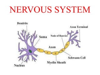
- 12. NEURON • Neuron is defined as the structural and functional unit of the nervous system. • It is like any other cell in the body having nucleus and all the organelles in cytoplasm. • Neuron is different from other cell by two ways: 1. Neuron has branches or process called axon & dendrites. 2.Neuron does not have centrosome so it does not undergo division.
- 14. Structure of Neuron • Neuron is made up of three parts: -Nerve cell body -Dendrites - Axon Nerve cell body -Nucleus - Nissl granules -Neurofibrils - Mitochondria -Golgi Apparatus Dendrites Axon
- 16. Schwann Cells Axon Nodes of Ranvier
- 17. bipolar eye, ear, & olfactory unipolar multipolar most abundant type in CNSDorsal root ganglion cells
- 18. Protein channels Tube shaped channels extend in the cell membrane from extracellular to the intracellular ends. Properties Selective permeability Gating mechanism Selective permeability Permit only one type of ion to pass through it. Due to characteristics of channel itself such as diameter, shape & nature of charge on it. E.g. Sodium channels Potassium channels
- 19. Gating mechanism Equipped with gate like extensions . Controlled by 3 principal pathways • Voltage gated channel Responds when there is change in the electrical potentials. E.g . Ca channels in NMJ. • Ligand gated channels (chemical gating) Extracellular ligands- are first messenger E.g . Acetylcholine . Intracellular ligands are second messenger E.g . Ca+, cAMP, G protein. • Mechanically gated channels Opening of channel by mechanical stretch. E.g. pressure receptors, receptor cell of organ of corti
- 20. sodium-potassium pump • The sodium-potassium pump is a protein complex that continually pumps three sodium ions out of the cells while drawing two potassium ions into the cell. – helps to maintain the electrical gradient. • The electrical gradient and the concentration gradient work to pull sodium ions into the cell. • The electrical gradient tends to pull potassium ions into the cells.
- 21. Fig. 2-15, p. 41 Figure 2.15: The sodium and potassium gradients for a resting membrane. Sodium ions are more concentrated outside the neuron; potassium ions are more concentrated inside. Protein and chloride ions (not shown) bear negative charges inside the cell. At rest, very few sodium ions cross the membrane except by the sodium- potassium pump. Potassium tends to flow into the cell because of an electrical gradient but tends to flow out because of the concentration gradient.
- 22. Carrier protein consists of 2 subunits- a) α subunit b) β subunit α subunit – concerned with Na and K transport whereas function of β subunit is unknown. α subunit has 6 sites- I. 3 receptor sites for binding Na ions. II. 2 receptor sites for K ions. III. 1 receptor site for binding of enzyme ATPase. Na+-k+ pump
- 23. Mechanism of Na+–K+ pump • Enzymes ATPase • 3 Na and 2 K bind to the respective receptor site of carrier protein. • Activates enzyme ATPase. • Breakdown of ATP in to ADP – relaese of one high energy phosphate. • Conformational change in the carrier protein – extruding Na into ECF and K into cytoplasm.
- 24. Na+– k+ pump K+ is released and Na+ sites are ready to bind Na+ again; the cycle repeats. Binding of cytoplasmic Na+ to the pump protein stimulates phosphorylation by ATP. Phosphorylation causes the protein to change its shape. The shape change expels Na+ to the outside, and extracellular K+ binds. K+ binding triggers release of the phosphate group. Loss of phosphate restores the original conformation of the pump protein. Extracellular fluid Cytoplasm 1 2 3 4 5 Concentration gradients of K+ and Na+
- 25. • Functions of Na+– K+ pump - Controlling cell volume - Electrogenic activity • Regulation of Na+ – K+ pump Increased by – cAMP, DAG, Thyroid hormone, Aldosterone, Insulin. inhibited by – Low temp. , Lack of O2, dopamine and digitalis. Ca++ Pump Calcium pump helps in maintaining extremely low conc. Of Ca in ICF. E.g. 1) cell membrane- extrudes Ca out of cell.
- 26. K+- H+ PUMP • Operates through K+-H+ ATPase activity. • Present at 2 places in human body- 1) Parietal cells of gastric gland – H+ Is actively transported into lumen of gastric gland and K+ in to the cell. 2) Renal tubules – located at DCT and collecting ducts. Excretes large amount of H+ in to the tubules to control blood pH.
- 27. Resting Membrane Potential(RMP) • It is electrical potential difference (voltage) across the cell membrane (between inside and outside of cell) under resting condition. • It is also called membrane potential/ transmembrane potential/ transmembrane potential difference or transmembrane potential gradient. • RMP is measured by cathode ray oscilloscope. • There is negativity inside and positivity outside the membrane.
- 28. Fig. 2-13, p. 40 Figure 2.13: Methods for recording activity of a neuron. (a) Diagram of the apparatus and a sample recording. (b) A microelectrode and stained neurons magnified hundreds of times by a light microscope.
- 29. Action potential • Action potential is created in excitable cell membrane . • It is electrochemical fluctuation in excitable cell membrane which spreads over membrane and act as signaling mechanism in our body. CHANGES are: Electrical changes Chemical changes