Recombinant DNA technology (Immunological screening)
December Highlights
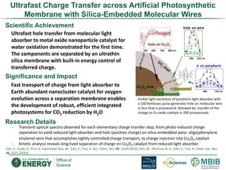
1. Scientific Achievement
Ultrafast hole transfer from molecular light
absorber to metal oxide nanoparticle catalyst for
water oxidation demonstrated for the first time.
The components are separated by an ultrathin
silica membrane with built-in energy control of
transferred charge.
Significance and Impact
Fast transport of charge from light absorber to
Earth abundant nanocluster catalyst for oxygen
evolution across a separation membrane enables
the development of robust, efficient integrated
photosystems for CO2 reduction by H2O
Research Details
Visible light excitation of porphyrin light absorber with
a 150 femtosec pulse generates hole on molecular wire
in less than a picosecond, followed by transfer of the
charge to Co oxide catalyst in 200 picoseconds
Edri, E.; Guldi, D.; Frei, H. Submitted. Kim, W.; Edri, E.; Frei, H. Acc. Chem. Res. 49, 1634 (2016). Kim, W.; McClure, B. A.; Edri, E.; Frei, H. Chem. Soc. Rev.
46, 3221 (2016).
Ultrafast Charge Transfer across Artificial Photosynthetic
Membrane with Silica-Embedded Molecular Wires
- Transient optical spectra observed for each elementary charge transfer step, from photo-induced charge
separation to yield reduced light absorber and hole (positive charge) on silica-embedded para- oligo(phenylene
vinylene) wire that accomplishes tightly controlled charge transport, to charge injection into Co3O4 catalyst
- Kinetic analysis reveals long lived separation of charge on Co3O4 catalyst from reduced light absorber.
2. Evolution of an atypical VDE in green algae
Li et al. (2016) Nature Plants 2, 16140.
Scientific Achievement
Using a genetic and biochemical approach, we
identified a novel green algal violaxanthin
de-epoxidase (CVDE).
Significance and Impact
CVDE can be used to alter photoprotection
and improve photosynthesis in plants.
Research Details
– VDE is involved in regulating photosynthetic light
harvesting in all plants and most eukaryotic algae.
Surprisingly, some green algae including
Chlamydomonas have an atypical VDE (CVDE) that
is unrelated to the VDE found in plants.
– CVDE can replace the function of the typical VDE
in plants, despite being located on the opposite
side of the photosynthetic membrane.
– This finding raises interesting questions about the
regulation and structure of CVDE.
Functional expression of the
green algal CVDE protein in
Arabidopsis affects NPQ and
zeaxanthin accumulation.
3. Tracking Excitation Flow Through LHCII
Scientific Achievement
We demonstrated that two-dimensional electronic-
vibrational spectroscopy (2DEV) can track the flow
of excitation energy through light harvesting
complexes using local vibrations as proxies for
position. 2DEV spectroscopy was developed in our
laboratory and is currently a unique capability.
Significance and Impact
We have shown that it is possible to directly follow
the excitation energy using an experimental
approach that does not require a model.
Research Details
– 2DEV correlates the electronic excitation energy
with the vibrational degrees of freedom of a
system.
– By using localized, assignable vibrations we can
identify where in the complex the excitation is
located.
Lewis, N. H. C. et al. J. Phys. Chem. Lett. 4197–4206 (2016).
The four singular vectors that describe the 2DEV data.
The fastest time constant is 50fs. Note the clear
intermediate state (purple trace).
4. Biological Nanomachine Architecture and Designed Function
with SAXS through DOE/BER:IDAT
“Designing and defining dynamic protein cage
nanoassemblies in solution” Science Advances Dec 14th 2016
IDAT: Greg L. Hura & John A. Tainer collaboration with
UCLA-DOE Institute: Yen Ting Lai and Todd O. Yeates
Emergent DOE/BER methodologies for
using SAXS to inform on protein design
5. Structure and Mechanisms of Complex Biological Pathways
with Combined X-ray Methods through DOE/BER:IDAT
Polycomb repressive complex 2 structure with inhibitor reveals a mechanism of
activation and drug resistance NATURE COMMUNICATIONS Industrial
Collaborators: Pfizer FEB 10 2016
Scaling Atomic to Cellular Distances
for Microbial Epigenetic Control
Cellular X-ray Tomography
Mutant 1
Mutant 2
Hammel M, Larabell C. Tainer JA et al
Science Advances July 2016
Molecular architecture of the human sperm IZUMO1 and egg
JUNO fertilization complex
NATURE By: Aydin, Halil; et al. JUN 23 2016
Biology requires complex
protein and DNA
assemblies that can only
be characterized by
multiple techniques at
multiple scales
An Intrinsically Disordered APLF Links Ku, DNA-
PKcs and XRCC4-DNA Ligase IV in an Extended
Flexible Non-Homologous End Joining Complex.
JBC By:Hammel M, Tainer JA, et al NOV 14 2016
Notas do Editor
Ultrafast photo-induced charge transfer (holes) from a molecular light absorber (free-base porphyrin) to a robust Earth abundant metal oxide catalyst for water oxidation (Co3O4) was achieved for the first time. The fast transfer is enabled by linkage of light absorber and catalyst by molecular wires embedded in a 2 nm thin, dense phase silica layer (p-oligo(phenylene vinylene) featuring 3 aryl units). Elementary charge transfer steps were detected by ultrafast optical absorption spectra of transient intermediates. Visible light induced charge injection into the wire molecule is complete within less than one picosecond as monitored by the characteristic absorption spectra of a hole residing on the wire molecule (500-600 nm) and the concurrently generated reduced light absorber (700 nm), shown in the Figure. Subsequent forward transfer of the charge from the wire to the Co oxide nanoparticle catalyst occurs in 200 ps (bleach at 485 nm), exceeding previously reported hole transfer rates from anchored molecular light absorber to metal oxide nanoparticle catalyst by six orders of magnitude. The experiments were conducted with aqueous colloidal solutions of spherical Co3O4-SiO2/wire nanoparticles.
Ultrafast hole transfer from molecular light absorbers to metal oxide catalysts for water oxidation is essential for achieving high quantum efficiency of photo-driven water oxidation. The fast transfer is achieved here under separation of light absorber and oxygen-evolving catalyst by a 2 nm thick silica membrane that we have shown to function as proton conducting, O2 impermeable membrane. Co oxide-silica core-shell constructs in the form of open nanotubes (assembled as square inch sized core-shell nanotube arrays) currently developed in our lab under this program offer a geometry for complete artificial systems for closing the photosynthetic cycle for CO2 reduction by H2O on the nanoscale under membrane separation of the products.
Plants, algae and cyanobacteria need to regulate photosynthetic light harvesting in response to the constantly changing light environment. Rapid adjustments are required to maintain fitness because of a tradeoff between efficient solar energy conversion and photoprotection. The xanthophyll cycle, in which the carotenoid pigment violaxanthin is reversibly converted into zeaxanthin, is ubiquitous among green algae and plants and is necessary for the regulation of light harvesting, protection from oxidative stress, and adaptation to different light conditions. Violaxanthin de-epoxidase (VDE) is the key enzyme responsible for zeaxanthin synthesis from violaxanthin under excess light. Here we show that the CVDE gene from the model green alga Chlamydomonas reinhardtii encodes an atypical VDE. This protein is not homologous to the VDE found in plants and is instead related to a lycopene cyclase from photosynthetic bacteria. Unlike the plant-type VDE that is located in the thylakoid lumen, the Chlamydomonas CVDE protein is located on the stromal side of the thylakoid membrane. Phylogenetic analysis suggests that CVDE evolved from an ancient de-epoxidase that was present in the common ancestor of green algae and plants, providing evidence of unexpected diversity in photoprotection in the green lineage.
Time resolved electronic spectroscopy, particularly two-dimensional Fourier transform spectroscopy is capable of tracking the evolution in time of the energy levels of a complex system. For example in a photosynthetic light harvesting complex that has a set of exciton levels, 2D electronic spectroscopy can track the relaxation from higher lying excitons to the lowest excited energy levels of the complex. Converting the resultant picture to relaxation in real space – how the excitation moves through the complex – requires an accurate model of the electronic structure of the complex, a highly complex undertaking of limited accuracy. Yet a picture in real space is surely what is wanted to relate the protein structure to function and to communicate the results to broader audiences. Accordingly we sought a method that would provide a model free mapping of the temporal evolution of an exciton manifold directly into the spatial domain. We developed the two-dimensional electronic vibrational (2DEV) spectroscopy method partly for this purpose. Because chlorophyll vibrations have frequencies sensitive to the specific site in the protein in which they are bound, but not particularly sensitive to the excitation of the chlorophyll, by combining electronic excitation with mid infrared detection in a 2D experiment we have the local tag required. The slide shows our first photosynthetic application of this method to LHCII. N. Lewis et al. J Phys Chem Lett, 7, 4197-4206 (2016)
The DOE/BER funded IDAT (Integrated Diffraction Analysis Technologies) program supports combining two biophysical techniques of SAXS (small angle X-ray scattering) and MX (Macromolecular Crystallography). When IDAT started SAXS was a niche technique but because we made use of significant and wide spread innovation in MX for SAXS, we have made SAXS accessible to hundreds of groups. A recently demonstrated “killer app” of SAXS is in the realm of macromolecular engineering. In Science, Nature and other journals this year SAXS we and IDAT supported users are creating novel structures with profound impact. Many constructs are also being patented with SAXS data. An example is the large (600kDa) self- assembling pyramid that expands, collapses or disassembles in response to solution conditions. We will build off of this structure to carry payloads for a variety of applications.
The joint application of SAXS, MX and other technologies is at the core of IDAT. It is well recognized that biology spans many scales. By combining MX and SAX we span the atomic to intra-cellular scale as demonstrated in many publications where IDAT collaborated with users. We have gone further and added X-ray Tomography with another synchrotron beamline (BER funded NCXT) to visualize the impact of changes to one protein in bacteria. The protein, HU, is modular in that it can either bind to an identical copy of itself forming an HU or it can bind to a different form making HU. These two forms appear during different stages of bacterial growth, creating different degrees of compaction in its DNA. We can visualize this compaction and atomically identify the mechanism using the combined techniques.