Whole brain optical imaging
•Transferir como PPTX, PDF•
2 gostaram•3,349 visualizações

Presentation by Leonardo Sacconi, LENS, at RDA National event in Florence, Italy, 14-15 November 2016

Recomendados
Recomendados
Mais conteúdo relacionado
Mais procurados
Mais procurados (19)
Semelhante a Whole brain optical imaging
Semelhante a Whole brain optical imaging (20)
Mais de Research Data Alliance
Mais de Research Data Alliance (20)
Último
Último (20)
Whole brain optical imaging
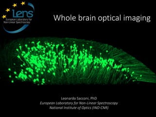
- 1. Whole brain optical imaging Leonardo Sacconi, PhD European Laboratory for Non-Linear Spectroscopy National Institute of Optics (INO-CNR)
- 4. The investigation of the brain requires several challenges in imaging technology, since brain activity spans many orders of magnitude both spatially (from nanometers to centimeters) and temporally (from milliseconds to months). Thanks to their flexibility in terms of spatial and temporal resolution optical methodologies have become very useful in neuroscience research. Whole mouse brain cm scale Pyramidal neuron mm scale Synapses μm scale Optical techniques to explore the brain
- 5. Light sheet microscopy (LSM) Key advantages: from J. Huisken and D. Y. Stainier (Development, 2009) Optical sectioning with wide field detection scheme Fast high resolution 3D imaging Optical sectioning with low-NA optics (having longer WD) Imaging of large specimens without sample sectioning. Only the observed plane is illuminated Reduced photobleaching.
- 6. Traditional clearing protocols are based on the substitution of water with a refractive-index- matching liquid, as Benzyl Alcohol/Benzyl Benzoate (BABB) or Dibenzyl Ether (DBE) [Becker et al. 2012]. Chemical clearing of entire brains Silvestri et al. JoVe 2013. In large specimens as whole mouse brains, a substantial amount of light scattering persist even after clearing. This expands the illumination beam, leading to out-of-focus background fluorescence and blurring of images.
- 7. We recently developed a new implementation of confocal slit detection (confocal light sheet microscopy, CLSM) in which the out-of-focus background rejection is assured by a spatial filter. Confocal light sheet microscopy (CLSM) Silvestri et al. Optics Express 2012
- 8. SV CV TV 10× 10× Cerebellum from a P10 L7-GFP mouse cleared in BABB Total volume 73 mm³, voxel size 0.8×0.8×1 µm³, acquisition time ≈ 24 h (1.3 MegaVoxels/s) Whole brain imaging
- 9. PV: PV-dTomato mouse (parvalbuminergic neurons labeled) GAD: GAD-dTomato mouse (GABAergic neurons labeled) PI: propidium iodide staining (all nuclei labeled) Whole brain imaging Mulleibroich et al. Neurophotonics 2013
- 10. Correlative two-photon and light sheet microscopy
- 11. Ex vivo light sheetIn vivo reflectanceIn vivo two-photon Light sheet microscopy of mouse brains is essentially an ex vivo technique. It can be combined with in vivo two-photon imaging to gain a more comprehensive multi-scale view of the brain. To find back in the cleared brain the same field of view imaged in vivo, blood vessels can be used as a reference map. Correlative two-photon and light sheet microscopy
- 12. The same neuron imaged in vivo with two-photon microscopy can be found again in the large-scale optical tomography obtained with CLSM The side of the red cube is 100 μm Correlative two-photon and light sheet microscopy Silvestri et al. Methods 2014
- 13. Correlative two-photon and light sheet microscopy
- 15. A 2 mm thick block of a formalin- fixed tissue of a patient with hemimegalencephaly (HME), treated with passive CLARITY protocol immunostained with different antibody and cleared with 47% TDE/PBS 0 200 400 µm 600 µm 800 µm 1000 µm Human Brain Imaging Costantinie et al. Sci Report 2015
- 16. Adapted from Kasthuri and Lichtman (2007) A data flood Data production: about 5 TB per week - 10 Gb/s dedicated connection from LENS to CINECA - Connection from LENS to Juelich via CINECA (using PRACE infrastructure)
- 17. To image an entire brain many parallel stacks of images are acquired. They are subsequently merged with a custom-made software suited to work with very large data sets (~ 1 TB) Tera Stitcher ® Bria et al., BMC Bioinformatics 2012
- 18. TeraFly ® Bria et al., Nat. Meth. (2016)
- 19. Very large samples (cm-sized) Large variability of contrast in different areas TB-sized datasets Naïve methods (as thresholding or clustering) are poorly effective as usually depend on sensitive parameters Advanced methods usually requires the calculation of multiple features per each image voxel, leading to a data multiplication which is not manageable with large datasets Automatic 3D cell localization
- 20. Semantic deconvolution uses a supervised neural network, to enhance selected features of the image reducing the intensity of other structures. This method creates a more uniform image where significant structures (hence the name semantic) are well visible. In the deconvolved image, easy low- cost localization algorithms (e.g. clustering) can achieve very good performances. Original image Ideal image Deconvolved image Manualsoma localization Neural network with 2 hidden layers Supervision Only on a small subset Semantic deconvolution Frasconi et al., Bioinformatics 2014
- 21. 224’222 Purkinje cells automatically localized in the cerebellum of an L7-GFP mouse. Precision (true positives/all localized cells) is 95%, recall (true positives/all real cells) 97% ~ 1 day of computation on a 16-cores workstation to analyze 120 Gvoxels Whole brain quantitative neuroanatomy Silvestri et al., Frontiers in Neuroanatomy 2015
- 22. Acknowledgements Ludovico Silvestri Irene costantini Francesco S. PavoneAntonino Paolo Di Giovanna
Notas do Editor
- 1
- 4
- 5
- 6
- 7
- 8
- 10
- 11
- 12
- 13