Bacterial Morphology.ppt
•
0 gostou•1,300 visualizações

Bacterial Morphology.ppt by Dr. Rakesh Prasad Sah

Recomendados
Recomendados
Mais conteúdo relacionado
Mais procurados
Mais procurados (20)
Semelhante a Bacterial Morphology.ppt
Semelhante a Bacterial Morphology.ppt (20)
Mais de Dr. Rakesh Prasad Sah
Mais de Dr. Rakesh Prasad Sah (20)
Último
Último (20)
Bacterial Morphology.ppt
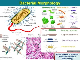
- 1. Bacterial Morphology By Dr. Rakesh Prasad Sah Associate Professor, Microbiology
- 2. Introduction • Microorganisms are living structures of microscopic in size. • Bacteria belongs to group Prokaryotes. S.No. Structure Prokaryotes Eukaryotes Nucleus 1. Nuclear membrane Absent Present 2 Nucleolus Absent Present 3 Chromosome One > One 4 Deoxyribonucleoprotein Absent Present 5 Division Binary fission Mitosis Cytoplasma 6 Mitochondria, Golgi apparatus, Lysosomes, Endoplasmic reticulum Absent Present Chemical Composition 7 Sterols Absent Present 8 Muramic acid Present Absent
- 3. SIZE OF BACTERIA • Unit for measurement : Micron or micrometer,μm: 1μm=10-3mm • Size: Varies with kinds of bacteria, and also related to their age and external environment. • Cocci: sphere, 1μm • Bacilli: rods , 0.5-1 μm in width -3 μm in length • Spiral bacteria: 1~3 μm in length and 0.3-0.6 μm in width
- 5. Bacterial Cell • Prokaryotes – No true nucleus – No organelles • Divide-binary fission
- 6. Parts of Cell • Cell Envelope – Cell wall – Cell membrane-plasma membrane, cytoplasmic membrane – Capsule • Cytoplasm – Nucleoid – Ribosomes – Granules/Inclusion bodies – Mesosomes • Spores • Plasmids • Appendages – Pilli – Flagella
- 7. • Is a tough & rigid structure surrounding the bacterium like a shell. • Weighs about 20-25% of the dry weight of the cell. • Functions – Accounts for shape of cell – Provides protection to the cell against osmotic damage. – Confers rigidity upon bacteria – Takes part in cell division. – Possesses target site for antibiotics, lysozymes and bacteriophages. – Carries bacterial antigens that are important in virulence and immunity. Cell Wall
- 8. • Rigid part of cell wall peptidoglycan mucopeptide (murein) composed of N-acetyl muramic acid and N-acetyl glucosamine alternating in chains, cross linked by peptide subunits.
- 10. Cell Wall S.N o. Character Gram Positive Gram Negative 1 Peptidoglycan Thicker (50-100 layers, 16-80nm) Thinner (1-2 layers, 2nm) 2 Teichoic acid Present Absent 3 Lipids Absent or Scanty (2-5%) Present (15-20%) 4 Periplasmic space Absent Present
- 11. Gram Positive Cell Wall • Peptidoglycan:- thicker (16-80nm) than gram negative bacteria (2nm). • Teichoic acid – Contains in a significant amount which is absent in gram negative. – Constitute major surface Ags – Two types (wall teichoic acid and lipoteichoic acid)
- 12. Special components of Gram positive cell wall Teichoic acid
- 13. Gram Negative Cell Wall • Gram negative cell wall complex structure with following components – Lipoprotein layer – Outer membrane – LPS (Lipopolysaccharide):- • constitutes endotoxin of GNB • Determine major surface Ag • Toxicity (pyrogenicity, lethal effect, tissue necrosis) – Periplasmic space – Peptidoglycan
- 14. Gram Negative Cell Wall • Lipoprotein Layer – Connects the peptidoglycan to outer membrane • Outer membrane – Contains certain proteins called as OMP (outer membrane protein). – Target sites for antibiotics and bacteriocins. • Lipopolysaccharides (LPS) – Consist of lipid A attached to a polysaccharide – Constitutes the endotoxin of GNB. • Periplasmic space – Space between inner and outer membrane – Contains various binding proteins for specific substrates. • Peptidoglycan
- 15. Special Components of Gram negative cell wall
- 16. • Rigid part of cell wall peptidoglycan mucopeptide (murein) composed of N-acetyl muramic acid and N-acetyl glucosamine alternating inchains, cross linked by peptide subunits.
- 17. Demonstration of Cell Wall • Plasmolysis – Bacteria placed in hypertonic saline, shrinkage of cytoplasm occurs while cell wall retains original shape and size. • Microdissection • Differential staining • Reaction with specific Ab • Electron microscopy
- 18. • Bacteria without cell walls or with deficient cell walls are of four types:- • Mycoplasma – Naturally occurring bacteria without cell wall. • L-forms – Kleineberger-Nobel studying Streptobacillus moniliformis in Listure institute London observed abnormal forms of the bacteria and named them L-forms after Lister institute. – Develop either spontaneously or in the +ce of penicillin due to interference with synthesis of cell wall. • Protoplast • Spheroplast Bacteria with Defective Cell Wall
- 19. Bacteria with Defective Cell Wall • Synthesis of cell wall interfered or inhibited by many factors :- – Antibiotics – Bacteriophage – Lysozyme Enzyme +nt in many tissue fluids Lyses succeptible bacteria by splitting linkage of peptidoglycan in the cell wall. Lysozyme Lysozyme Lysozyme Gram Positive Bacteria Gram Negative Bacteria In Hypertonic Solution Protoplast Spheroplast
- 20. Cytoplamsic Membrane • Is 5-10nm thick elastic semipermeable layer lies beneath cell wall separating it from cell cytoplasm. – Acts as an osmotic barrier. – Acts as a semipermeable membrane controlling the inflow and outflow of metabolites to and from the protoplasm. – Contains enzymes necessary for cell wall synthesis (cytochrome oxidase, enzymes of tricaboxylic acid).
- 21. Cytoplasm • Is a colloidal system containing variety of organic and inorganic solutes in a viscous watery solution. • Lacks – Mitochondria – Endoplasmic reticulum • Contains – 70% water of bacterial mass – Ribosomes (site for Protein synthesis) – Mesosomes (Bacterial respiration, cell wall formation and chromosome replication) – Vacuoles – Inclusions bodies (tiny particles found freely suspended and floating within the cytoplasmic matrix) – Nucleoid
- 22. Ribosomes • Centre for protein synthesis. • Are composed of – Ribosomal RNA (rRNA) and – Ribosomal proteins. Intracytoplasmic inclusions • Source of stored energy • Are grown under conditions of nutritional deficiency and disappear when deficient nutrients are supplied. • Volutin or metachromatic granules are +nt in C. diptheria. Metachromatic granules of C. diptheria by Albert Stain
- 23. Nucleoid • Bacteria don't have true nucleus and there is no nuclear membrane or nucleolus. • Nucleiod is present irregularly shaped region containing DNA. • Bacterial DNA is haploid replicates by simple binary fission and maintains bacterial genetic characteristics. • Some bacteria may possess extra-nuclear genetic material in the cytoplasm consisting of DNA named as Plasmids or episomes.
- 24. Capsule and Slime layer • Is amorphous viscid bacterial secretion surrounds outermost layer of bacteria when diffuses into surrounding medium and remains as a loose undermarcated secretion as Leuconostoc (made up of Cellulose contains Glucoses & fructose) known as “Slime layer” when it is organized into a defined structure known as “Capsule”. E.g. Streptococcus pneumoniae. • Capsule which are very thin and can not be demonstrated under light microscope are called “Microcapsule”. E.g. Neisseria meningitidis. • Is polysaccharide in nature.
- 26. Functions • Antiphagocytic in nature. • Antigenic • Virulence Demonstration of capsules • India ink staining (negative staining) • Serological method – Quellung phenomenon
- 27. Capsule (Polysaccharides) colonies are mucoid
- 28. Flagella • Cytoplasmic appendages protruding through cell wall. • Composed of a protein (flagellin) (5-20µm in length and 0.01- 0.02µm in diameter.) • Organ of locomotion. • All motile bacteria contains either one or more flagella. (except spirochetes).
- 29. Parts and composition • Three parts – Filament – Hook – Basal body
- 30. Arrangements/Types • Monotrichous • Lophotrichous • Amphitrichous • Peritrichous
- 31. Demonstration • Dark ground illumination • Electron microscopy • Indirect methods – Spreading type of growth on a medium e.g. swarming growth of Proteus sp. – Motility under microscope e.g. hanging drop preparation – Spreading of bacteria in semisolid agar e.g. Craigie’s tube method.
- 32. Fimbriae • Hair like appendage projecting from cell surface as straight filaments. • Also called as “Pili”. • Composed of protein called Pilin. • Unrelated to motility. • Types – Common pili – Sex or F pili – Col I pili
- 33. • Functions – Adhesion – Transfer of genetic material (sex pili is +nt in male bacteria. Help the male cells to attach with female cells in forming conjugation tubes through which genetic materials is believed to be transferred from male to female; thus helps in bacterial conjugation) • Detection – Electron Microscopy – Haemagglutination • Clinical Significance – Sex pilli has role in conjugation helps in transfer of genetic material; Transfer of R factor by conjugation is very important method of drug resistance in bacteria.
- 34. Endospores • Are highly resistant resting stage formed in unfavorable environmental conditions – Depletion of nutrients. • Sporulation is not a method of reproduction as bacteria is not divide during sporulation. • In the process of sporulation each vegetative cells one spore and during subsequent germination each spore one vegetative bacterium.
- 35. Morphology of Spore • Core • Spore membrane • Spore cortex • Spore coat • Exosporium
- 36. Shape and Position of Spores • Position: – Central – Sub-terminal or – Terminal. • Shape: Oval or spherical. • Width: – Non-bulging spore, or – Bulging spore.
- 37. Resistance • Are extremely resistant to – Ordinary boiling – Disinfectant – Heating • Resistance of spore is due to high content of “Calcium and Dipicolinic acid” • Are destroyed by autoclaving at 1210C for 15mins. • Demonstration – Modified ZN-staining (0.25-0.5% Sulphuric acid instead of 20%)
- 38. • Uses – Biological indicator to check the sterilization process e.g. Autoclave, Hot air oven, Ethylene oxide sterilizer. • Spore forming bacteria – B. anthracis, B. subtilis, Cl. tetani, Cl. welchii, Cl. botulinum
- 40. MCQs 1. Periplasmic space is absent in………………. 2. Teichoic acid is present in ……………………… 3. Gram positive cell wall have ………………peptidoglycan layer. 4. Bacteria divide by……………… 5. Lysozyme acting on Gram positive bacteria in hypertonic solution leads to formation of……………….. 6. Lysozyme acting on Gram negative bacteria in hypertonic solution leads to formation of………………..
- 41. 7. Intracytoplasmic inclusion bodies will formed under………………………conditions and they are source of ………… of energy. 8. The main function of Capsule is to……………………………… 9. Demonstration of capsule can be done by………………..and …………….reaction. 10.Bacterial colonies are mucoid due to presence of…………… 11.Organ of locomotion in bacteria is……………….
- 42. 1. All motile bacteria posses one or more flagella except……………….. 2. Flagella is made up of a protein called…………….. 3. The arrangement of Flagella all around the body of bacteria is called…………….. 4. A tuft of flagella at one end is called………….. 5. A tuft of flagella at both end is called………. 6. Endospores are highly resistant due to presence of …………&…………….