SlideShare uma empresa Scribd logo
1 de 1
Baixar para ler offline
Establishment of the Local Dose Reference Levels for
Direct Digital Radiology protocols at the
Nicosia General Hospital
Stelios Christofides1, Georgiana Kokona1, Constantinos Demetroullas2,
Omiros Omirou2, Nicoletta Nicolaou3
1. Medical Physics Department - Nicosia General hospital
2. Radiology Department – Arch. Makarios III Hospital
3. Physics Department, University of Cyprus, Nicosia, Cyprus.
4. School of Physics and Astronomy, University of Birmingham, U.K.
1. INTRODUCTION
Local Dose Reference Levels (LDRLs) of Nicosia General Hospital (NGH) were established in
2008 from four Direct Digital Radiography Systems MECALL EIDOS 3000 nine most common
examination protocols. Recently an effort is being made to optimise these protocols. So far
four of these have been optimized taking into account the weight of the patients and LDRLs
have been revised. Evaluation of the above protocols carried out from radiologists according
the EUR 162601.
2. MATERIALS AND METHODS
The original LDRLs have been established based on Dose Area Product (DAP) and Entrance
Surface Dose (ESD) measurements. DAP values of randomly selected patients were retrieved
from the Picture Archiving and Communications System (PACS). ESD measurements were
derived from the X-ray tube output measurements and X-ray exposure measurements for
each radiology system2.
Four protocols (Chest PA, Lumbar Spine AP, Lumbar Spine Lat, and Abdomen AP) were
chosen and data were collected from 10 patients weighted in the range of the 70 ± 10 kg3.
The ESD doses were derived following the aforementioned procedure. At the same time for
each measurement DAPs readings was also recorded.
Examination protocols established in the previous stage (Chest PA, Lumbar Spine AP, Pelvis)
were evaluated and optimized following the below procedure. ESD and DAP measurements
were assessed for the above protocols after small changes at the tube voltage (±10 kV with
±5kV pace). The images of three protocols were chosen and two radiologists evaluated
them by scoring them according to the EUR 16260.The scoring criteria were divided in three
groups relating the image quality with the tube output, the good positioning and the
preparation of the patient. The best protocols were chosen after a compromise between
lower doses and highest scored quality criteria.
3.ESTABLISHMENT OF LOCAL DRLS
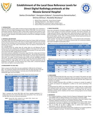
Table 1: NGH LDRLs compared to the UK NDRLs. Patients were selected randomly
4. IMAGE EVALUATION
Table 2: Revised NGH LDRLs compared to the original LDRLs. Patients were
selected in the range of the 70 ± 10 kg
6. REFERENCES
1. CEC. European Guidelines on Quality Criteria for Diagnostic Radiographic Images. Report
EUR 16260EN. Office for Official Publication of the European Community (1996)
2. Wall, B. (eds), European Guidance on Estimating Population Doses from Medical X-Ray
Procedures, Radiation Protection No 154, European Commission 2008.
3. International Commission for Radiation Units and Measurements (ICRU) (1998). Patient
dosimetry for x-rays used in medical imaging. J ICRU 2005; 5: iv–vi, Petoussi-Henss et al.
4. Hart, D. & Hillier, M. & Wall, B. (2002), Doses to patients from medical X-ray
examinations in the UK- 2000 review, NRPB-W4.
5. Irish X-ray departments demonstrate varying levels of adherence to European guidelines
on good radiographic technique. The British Journal of Radiology, 75 (2002).
5. CONCLUSIONS
The original established NGH ESD LDRLs were lower as the weight of the patients was taken
into account. It was revealed that radiographers after the acquisition of the image are often
trimming it to the correct area electronically and therefore the NGH DAP LDRLs (original and
revised) are not reliable5.
In order to establish effectively LDRLs and NDRLs it is necessary to consider the weight of
the patient and educate the radiographers to adhere to the correct positioning of the
patient.
In implementing EUR 16260 it was observed that poor quality image was strongly related
with inadequate patient positioning, and preparation. In addition doctors should further
familiarize themselves with evaluation criteria. It is recommended that this study be
extended to greater scale for reliability and that relevant training programs must be initiated
to all radiology staff.
Examination
Prorocol
ESD (mGy) DAP (mGy.cm2)
NGH LDRs UK NDRLs NGH LDRLs UK NDRLs
Skull AP 2.78 3.00 795.21 N/A
Skull Lat 1.71 1.50 693.48 N/A
Cervical AP 1.17 N/A 226.31 400.00
Cervical Lat 1.17 N/A 157.27 400.00
Chest PA 0.39 0.20 124.84 120.00
Lumbar Spine AP 4.00 6.00 2228.36 1600.00
Lumbar Spine Lat 10.49 14.00 3715.17 3000.00
Abdomen AP 4.50 6.00 3126.08 3000.00
Pelvis AP 3.83 4.00 2714.73 3000.00
Table 1 compares the NGH LDRLs that were established in 2008 with the UK National
DRLs4. This comparison indicates that the ESD NGH LDRLs are lower that those of the UK
NDRLs except for the Chest PA protocol. Contrarily, most of the NGH DAP LDRLs are
substantially higher than the UK NDRLs.
Examination
Prorocol
ESD (mGy) DAP (mGy.cm2)
Original Revised Original Revised
Chest PA 0.39 0.11 124.84 121,08
Lumbar Spine AP 4.00 4,96 2228.36 2121.61
Lumbar Spine Lat 10.49 8,49 3715.17 4399,66
Pelvis AP 3.83 3,59 2714.73 3583,61
Table 2 compares the revised NGH LDRLs with those originally established. It is
evident that most of the revised ESD LDRLs show improvement. This is not true for
the DAP LDRLs that do not show any improvement.
Doses were calculated for 10 patients weighted in the range of the 70 ± 10 kg for different
values of tube output for the below examination protocols. Two doctors evaluated the
image quality with different tube output values, good positioning and preparation of the
patient according to the EUR 16260. Table 3 summarizes only the tube output values that
give the best quality scoring . There is a strong correlation between good positioning and
preparation of the patient with poor image quality.
Table 4 presents the tube output values that result in the maximum dose minimization.
These protocols were chosen as the optimum protocol after the compromise between
lower doses and highest scored quality criteria.
Examination
Prorocol
Tube output-image quality
Good
positiong
Good
preparation
Tube voltage Scores (%) Dose(mGy) Scores(%) Scores(%)
Chest PA 115 99 0,04 77 N/A
Lumbar Spine AP 80 93 2,66 93 N/A
Pelvis AP 85 100 1,59 89,5 61
Examination
Prorocol
Before optimization After optimization
Difference
In
Dose
Tube voltage Dose(mGy) Tube voltage Dose(mGy) %
Chest PA 100 0,048 115 0,037 54
Lumbar Spine
AP
75 4,54 80 2,66 41
Pelvis AP 75 3,5 85 1,59 23
Table 4:Protocls that were chosen after the compromise between lower dose and highest quality scored
Table 3: Scores with best scored tube output value , good positioning and preparation of the patient

Mais conteúdo relacionado

Mais procurados

Use of pre treatment protocols
Use of pre treatment protocols   Use of pre treatment protocols
Use of pre treatment protocols Bartosz Bąk
 
Management dell’organ motion nei trattamenti stereo-RT e radiochirurgici:
Management dell’organ motion nei trattamenti stereo-RT e radiochirurgici: Management dell’organ motion nei trattamenti stereo-RT e radiochirurgici:
Management dell’organ motion nei trattamenti stereo-RT e radiochirurgici: Gemelli Advanced Radiation Therapy
 
The vmat vs other recent radiotherapy techniques
The vmat vs other recent radiotherapy techniquesThe vmat vs other recent radiotherapy techniques
The vmat vs other recent radiotherapy techniquesM'dee Phechudi
 
San Antonio Breast Cancer Symposium 2007 Highlights – Radiotherapy
San Antonio Breast Cancer Symposium 2007 Highlights – RadiotherapySan Antonio Breast Cancer Symposium 2007 Highlights – Radiotherapy
San Antonio Breast Cancer Symposium 2007 Highlights – Radiotherapyfondas vakalis
 
Guidelines for the prescribed activity for 131I remnant ablation on different...
Guidelines for the prescribed activity for 131I remnant ablation on different...Guidelines for the prescribed activity for 131I remnant ablation on different...
Guidelines for the prescribed activity for 131I remnant ablation on different...Michael
 
Niveles de Referencia de Diagnóstico en Radiología Pediátrica
Niveles de Referencia de Diagnóstico en Radiología PediátricaNiveles de Referencia de Diagnóstico en Radiología Pediátrica
Niveles de Referencia de Diagnóstico en Radiología PediátricaEduardo Medina Gironzini
 
RADIOTHERAPY FOR BREAST CANCER
RADIOTHERAPY FOR BREAST CANCERRADIOTHERAPY FOR BREAST CANCER
RADIOTHERAPY FOR BREAST CANCERfondas vakalis
 
Accelerated partial breast irradiation
Accelerated partial breast irradiationAccelerated partial breast irradiation
Accelerated partial breast irradiationBharti Devnani
 
Physical Models For Time Dose & Fractionation
Physical Models For Time Dose & FractionationPhysical Models For Time Dose & Fractionation
Physical Models For Time Dose & FractionationIsha Jaiswal
 
CDH Proton Research Poster
CDH Proton Research PosterCDH Proton Research Poster
CDH Proton Research PosterRachel Rendall
 
MINIMALLY INVASIVE SURGERY IN TOTAL HIP ARTHROPLASTY: A COSTEFFECTIVENESS ANA...
MINIMALLY INVASIVE SURGERY IN TOTAL HIP ARTHROPLASTY: A COSTEFFECTIVENESS ANA...MINIMALLY INVASIVE SURGERY IN TOTAL HIP ARTHROPLASTY: A COSTEFFECTIVENESS ANA...
MINIMALLY INVASIVE SURGERY IN TOTAL HIP ARTHROPLASTY: A COSTEFFECTIVENESS ANA...ELISA HERNANDEZ TORRES
 
Evolution of Intracavitary brachytherapy for carcinoma of cervix
Evolution of Intracavitary brachytherapy for carcinoma of cervixEvolution of Intracavitary brachytherapy for carcinoma of cervix
Evolution of Intracavitary brachytherapy for carcinoma of cervixAjeet Gandhi
 
SBRT in head and neck cancer
SBRT in  head and neck cancerSBRT in  head and neck cancer
SBRT in head and neck cancerDr Rushi Panchal
 
Breast Cancer Radiotherapy
Breast Cancer RadiotherapyBreast Cancer Radiotherapy
Breast Cancer Radiotherapyfondas vakalis
 
Adjuvant radiotherapy in cancer breast pptx
Adjuvant radiotherapy in cancer breast pptxAdjuvant radiotherapy in cancer breast pptx
Adjuvant radiotherapy in cancer breast pptxGopa Ghosh
 
CT radiation dose concepts and radiation dose optimization- Avinesh Shrestha
CT radiation dose concepts and radiation dose optimization- Avinesh Shrestha CT radiation dose concepts and radiation dose optimization- Avinesh Shrestha
CT radiation dose concepts and radiation dose optimization- Avinesh Shrestha Avinesh Shrestha
 

Mais procurados (20)

Use of pre treatment protocols
Use of pre treatment protocols   Use of pre treatment protocols
Use of pre treatment protocols
 
Management dell’organ motion nei trattamenti stereo-RT e radiochirurgici:
Management dell’organ motion nei trattamenti stereo-RT e radiochirurgici: Management dell’organ motion nei trattamenti stereo-RT e radiochirurgici:
Management dell’organ motion nei trattamenti stereo-RT e radiochirurgici:
 
The vmat vs other recent radiotherapy techniques
The vmat vs other recent radiotherapy techniquesThe vmat vs other recent radiotherapy techniques
The vmat vs other recent radiotherapy techniques
 
San Antonio Breast Cancer Symposium 2007 Highlights – Radiotherapy
San Antonio Breast Cancer Symposium 2007 Highlights – RadiotherapySan Antonio Breast Cancer Symposium 2007 Highlights – Radiotherapy
San Antonio Breast Cancer Symposium 2007 Highlights – Radiotherapy
 
Guidelines for the prescribed activity for 131I remnant ablation on different...
Guidelines for the prescribed activity for 131I remnant ablation on different...Guidelines for the prescribed activity for 131I remnant ablation on different...
Guidelines for the prescribed activity for 131I remnant ablation on different...
 
Brachytherapy Final
Brachytherapy FinalBrachytherapy Final
Brachytherapy Final
 
Niveles de Referencia de Diagnóstico en Radiología Pediátrica
Niveles de Referencia de Diagnóstico en Radiología PediátricaNiveles de Referencia de Diagnóstico en Radiología Pediátrica
Niveles de Referencia de Diagnóstico en Radiología Pediátrica
 
RADIOTHERAPY FOR BREAST CANCER
RADIOTHERAPY FOR BREAST CANCERRADIOTHERAPY FOR BREAST CANCER
RADIOTHERAPY FOR BREAST CANCER
 
Accelerated partial breast irradiation
Accelerated partial breast irradiationAccelerated partial breast irradiation
Accelerated partial breast irradiation
 
Physical Models For Time Dose & Fractionation
Physical Models For Time Dose & FractionationPhysical Models For Time Dose & Fractionation
Physical Models For Time Dose & Fractionation
 
CDH Proton Research Poster
CDH Proton Research PosterCDH Proton Research Poster
CDH Proton Research Poster
 
MINIMALLY INVASIVE SURGERY IN TOTAL HIP ARTHROPLASTY: A COSTEFFECTIVENESS ANA...
MINIMALLY INVASIVE SURGERY IN TOTAL HIP ARTHROPLASTY: A COSTEFFECTIVENESS ANA...MINIMALLY INVASIVE SURGERY IN TOTAL HIP ARTHROPLASTY: A COSTEFFECTIVENESS ANA...
MINIMALLY INVASIVE SURGERY IN TOTAL HIP ARTHROPLASTY: A COSTEFFECTIVENESS ANA...
 
Evolution of Intracavitary brachytherapy for carcinoma of cervix
Evolution of Intracavitary brachytherapy for carcinoma of cervixEvolution of Intracavitary brachytherapy for carcinoma of cervix
Evolution of Intracavitary brachytherapy for carcinoma of cervix
 
SBRT in head and neck cancer
SBRT in  head and neck cancerSBRT in  head and neck cancer
SBRT in head and neck cancer
 
Breast Cancer Radiotherapy
Breast Cancer RadiotherapyBreast Cancer Radiotherapy
Breast Cancer Radiotherapy
 
AAPM REPORT 51
AAPM REPORT 51AAPM REPORT 51
AAPM REPORT 51
 
Adjuvant radiotherapy in cancer breast pptx
Adjuvant radiotherapy in cancer breast pptxAdjuvant radiotherapy in cancer breast pptx
Adjuvant radiotherapy in cancer breast pptx
 
CT radiation dose concepts and radiation dose optimization- Avinesh Shrestha
CT radiation dose concepts and radiation dose optimization- Avinesh Shrestha CT radiation dose concepts and radiation dose optimization- Avinesh Shrestha
CT radiation dose concepts and radiation dose optimization- Avinesh Shrestha
 
2010 Poznan G Guidi Adaptive Rt Rcs Sbrt
2010 Poznan G Guidi Adaptive Rt Rcs Sbrt2010 Poznan G Guidi Adaptive Rt Rcs Sbrt
2010 Poznan G Guidi Adaptive Rt Rcs Sbrt
 
Hypofractionation in breast cancer
Hypofractionation in breast cancerHypofractionation in breast cancer
Hypofractionation in breast cancer
 

Destaque

100 เรื่องเด่น ความรู้เพื่อส่งเสริมสุขภาพ
100 เรื่องเด่น ความรู้เพื่อส่งเสริมสุขภาพ100 เรื่องเด่น ความรู้เพื่อส่งเสริมสุขภาพ
100 เรื่องเด่น ความรู้เพื่อส่งเสริมสุขภาพอลงกรณ์ อารามกูล
 
Radiology in planning
Radiology in planningRadiology in planning
Radiology in planningDorogi Gábor
 
Introduction to radiologic technology
Introduction to radiologic technologyIntroduction to radiologic technology
Introduction to radiologic technologyRad Tech
 
Ultrasound 2
Ultrasound 2Ultrasound 2
Ultrasound 2Rad Tech
 
Basics of radiology
Basics of radiologyBasics of radiology
Basics of radiologyEhab Abdu
 
Chapter20 radiology and diagnostic imaging terminology
Chapter20 radiology and diagnostic imaging terminologyChapter20 radiology and diagnostic imaging terminology
Chapter20 radiology and diagnostic imaging terminologyRad Tech
 

Destaque (8)

Smr1
Smr1Smr1
Smr1
 
Show me how 500 things you should know instructions for life from the every...
Show me how 500 things you should know   instructions for life from the every...Show me how 500 things you should know   instructions for life from the every...
Show me how 500 things you should know instructions for life from the every...
 
100 เรื่องเด่น ความรู้เพื่อส่งเสริมสุขภาพ
100 เรื่องเด่น ความรู้เพื่อส่งเสริมสุขภาพ100 เรื่องเด่น ความรู้เพื่อส่งเสริมสุขภาพ
100 เรื่องเด่น ความรู้เพื่อส่งเสริมสุขภาพ
 
Radiology in planning
Radiology in planningRadiology in planning
Radiology in planning
 
Introduction to radiologic technology
Introduction to radiologic technologyIntroduction to radiologic technology
Introduction to radiologic technology
 
Ultrasound 2
Ultrasound 2Ultrasound 2
Ultrasound 2
 
Basics of radiology
Basics of radiologyBasics of radiology
Basics of radiology
 
Chapter20 radiology and diagnostic imaging terminology
Chapter20 radiology and diagnostic imaging terminologyChapter20 radiology and diagnostic imaging terminology
Chapter20 radiology and diagnostic imaging terminology
 

Semelhante a Datamed Poster DDR DRLS-Final

Feasibility of CT scan studies with triple split bolus intravenous contrast ...
Feasibility of CT scan studies with triple split bolus  intravenous contrast ...Feasibility of CT scan studies with triple split bolus  intravenous contrast ...
Feasibility of CT scan studies with triple split bolus intravenous contrast ...Jhon Arriaga Cordova
 
Diagnostic Reference Level in Lumbar Radiography in Abidjan, Côte d’ivoire
Diagnostic Reference Level in Lumbar Radiography in Abidjan, Côte d’ivoireDiagnostic Reference Level in Lumbar Radiography in Abidjan, Côte d’ivoire
Diagnostic Reference Level in Lumbar Radiography in Abidjan, Côte d’ivoiretheijes
 
Berbeco et al, Low Z target switching to increase tumor endothelial cell dose...
Berbeco et al, Low Z target switching to increase tumor endothelial cell dose...Berbeco et al, Low Z target switching to increase tumor endothelial cell dose...
Berbeco et al, Low Z target switching to increase tumor endothelial cell dose...David Parsons
 
J0362069073
J0362069073J0362069073
J0362069073theijes
 
Presentation 215 a j_mark_barch_etco2 monitoring generic
Presentation 215 a j_mark_barch_etco2 monitoring genericPresentation 215 a j_mark_barch_etco2 monitoring generic
Presentation 215 a j_mark_barch_etco2 monitoring genericThe ALS Association
 
SPECT/CT: HOW Much Radiation Dose CT Constitute
SPECT/CT: HOW Much Radiation Dose CT ConstituteSPECT/CT: HOW Much Radiation Dose CT Constitute
SPECT/CT: HOW Much Radiation Dose CT ConstituteShahid Younas
 
CT Dose Issues.pptx on the factors to be considered on radiation protection
CT Dose Issues.pptx on the factors to be considered on radiation protectionCT Dose Issues.pptx on the factors to be considered on radiation protection
CT Dose Issues.pptx on the factors to be considered on radiation protectionsanyengere
 
Validation of pediatric thyroid phantom using Single-Energy and Dual-Energy CT
Validation of pediatric thyroid phantom using Single-Energy and Dual-Energy CTValidation of pediatric thyroid phantom using Single-Energy and Dual-Energy CT
Validation of pediatric thyroid phantom using Single-Energy and Dual-Energy CTMOAYYAD ALSSABBAGH
 
Dual energy CT in radiotherapy: Current applications and future outlook
Dual energy CT in radiotherapy: Current applications and future outlookDual energy CT in radiotherapy: Current applications and future outlook
Dual energy CT in radiotherapy: Current applications and future outlookWookjin Choi
 
FINAL CTCA CAMRT presentation - Yili
FINAL CTCA CAMRT presentation - YiliFINAL CTCA CAMRT presentation - Yili
FINAL CTCA CAMRT presentation - YiliYili Wang
 
Novel Functional Delta-Radiomics for Predicting Overall Survival in Lung Canc...
Novel Functional Delta-Radiomics for Predicting Overall Survival in Lung Canc...Novel Functional Delta-Radiomics for Predicting Overall Survival in Lung Canc...
Novel Functional Delta-Radiomics for Predicting Overall Survival in Lung Canc...Wookjin Choi
 
Multispectral imaging of normal bladder mucous membrane and clinical stage of...
Multispectral imaging of normal bladder mucous membrane and clinical stage of...Multispectral imaging of normal bladder mucous membrane and clinical stage of...
Multispectral imaging of normal bladder mucous membrane and clinical stage of...Clinical Surgery Research Communications
 
Design and development of nanomaterials for biomolecular detection and cancer...
Design and development of nanomaterials for biomolecular detection and cancer...Design and development of nanomaterials for biomolecular detection and cancer...
Design and development of nanomaterials for biomolecular detection and cancer...Arun kumar
 
Lab on a chip 2013 qian liu
Lab on a chip 2013 qian liuLab on a chip 2013 qian liu
Lab on a chip 2013 qian liuQian Liu, phD
 
Reduced Radiation Exposure in Dual-Energy Computed Tomography of the Chest: ...
Reduced Radiation Exposure in Dual-Energy  Computed Tomography of the Chest: ...Reduced Radiation Exposure in Dual-Energy  Computed Tomography of the Chest: ...
Reduced Radiation Exposure in Dual-Energy Computed Tomography of the Chest: ...MehranMouzam
 
Radiation dose to lens in CT
Radiation dose to lens in CTRadiation dose to lens in CT
Radiation dose to lens in CTKalpana Parajuli
 

Semelhante a Datamed Poster DDR DRLS-Final (20)

Feasibility of CT scan studies with triple split bolus intravenous contrast ...
Feasibility of CT scan studies with triple split bolus  intravenous contrast ...Feasibility of CT scan studies with triple split bolus  intravenous contrast ...
Feasibility of CT scan studies with triple split bolus intravenous contrast ...
 
IGRT APP.pdf
IGRT APP.pdfIGRT APP.pdf
IGRT APP.pdf
 
Diagnostic Reference Level in Lumbar Radiography in Abidjan, Côte d’ivoire
Diagnostic Reference Level in Lumbar Radiography in Abidjan, Côte d’ivoireDiagnostic Reference Level in Lumbar Radiography in Abidjan, Côte d’ivoire
Diagnostic Reference Level in Lumbar Radiography in Abidjan, Côte d’ivoire
 
Berbeco et al, Low Z target switching to increase tumor endothelial cell dose...
Berbeco et al, Low Z target switching to increase tumor endothelial cell dose...Berbeco et al, Low Z target switching to increase tumor endothelial cell dose...
Berbeco et al, Low Z target switching to increase tumor endothelial cell dose...
 
J0362069073
J0362069073J0362069073
J0362069073
 
Presentation 215 a j_mark_barch_etco2 monitoring generic
Presentation 215 a j_mark_barch_etco2 monitoring genericPresentation 215 a j_mark_barch_etco2 monitoring generic
Presentation 215 a j_mark_barch_etco2 monitoring generic
 
SPECT/CT: HOW Much Radiation Dose CT Constitute
SPECT/CT: HOW Much Radiation Dose CT ConstituteSPECT/CT: HOW Much Radiation Dose CT Constitute
SPECT/CT: HOW Much Radiation Dose CT Constitute
 
CT Dose Issues.pptx on the factors to be considered on radiation protection
CT Dose Issues.pptx on the factors to be considered on radiation protectionCT Dose Issues.pptx on the factors to be considered on radiation protection
CT Dose Issues.pptx on the factors to be considered on radiation protection
 
Validation of pediatric thyroid phantom using Single-Energy and Dual-Energy CT
Validation of pediatric thyroid phantom using Single-Energy and Dual-Energy CTValidation of pediatric thyroid phantom using Single-Energy and Dual-Energy CT
Validation of pediatric thyroid phantom using Single-Energy and Dual-Energy CT
 
Dual energy CT in radiotherapy: Current applications and future outlook
Dual energy CT in radiotherapy: Current applications and future outlookDual energy CT in radiotherapy: Current applications and future outlook
Dual energy CT in radiotherapy: Current applications and future outlook
 
FINAL CTCA CAMRT presentation - Yili
FINAL CTCA CAMRT presentation - YiliFINAL CTCA CAMRT presentation - Yili
FINAL CTCA CAMRT presentation - Yili
 
CT dose reduction
CT dose reductionCT dose reduction
CT dose reduction
 
Novel Functional Delta-Radiomics for Predicting Overall Survival in Lung Canc...
Novel Functional Delta-Radiomics for Predicting Overall Survival in Lung Canc...Novel Functional Delta-Radiomics for Predicting Overall Survival in Lung Canc...
Novel Functional Delta-Radiomics for Predicting Overall Survival in Lung Canc...
 
Multispectral imaging of normal bladder mucous membrane and clinical stage of...
Multispectral imaging of normal bladder mucous membrane and clinical stage of...Multispectral imaging of normal bladder mucous membrane and clinical stage of...
Multispectral imaging of normal bladder mucous membrane and clinical stage of...
 
Design and development of nanomaterials for biomolecular detection and cancer...
Design and development of nanomaterials for biomolecular detection and cancer...Design and development of nanomaterials for biomolecular detection and cancer...
Design and development of nanomaterials for biomolecular detection and cancer...
 
Radiation dose reduction
Radiation dose reduction Radiation dose reduction
Radiation dose reduction
 
Lab on a chip 2013 qian liu
Lab on a chip 2013 qian liuLab on a chip 2013 qian liu
Lab on a chip 2013 qian liu
 
Reduced Radiation Exposure in Dual-Energy Computed Tomography of the Chest: ...
Reduced Radiation Exposure in Dual-Energy  Computed Tomography of the Chest: ...Reduced Radiation Exposure in Dual-Energy  Computed Tomography of the Chest: ...
Reduced Radiation Exposure in Dual-Energy Computed Tomography of the Chest: ...
 
Radiation dose to lens in CT
Radiation dose to lens in CTRadiation dose to lens in CT
Radiation dose to lens in CT
 
Xerostomia level
Xerostomia levelXerostomia level
Xerostomia level
 

Datamed Poster DDR DRLS-Final

  • 1. Establishment of the Local Dose Reference Levels for Direct Digital Radiology protocols at the Nicosia General Hospital Stelios Christofides1, Georgiana Kokona1, Constantinos Demetroullas2, Omiros Omirou2, Nicoletta Nicolaou3 1. Medical Physics Department - Nicosia General hospital 2. Radiology Department – Arch. Makarios III Hospital 3. Physics Department, University of Cyprus, Nicosia, Cyprus. 4. School of Physics and Astronomy, University of Birmingham, U.K. 1. INTRODUCTION Local Dose Reference Levels (LDRLs) of Nicosia General Hospital (NGH) were established in 2008 from four Direct Digital Radiography Systems MECALL EIDOS 3000 nine most common examination protocols. Recently an effort is being made to optimise these protocols. So far four of these have been optimized taking into account the weight of the patients and LDRLs have been revised. Evaluation of the above protocols carried out from radiologists according the EUR 162601. 2. MATERIALS AND METHODS The original LDRLs have been established based on Dose Area Product (DAP) and Entrance Surface Dose (ESD) measurements. DAP values of randomly selected patients were retrieved from the Picture Archiving and Communications System (PACS). ESD measurements were derived from the X-ray tube output measurements and X-ray exposure measurements for each radiology system2. Four protocols (Chest PA, Lumbar Spine AP, Lumbar Spine Lat, and Abdomen AP) were chosen and data were collected from 10 patients weighted in the range of the 70 ± 10 kg3. The ESD doses were derived following the aforementioned procedure. At the same time for each measurement DAPs readings was also recorded. Examination protocols established in the previous stage (Chest PA, Lumbar Spine AP, Pelvis) were evaluated and optimized following the below procedure. ESD and DAP measurements were assessed for the above protocols after small changes at the tube voltage (±10 kV with ±5kV pace). The images of three protocols were chosen and two radiologists evaluated them by scoring them according to the EUR 16260.The scoring criteria were divided in three groups relating the image quality with the tube output, the good positioning and the preparation of the patient. The best protocols were chosen after a compromise between lower doses and highest scored quality criteria. 3.ESTABLISHMENT OF LOCAL DRLS Table 1: NGH LDRLs compared to the UK NDRLs. Patients were selected randomly 4. IMAGE EVALUATION Table 2: Revised NGH LDRLs compared to the original LDRLs. Patients were selected in the range of the 70 ± 10 kg 6. REFERENCES 1. CEC. European Guidelines on Quality Criteria for Diagnostic Radiographic Images. Report EUR 16260EN. Office for Official Publication of the European Community (1996) 2. Wall, B. (eds), European Guidance on Estimating Population Doses from Medical X-Ray Procedures, Radiation Protection No 154, European Commission 2008. 3. International Commission for Radiation Units and Measurements (ICRU) (1998). Patient dosimetry for x-rays used in medical imaging. J ICRU 2005; 5: iv–vi, Petoussi-Henss et al. 4. Hart, D. & Hillier, M. & Wall, B. (2002), Doses to patients from medical X-ray examinations in the UK- 2000 review, NRPB-W4. 5. Irish X-ray departments demonstrate varying levels of adherence to European guidelines on good radiographic technique. The British Journal of Radiology, 75 (2002). 5. CONCLUSIONS The original established NGH ESD LDRLs were lower as the weight of the patients was taken into account. It was revealed that radiographers after the acquisition of the image are often trimming it to the correct area electronically and therefore the NGH DAP LDRLs (original and revised) are not reliable5. In order to establish effectively LDRLs and NDRLs it is necessary to consider the weight of the patient and educate the radiographers to adhere to the correct positioning of the patient. In implementing EUR 16260 it was observed that poor quality image was strongly related with inadequate patient positioning, and preparation. In addition doctors should further familiarize themselves with evaluation criteria. It is recommended that this study be extended to greater scale for reliability and that relevant training programs must be initiated to all radiology staff. Examination Prorocol ESD (mGy) DAP (mGy.cm2) NGH LDRs UK NDRLs NGH LDRLs UK NDRLs Skull AP 2.78 3.00 795.21 N/A Skull Lat 1.71 1.50 693.48 N/A Cervical AP 1.17 N/A 226.31 400.00 Cervical Lat 1.17 N/A 157.27 400.00 Chest PA 0.39 0.20 124.84 120.00 Lumbar Spine AP 4.00 6.00 2228.36 1600.00 Lumbar Spine Lat 10.49 14.00 3715.17 3000.00 Abdomen AP 4.50 6.00 3126.08 3000.00 Pelvis AP 3.83 4.00 2714.73 3000.00 Table 1 compares the NGH LDRLs that were established in 2008 with the UK National DRLs4. This comparison indicates that the ESD NGH LDRLs are lower that those of the UK NDRLs except for the Chest PA protocol. Contrarily, most of the NGH DAP LDRLs are substantially higher than the UK NDRLs. Examination Prorocol ESD (mGy) DAP (mGy.cm2) Original Revised Original Revised Chest PA 0.39 0.11 124.84 121,08 Lumbar Spine AP 4.00 4,96 2228.36 2121.61 Lumbar Spine Lat 10.49 8,49 3715.17 4399,66 Pelvis AP 3.83 3,59 2714.73 3583,61 Table 2 compares the revised NGH LDRLs with those originally established. It is evident that most of the revised ESD LDRLs show improvement. This is not true for the DAP LDRLs that do not show any improvement. Doses were calculated for 10 patients weighted in the range of the 70 ± 10 kg for different values of tube output for the below examination protocols. Two doctors evaluated the image quality with different tube output values, good positioning and preparation of the patient according to the EUR 16260. Table 3 summarizes only the tube output values that give the best quality scoring . There is a strong correlation between good positioning and preparation of the patient with poor image quality. Table 4 presents the tube output values that result in the maximum dose minimization. These protocols were chosen as the optimum protocol after the compromise between lower doses and highest scored quality criteria. Examination Prorocol Tube output-image quality Good positiong Good preparation Tube voltage Scores (%) Dose(mGy) Scores(%) Scores(%) Chest PA 115 99 0,04 77 N/A Lumbar Spine AP 80 93 2,66 93 N/A Pelvis AP 85 100 1,59 89,5 61 Examination Prorocol Before optimization After optimization Difference In Dose Tube voltage Dose(mGy) Tube voltage Dose(mGy) % Chest PA 100 0,048 115 0,037 54 Lumbar Spine AP 75 4,54 80 2,66 41 Pelvis AP 75 3,5 85 1,59 23 Table 4:Protocls that were chosen after the compromise between lower dose and highest quality scored Table 3: Scores with best scored tube output value , good positioning and preparation of the patient