
Recomendados
Mais conteúdo relacionado
Mais procurados
Mais procurados (20)
Semelhante a Compound microscope
Semelhante a Compound microscope (20)
Mais de NithyaNandapal
Mais de NithyaNandapal (20)
Último
Último (20)
Compound microscope
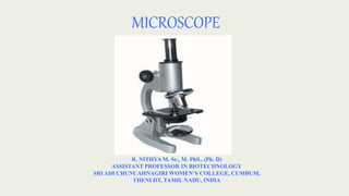
- 1. MICROSCOPE R. NITHYA M. Sc., M. Phil., (Ph. D) ASSISTANT PROFESSOR IN BIOTECHNOLOGY SRI ADI CHUNCAHNAGIRI WOMEN’S COLLEGE, CUMBUM, THENI DT, TAMIL NADU, INDIA
- 2. MICROSCOPE An instrument designed for the study of objects which cannot be seen with naked eyes. Microscope (micros = small; skopein = to see/ to look) means an instrument to see small things. It is an inevitable instrument for clinical and research laboratories.
- 3. The first microscope was designed by Jenssen and Hans Lippershey.
- 4. MICROSCOPY The field of designing and using microscopes to view objects that cannot be seen with unaided eyes is known as microscopy. Microscopy has three distinct branches. They are Optical microscopy Electron microscopy Scanning probe microscopy.
- 5. TYPES OF MICROSCOPE The microscopes are classified into three types based on the source of illumination. They are Light microscope Electron microscope X-ray microscope
- 6. LIGHT MICROSCOPE In light microscopes, light is the source of illumination. They are subdivided into following types. Simple microscope Compound microscope Binocular microscope Phase contract microscope Interference microscope Polarizing microscope Dark field microscope Ultraviolet microscope Fluorescence microscope Cinematography
- 7. Compound Microscope It is an optical microscope. It is formed by the combination of two simple microscopes (a simple microscope is formed of only one lens). Principle The lenses magnify objects. It has light source, a diaphragm, an object, an objective lens and an eye piece. The light passes through the diaphragm. The diaphragm gathers the light on the object. The objective lens produces a real (actual), inverted (upturned – upside down) magnified image of the object. The magnified image acts as an object for the eye piece. The eyepiece produces a virtual (effective, actual) inverted and magnified image of the object.
- 8. MAGNIFICATION POWER The ratio of magnified image to that formed in retina of unaided eye is called magnification. Magnification power = Size of retinal image seen with a microscope Size of retinal image seen with unaided eye The magnifying power of a compound microscope is the ratio of size of the final image to the size of the object. M = Size of final image/ Size of the object
- 11. RESOLVING POWER The ability of the microscope to distinguish two very small and closely spaced objects as separate entities is called resolving power of the microscope. Resolving power of microscopes depends on the wavelength of rays and numerical aperture (NA) of objective lens. The RP of a microscope can be calculated by using the formula – Resolving power (RP) = ʎ 2 x NA
- 13. NUMERICAL APERTURE The resolving power of microscope can be increased by increasing the size of numerical aperture. The NA is the function of the objective lens in relation to its focal length. It can be calculated by using the formula Numerical aperture (NA) = ƞ sin Ɵ
- 15. SOURCE OF ILLUMINATION These two microscopes mainly differ in the source of light they use. One uses a mirror to converge sunlight while other uses directly an illuminator.
- 16. STRUCTURE The compound microscope has the following parts. REFLECTING MIRROR It reflects light on the object.
- 17. CONDENSER It gathers and focuses the reflected light on the object. The condenser has a diaphragm. It allows the required intensity (power, concentration, strength) of light to pass through.
- 18. DIAPHRAGM. Diaphragm is a five holed disk placed under the stage. Each hole is of a different diameter. By turning it, you can vary the amount of light passing through the stage opening.
- 19. NOSEPIECE AND APERTURE •Nosepiece is a rotating turret that holds the Objective lenses. The viewer spins the Nosepiece to select different Objective lenses. The Aperture is the middle of the stage that allows light from the Illuminator to reach the specimen.
- 20. OBJECTIVE LENS Metal cylinders attached below the nosepiece and contains especially ground and polished lenses It magnifies the object. Onion cells
- 21. TYPES OF OBJECTIVE LENSES LPO / Low Power Objective Gives the lowest magnification, usually 10x HPO / High Power Objective Gives higher magnification usually 40x or 43x OIO / Oil Immersion Objective Gives the highest magnification, usually 97x or 100x, and is used cedar wood oil or synthetic oil for better resolution.
- 22. EYE PIECE It magnifies the image produced by the objective lens.
- 23. BODY TUBE It is a tube with the objective lens at the lower end and the eye piece lens at the upper end.
- 24. COARSE ADJUSTMENT It moves the body tube up and down rapidly to correct the distance from the object to get focusing. FINE ADJUSTMENT It moves the body tube up and down slowly to make exact focusing.
- 25. SPECIMEN STAGE It is a platform with a hole in the centre. The light falls on the object through the hole. The slide is placed on the stage.
- 26. STAGE CLIPS Stage clips hold the slide firmly on the stage
- 27. NOSE PIECE It is in the form of rotating disc having holes for fitting the objective lenses.
- 28. INCLINATION JOINT It permits tilting of upper part of the microscope to adjust to the eye level.
- 29. BASE OR FOOT The foot keeps the body in position.
- 30. USES OF COMPOUND LIGHT MICROSCOPE. • Compound Microscope is used in pathology labs to identify diseases. • In Forensic laboratories, Compound Light Microscopes are used to identify presence of minerals or metals in human cells so as to solve criminal cases. • Forensic Experts can also find out the origin of a drug by viewing its component particles under a Microscope.
- 31. Blood cells Human cell Plant cell Paramecia COMPOUND MICROSCOPE IMAGES