SlideShare uma empresa Scribd logo
1 de 84
Essentials of Human Anatomy & Physiology
Seventh Edition
Slides 16.1 – 16.20
Lecture Slides in PowerPoint by Jerry L. Cook
Copyright © 2003 Pearson Education, Inc. publishing as Benjamin Cummings
The Reproductive System
Slide 16.1
Copyright © 2003 Pearson Education, Inc. publishing as Benjamin Cummings
• Gonads – primary sex organs
• Testes in males
• Ovaries in females
• Gonads produce gametes (sex cells) and
secrete hormones
• Sperm – male gametes
• Ova (eggs) – female gametes
Male Reproductive System
• Testes
• Duct system
• Epididymis
• Ductus deferens
• Urethra
Slide 16.2a
Copyright © 2003 Pearson Education, Inc. publishing as Benjamin Cummings
Male Reproductive System
• Accessory organs
• Seminal vesicle
• Prostate gland
• Bulbourethral gland
• External genitalia
• Penis
• Scrotum
Slide 16.2b
Copyright © 2003 Pearson Education, Inc. publishing as Benjamin Cummings
Male Reproductive System
Slide 16.2c
Copyright © 2003 Pearson Education, Inc. publishing as Benjamin Cummings
Figure 16.2
Testes
• Coverings of
the testes
• Tunica
albuginea –
capsule that
surrounds
each testis
Slide 16.3a
Figure 16.1
Copyright © 2003 Pearson Education, Inc. publishing as Benjamin Cummings
Testes
• Coverings of
the testes
(continued)
• Septa –
extensions of
the capsule that
extend into the
testis and divide
it into lobules
Slide 16.3b
Figure 16.1
Copyright © 2003 Pearson Education, Inc. publishing as Benjamin Cummings
Testes
• Each lobule contains one to four
seminiferous tubules
• Tightly coiled structures
• Function as sperm-forming factories
• Empty sperm into the rete testis
• Sperm travels through the rete testis to
the epididymis
• Interstitial cells produce androgens such
as testosterone
Slide 16.4
Copyright © 2003 Pearson Education, Inc. publishing as Benjamin Cummings
Epididymis
• Comma-shaped, tightly coiled tube
• Found on the superior part of the testis
and along the posterior lateral side
• Functions to mature and store sperm
cells (at least 20 days)
• Expels sperm with the contraction of
muscles in the epididymis walls to the
vas deferens
Slide 16.5
Copyright © 2003 Pearson Education, Inc. publishing as Benjamin Cummings
Ductus Deferens (Vas Deferens)
• Carries sperm from the epididymis to the
ejaculatory duct
• Passes through the inguinal canal and
over the bladder
• Moves sperm by peristalsis
• Spermatic cord – ductus deferens, blood
vessels, and nerves in a connective
tissue sheath
Slide 16.6a
Copyright © 2003 Pearson Education, Inc. publishing as Benjamin Cummings
Ductus Deferens (Vas Deferens)
• Ends in the ejaculatory duct which
unites with the urethra
• Vasectomy – cutting of the ductus
deferens at the level of the testes to
prevent transportation of sperm
Slide 16.6b
Copyright © 2003 Pearson Education, Inc. publishing as Benjamin Cummings
Urethra
• Extends from the base of the urinary
bladder to the tip of the penis
• Carries both urine and sperm
• Sperm enters from the ejaculatory duct
Slide 16.7a
Copyright © 2003 Pearson Education, Inc. publishing as Benjamin Cummings
Urethra
Slide 16.7b
Copyright © 2003 Pearson Education, Inc. publishing as Benjamin Cummings
• Regions of the urethra
•Prostatic urethra –surrounded by
prostate
•Membranous urethra – from prostatic
urethra to penis
•Spongy (penile) urethra – runs the
length of the penis
Seminal Vesicles
• Located at the base of the bladder
• Produces a thick, yellowish secretion
(60% of semen)
• Fructose (sugar)
• Vitamin C
• Prostaglandins
• Other substances that nourish and activate
sperm
Slide 16.8
Copyright © 2003 Pearson Education, Inc. publishing as Benjamin Cummings
Prostate Gland
• Encircles the upper part of the urethra
• Secretes a milky fluid
• Helps to activate sperm
• Enters the urethra through several small
ducts
Slide 16.9
Copyright © 2003 Pearson Education, Inc. publishing as Benjamin Cummings
Bulbourethral Glands
• Pea-sized gland inferior to the prostate
• Produces a thick, clear mucus
• Cleanses the urethra of acidic urine
• Serves as a lubricant during sexual
intercourse
• Secreted into the penile urethra
Slide 16.10
Copyright © 2003 Pearson Education, Inc. publishing as Benjamin Cummings
Semen
• Mixture of sperm and accessory gland
secretions
• Advantages of accessory gland
secretions
• Fructose provides energy for sperm cells
• Alkalinity of semen helps neutralize the
acidic environment of vagina
• Semen inhibits bacterial multiplication
• Elements of semen enhance sperm motility
Slide 16.11
Copyright © 2003 Pearson Education, Inc. publishing as Benjamin Cummings
External Genitalia
• Scrotum
• Divided sac of skin outside the abdomen
• Maintains testes at 3°C lower than normal
body temperature to protect sperm viability
Slide 16.12
Copyright © 2003 Pearson Education, Inc. publishing as Benjamin Cummings
External Genitalia
• Penis
• Delivers sperm into the female reproductive
tract
• Regions of the penis
• Shaft
• Glans penis (enlarged tip)
• Prepuce (foreskin)
• Folded cuff of skin around proximal end
• Often removed by circumcision
Slide 16.13a
Copyright © 2003 Pearson Education, Inc. publishing as Benjamin Cummings
External Genitalia
• Internally there are three areas of spongy
erectile tissue around the urethra
Slide 16.13b
Copyright © 2003 Pearson Education, Inc. publishing as Benjamin Cummings
Spermatogenesis
• Production of sperm cells
• Begins at puberty and continues
Slide 16.14
Copyright © 2003 Pearson Education, Inc. publishing as Benjamin Cummings
throughout life
• Occurs in the seminiferous tubules
Processes of Spermatogenesis
Slide 16.15a
Copyright © 2003 Pearson Education, Inc. publishing as Benjamin Cummings
• Spermatogonia (stem cells) undergo
rapid mitosis to produce more stem cells
before puberty
• Follicle stimulating hormone (FSH)
modifies spermatogonia division
•One cell produced is a stem cell
•The other cell produced becomes a
primary spermatocyte
Processes of Spermatogenesis
Slide 16.15b
Copyright © 2003 Pearson Education, Inc. publishing as Benjamin Cummings
• Primary spermatocytes undergo meiosis
• Haploid spermatids are produced
Processes of Spermatogenesis
• Spermiogenesis
• Late spermatids are produced with distinct
regions
• Head – contains DNA covered by the
acrosome
• Midpiece
• Tail
• Sperm cells result after maturing of
spermatids
• Spermatogenesis takes 64 to 72 days
Slide 16.16
Copyright © 2003 Pearson Education, Inc. publishing as Benjamin Cummings
Processes of
Spermatogenesis
Figure 16.3
Slide 16.17
Copyright © 2003 Pearson Education, Inc. publishing as Benjamin Cummings
Anatomy of a Mature Sperm Cell
• The only
human
flagellated cell
• DNA is found
in the head
Figure 16.5
Slide 16.18
Copyright © 2003 Pearson Education, Inc. publishing as Benjamin Cummings
Testosterone Production
• The most important hormone of the
testes
• Produced in interstitial cells
Slide 16.19a
Copyright © 2003 Pearson Education, Inc. publishing as Benjamin Cummings
Testosterone Production
• Functions of testosterone
• Stimulates reproductive organ development
• Underlies sex drive
• Causes secondary sex characteristics
• Deepening of voice
• Increased hair growth
• Enlargement of skeletal muscles
• Thickening of bones
Slide 16.19b
Copyright © 2003 Pearson Education, Inc. publishing as Benjamin Cummings
Regulation
of Male
Androgens
(Sex
Hormones)
Slide 16.20
Copyright © 2003 Pearson Education, Inc. publishing as Benjamin Cummings
Figure 16.6
Female Reproductive System
Slide 16.21a
Copyright © 2003 Pearson Education, Inc. publishing as Benjamin Cummings
• Ovaries
• Duct System
• Uterine tubes (fallopian tubes)
• Uterus
• Vagina
• External genitalia
Female Reproductive System
Slide 16.21b
Copyright © 2003 Pearson Education, Inc. publishing as Benjamin Cummings
Figure 16.8a
Ovaries
• Composed
of ovarian
follicles
(sac-like
structures)
• Structure of
an ovarian
follicle
• Oocyte
• Follicular cells Figure 16.7
Slide 16.22
Copyright © 2003 Pearson Education, Inc. publishing as Benjamin Cummings
Ovarian Follicle Stages
• Primary follicle – contains an immature
oocyte
• Graafian (vesicular) follicle – growing
follicle with a maturing oocyte
• Ovulation – when the egg is mature the
follicle ruptures
• Occurs about every 28 days
• The ruptured follicle is transformed into a
corpus luteum
Slide 16.23
Copyright © 2003 Pearson Education, Inc. publishing as Benjamin Cummings
Support for Ovaries
• Suspensory ligaments – secure ovary to
lateral walls of the pelvis
• Ovarian ligaments – attach to uterus
• Broad ligament – a fold of the
peritoneum, encloses suspensory
ligament
Slide 16.24a
Copyright © 2003 Pearson Education, Inc. publishing as Benjamin Cummings
Support for Ovaries
Slide 16.24b
Copyright © 2003 Pearson Education, Inc. publishing as Benjamin Cummings
Figure 16.8b
Uterine (Fallopian) Tubes
• Receive the ovulated oocyte
• Provide a site for fertilization
• Attaches to the uterus
• Does not physically attach to the ovary
• Supported by the broad ligament
Slide 16.25
Copyright © 2003 Pearson Education, Inc. publishing as Benjamin Cummings
Uterine Tube Function
• Fimbriae – finger-like projections at the
distal end that receive the oocyte
• Cilia inside the uterine tube slowly move
the oocyte towards the uterus
(takes 3–4 days)
• Fertilization occurs inside the uterine
tube
Slide 16.26
Copyright © 2003 Pearson Education, Inc. publishing as Benjamin Cummings
Uterus
• Located between the urinary bladder
and rectum
• Hollow organ
• Functions of the uterus
• Receives a fertilized egg
• Retains the fertilized egg
• Nourishes the fertilized egg
Slide 16.27
Copyright © 2003 Pearson Education, Inc. publishing as Benjamin Cummings
Support for the Uterus
Slide 16.28a
Copyright © 2003 Pearson Education, Inc. publishing as Benjamin Cummings
• Broad ligament – attached to the pelvis
•
• Uterosacral ligaments – anchored
posteriorly
Support for the Uterus
Slide 16.28b
Copyright © 2003 Pearson Education, Inc. publishing as Benjamin Cummings
Figure 16.8b
Regions of the Uterus
• Body – main portion
• Fundus – area where uterine tube
enters
• Cervix – narrow outlet that protrudes
into the vagina
Slide 16.29
Copyright © 2003 Pearson Education, Inc. publishing as Benjamin Cummings
Walls of the Uterus
• Endometrium
• Inner layer
• Allows for implantation of a fertilized egg
• Sloughs off if no pregnancy occurs
(menses)
• Myometrium – middle layer of smooth
muscle
• Serous layer – outer visceral
peritoneum
Slide 16.30
Copyright © 2003 Pearson Education, Inc. publishing as Benjamin Cummings
Vagina
• Extends from cervix to exterior of body
• Behind bladder and in front of rectum
• Serves as the birth canal
• Receives the penis during sexual
intercourse
• Hymen – partially closes the vagina until
it is ruptured
Slide 16.31
Copyright © 2003 Pearson Education, Inc. publishing as Benjamin Cummings
External Genitalia (Vulva)
Slide 16.32a
Copyright © 2003 Pearson Education, Inc. publishing as Benjamin Cummings
• Mons pubis
• Fatty area
overlying the
pubic symphysis
• Covered with
pubic hair after
puberty
Figure 16.9
External Genitalia (Vulva)
• Labia – skin folds
• Labia majora
• Labia minora
Slide 16.32b
Copyright © 2003 Pearson Education, Inc. publishing as Benjamin Cummings
Figure 16.9
External Genitalia
• Vestibule
• Enclosed by labia majora
• Contains opening of the urethra and the
greater vestibular glands (produce mucus)
• Clitoris
• Contains erectile tissue
• Corresponds to the male penis
Slide 16.33
Copyright © 2003 Pearson Education, Inc. publishing as Benjamin Cummings
Oogenesis
• The total supply of eggs are present at
birth
• Ability to release eggs begins at puberty
• Reproductive ability ends at menopause
• Oocytes are matured in developing
ovarian follicles
Slide 16.34
Copyright © 2003 Pearson Education, Inc. publishing as Benjamin Cummings
Oogenesis
• Oogonia – female stem cells found in a
developing fetus
• Oogonia undergo mitosis to produce
primary oocytes
• Primary oocytes are surrounded by cells
that form primary follicles in the ovary
• Oogonia no longer exist by the time of
birth
Slide 16.35
Copyright © 2003 Pearson Education, Inc. publishing as Benjamin Cummings
Oogenesis
• Primary oocytes are inactive until puberty
• Follicle stimulating hormone (FSH)
causes some primary follicles to mature
• Meiosis starts inside maturing follicle
• Produces a secondary oocyte and the first
polar body
• Meiosis is completed after ovulation only if
sperm penetrates
• Two additional polar bodies are produced
Slide 16.36
Copyright © 2003 Pearson Education, Inc. publishing as Benjamin Cummings
Oogenesis
Figure 16.10
Slide 16.37
Copyright © 2003 Pearson Education, Inc. publishing as Benjamin Cummings
Menstrual (Uterine) Cycle
• Cyclic changes of the endometrium
• Regulated by cyclic production of
estrogens and progesterone
• Stages of the menstrual cycle
• Menses – functional layer of the
endometrium is sloughed
• Proliferative stage – regeneration of
functional layer
• Secretory stage – endometrium increases
in size and readies for implantation
Slide 16.38
Copyright © 2003 Pearson Education, Inc. publishing as Benjamin Cummings
Hormonal Control of the Ovarian and Uterine
Cycles
Slide 16.39a
Copyright © 2003 Pearson Education, Inc. publishing as Benjamin Cummings
Figure 16.12a, b
Hormonal Control of the Ovarian and Uterine
Cycles
Slide 16.39b
Copyright © 2003 Pearson Education, Inc. publishing as Benjamin Cummings
Figure 16.12c, d
Hormone Production by the Ovaries
• Estrogens
• Produced by follicle cells
• Cause secondary sex characteristics
• Enlargement of accessory organs
• Development of breasts
• Appearance of pubic hair
• Increase in fat beneath the skin
• Widening and lightening of the pelvis
• Onset of menses
Slide 16.40
Copyright © 2003 Pearson Education, Inc. publishing as Benjamin Cummings
Hormone Production by the Ovaries
• Progesterone
• Produced by the corpus luteum
• Production continues until LH diminishes in
the blood
• Helps maintain pregnancy
Slide 16.41
Copyright © 2003 Pearson Education, Inc. publishing as Benjamin Cummings
Mammary Glands
• Present in both sexes, but only function
in females
• Modified sweat glands
• Function is to produce milk
• Stimulated by sex hormones (mostly
estrogens) to increase in size
Slide 16.42
Copyright © 2003 Pearson Education, Inc. publishing as Benjamin Cummings
Anatomy of Mammary Glands
• Areola – central pigmented area
• Nipple – protruding central area of
areola
• Lobes – internal structures that radiate
around nipple
• Alveolar glands – clusters of milk
producing glands within lobules
• Lactiferous ducts – connect alveolar
glands to nipple
Slide 16.43
Copyright © 2003 Pearson Education, Inc. publishing as Benjamin Cummings
Stages of Pregnancy and
Development
• Fertilization
•
• Fetal development
• Childbirth
Slide 16.44
Copyright © 2003 Pearson Education, Inc. publishing as Benjamin Cummings
Fertilization
• The oocyte is viable for 12 to 24 hours
after ovulation
• Sperm are viable for 12 to 48 hours
after ejaculation
• Sperm cells must make their way to the
uterine tube for fertilization to be
possible
Slide 16.45
Copyright © 2003 Pearson Education, Inc. publishing as Benjamin Cummings
Mechanisms of Fertilization
• Membrane receptors on an oocyte pulls
in the head of the first sperm cell to
make contact
• The membrane of the oocyte does not
permit a second sperm head to enter
• The oocyte then undergoes its second
meiotic division
• Fertilization occurs when the genetic
material of a sperm combines with that
of an oocyte to form a zygote
Slide 16.46
Copyright © 2003 Pearson Education, Inc. publishing as Benjamin Cummings
The Zygote
• First cell of a new individual
• The result of the fusion of DNA from
sperm and egg
• The zygote begins rapid mitotic cell
divisions
• The zygote stage is in the uterine tube,
moving toward the uterus
Slide 16.47
Copyright © 2003 Pearson Education, Inc. publishing as Benjamin Cummings
The Embryo
• Developmental stage from the start of
cleavage until the ninth week
• The embryo first undergoes division
without growth
• The embryo enters the uterus at the
16-cell state
• The embryo floats free in the uterus
temporarily
• Uterine secretions are used for
nourishment
Slide 16.48
Copyright © 2003 Pearson Education, Inc. publishing as Benjamin Cummings
The Blastocyst
• Ball-like circle of cells
• Begins at about the 100 cell stage
• Secretes human chorionic gonadotropin
(hCG) to produce the corpus luteum to
continue producing hormones
• Functional areas of the blastocyst
• Trophoblast – large fluid-filled sphere
• Inner cell mass
Slide 16.49
Copyright © 2003 Pearson Education, Inc. publishing as Benjamin Cummings
The Blastocyst
• Primary germ layers are eventually
formed
• Ectoderm – outside layer
• Mesoderm – middle layer
• Endoderm – inside layer
• The late blastocyst implants in the wall
of the uterus (by day 14)
Slide 16.50
Copyright © 2003 Pearson Education, Inc. publishing as Benjamin Cummings
Derivatives of Germ Layers
• Ectoderm
• Nervous system
• Epidermis of the skin
• Endoderm
• Mucosae
• Glands
• Mesoderm
• Everything else
Slide 16.51
Copyright © 2003 Pearson Education, Inc. publishing as Benjamin Cummings
Development from Ovulation to
Implantation
Figure 16.15
Slide 16.52
Copyright © 2003 Pearson Education, Inc. publishing as Benjamin Cummings
Development After Implantation
• Chorionic villi (projections of the
blastocyst) develop
• Cooperate with cells of the uterus to form
the placenta
• The embryo is surrounded by the
amnion (a fluid filled sac)
• An umbilical cord forms to attach the
embryo to the placenta
Slide 16.53
Copyright © 2003 Pearson Education, Inc. publishing as Benjamin Cummings
Development After Implantation
Figure 16.16
Slide 16.54
Copyright © 2003 Pearson Education, Inc. publishing as Benjamin Cummings
Functions of the Placenta
• Forms a barrier between mother and embryo
(blood is not exchanged)
• Delivers nutrients and oxygen
• Removes waste from embryonic blood
• Becomes an endocrine organ (produces
hormones) and takes over for the corpus
luteum
• Estrogen
• Progesterone
• Other hormones that maintain pregnancy
Slide 16.55
Copyright © 2003 Pearson Education, Inc. publishing as Benjamin Cummings
The Fetus (Beginning of the Ninth
Week)
• All organ systems are formed by the
end of the eighth week
• Activities of the fetus are growth and
organ specialization
• A stage of tremendous growth and
change in appearance
Slide 16.56
Copyright © 2003 Pearson Education, Inc. publishing as Benjamin Cummings
The Effects of Pregnancy on the
Mother
• Pregnancy – period from conception
until birth
• Anatomical changes
• Enlargements of the uterus
• Accentuated lumbar curvature
• Relaxation of the pelvic ligaments and
pubic symphysis due to production of
relaxin
Slide 16.57
Copyright © 2003 Pearson Education, Inc. publishing as Benjamin Cummings
Effects of Pregnancy on the Mother
• Physiological changes
• Gastrointestinal system
• Morning sickness is common due to
Slide 16.58a
Copyright © 2003 Pearson Education, Inc. publishing as Benjamin Cummings
elevated progesterone
• Heartburn is common because of organ
crowding by the fetus
• Constipation is caused by declining
motility of the digestive tract
Effects of Pregnancy on the Mother
Slide 16.58b
Copyright © 2003 Pearson Education, Inc. publishing as Benjamin Cummings
• Physiological changes
• Urinary System
• Kidneys have additional burden and
produce more urine
• The uterus compresses the bladder
Effects of Pregnancy on the Mother
Slide 16.59a
Copyright © 2003 Pearson Education, Inc. publishing as Benjamin Cummings
• Physiological changes
• Respiratory System
• Nasal mucosa becomes congested and
swollen
• Vital capacity and respiratory rate
increase
Effects of Pregnancy on the Mother
Slide 16.59b
Copyright © 2003 Pearson Education, Inc. publishing as Benjamin Cummings
• Physiological changes
• Cardiovascular system
• Body water rises
• Blood volume increases by 25 to 40
percent
• Blood pressure and pulse increase
• Varicose veins are common
Childbirth (Partition)
• Labor – the series of events that expel
the infant from the uterus
• Initiation of labor
• Estrogen levels rise
• Uterine contractions begin
• The placenta releases prostaglandins
• Oxytocin is released by the pituitary
• Combination of these hormones produces
contractions
Slide 16.60
Copyright © 2003 Pearson Education, Inc. publishing as Benjamin Cummings
Initiation of Labor
Figure 16.18
Slide 16.61
Copyright © 2003 Pearson Education, Inc. publishing as Benjamin Cummings
Stages of Labor
Slide 16.62a
Copyright © 2003 Pearson Education, Inc. publishing as Benjamin Cummings
• Dilation
• Cervix becomes dilated
• Uterine contractions begin and increase
• The amnion ruptures
Stages of Labor
• Expulsion
• Infant passes through the cervix and vagina
• Normal delivery is head first
• Placental stage
• Delivery of the placenta
Slide 16.62b
Copyright © 2003 Pearson Education, Inc. publishing as Benjamin Cummings
Stages of Labor
Slide 16.63
Copyright © 2003 Pearson Education, Inc. publishing as Benjamin Cummings
Figure 16.19
Developmental Aspects of the
Reproductive System
Slide 16.64a
Copyright © 2003 Pearson Education, Inc. publishing as Benjamin Cummings
• Gender is determined at fertilization
• Males have XY sex chromosomes
• Females have XX sex chromosomes
• Gonads do not begin to form until the
eighth week
Developmental Aspects of the
Reproductive System
• Testes form in the abdominal cavity and
descend to the scrotum one month
before birth
• The determining factor for gonad
differentiation is testosterone
Slide 16.64b
Copyright © 2003 Pearson Education, Inc. publishing as Benjamin Cummings
Developmental Aspects of the
Reproductive System
• Reproductive system organs do not
function until puberty
• Puberty usually begins between ages
10 and 15
• The first menses usually occurs about
two years after the start of puberty
• Most women reach peak reproductive
ability in their late 20s
Slide 16.65
Copyright © 2003 Pearson Education, Inc. publishing as Benjamin Cummings
Developmental Aspects of the
Reproductive System
• Menopause occurs when ovulation and
menses cease entirely
• Ovaries stop functioning as endocrine
organs
• There is a no equivalent of menopause
in males, but there is a steady decline in
testosterone
Slide 16.66
Copyright © 2003 Pearson Education, Inc. publishing as Benjamin Cummings

Mais conteúdo relacionado

Semelhante a Chapter 16 jk.pptx

Semelhante a Chapter 16 jk.pptx (20)

Unit 6 human reproduction
Unit 6  human reproductionUnit 6  human reproduction
Unit 6 human reproduction
 
Human reproduction
Human reproductionHuman reproduction
Human reproduction
 
Human reproduction
Human reproductionHuman reproduction
Human reproduction
 
Unit 6 human reproduction
Unit 6  human reproductionUnit 6  human reproduction
Unit 6 human reproduction
 
Unit 6 human reproduction
Unit 6  human reproductionUnit 6  human reproduction
Unit 6 human reproduction
 
Unit 6 human reproduction
Unit 6  human reproductionUnit 6  human reproduction
Unit 6 human reproduction
 
Unit 6 human reproduction
Unit 6  human reproductionUnit 6  human reproduction
Unit 6 human reproduction
 
Unit 6 human reproduction
Unit 6  human reproductionUnit 6  human reproduction
Unit 6 human reproduction
 
Unit 6 human reproduction
Unit 6  human reproductionUnit 6  human reproduction
Unit 6 human reproduction
 
Unit 6 human reproduction(1)
Unit 6  human reproduction(1)Unit 6  human reproduction(1)
Unit 6 human reproduction(1)
 
163 ch 19_lecture_presentation
163 ch 19_lecture_presentation163 ch 19_lecture_presentation
163 ch 19_lecture_presentation
 
UNIT 6 HUMAN REPRODUCTION.ppt
UNIT 6  HUMAN REPRODUCTION.pptUNIT 6  HUMAN REPRODUCTION.ppt
UNIT 6 HUMAN REPRODUCTION.ppt
 
human reproduction system
human reproduction systemhuman reproduction system
human reproduction system
 
11.reproduksi
11.reproduksi11.reproduksi
11.reproduksi
 
2 humanreproductionsystem-130922023408-phpapp01
2 humanreproductionsystem-130922023408-phpapp012 humanreproductionsystem-130922023408-phpapp01
2 humanreproductionsystem-130922023408-phpapp01
 
8692656.ppt
8692656.ppt8692656.ppt
8692656.ppt
 
urinary system.ppt
urinary system.ppturinary system.ppt
urinary system.ppt
 
Male reproductive system the sperm testes and the penis all included
Male reproductive system the sperm testes and the penis all includedMale reproductive system the sperm testes and the penis all included
Male reproductive system the sperm testes and the penis all included
 
Reproductive system
Reproductive systemReproductive system
Reproductive system
 
Pectoral region and breast surgical anatomy
Pectoral region and breast surgical anatomyPectoral region and breast surgical anatomy
Pectoral region and breast surgical anatomy
 

Mais de NehaPandey199

roleofnursemanager-201103175440.pptx
roleofnursemanager-201103175440.pptxroleofnursemanager-201103175440.pptx
roleofnursemanager-201103175440.pptxNehaPandey199
 
2016 Intro lec2 routes.ppt
2016 Intro lec2 routes.ppt2016 Intro lec2 routes.ppt
2016 Intro lec2 routes.pptNehaPandey199
 
bloodkb-160720181259 (1).pptx
bloodkb-160720181259 (1).pptxbloodkb-160720181259 (1).pptx
bloodkb-160720181259 (1).pptxNehaPandey199
 
RTforHealthCareProfessionals.pptx
RTforHealthCareProfessionals.pptxRTforHealthCareProfessionals.pptx
RTforHealthCareProfessionals.pptxNehaPandey199
 
Google-Keep-Document (1).ppt
Google-Keep-Document (1).pptGoogle-Keep-Document (1).ppt
Google-Keep-Document (1).pptNehaPandey199
 
presentation1-140615135842-phpapp02.pptx
presentation1-140615135842-phpapp02.pptxpresentation1-140615135842-phpapp02.pptx
presentation1-140615135842-phpapp02.pptxNehaPandey199
 
Lecture 5_M08_KARR120_10E_PPT_C08.ppt
Lecture 5_M08_KARR120_10E_PPT_C08.pptLecture 5_M08_KARR120_10E_PPT_C08.ppt
Lecture 5_M08_KARR120_10E_PPT_C08.pptNehaPandey199
 
NUR201-ModuleGSlides.pptx
NUR201-ModuleGSlides.pptxNUR201-ModuleGSlides.pptx
NUR201-ModuleGSlides.pptxNehaPandey199
 
ch08%20Integumentary%20System%20The%20Protective%20Covering.pptx
ch08%20Integumentary%20System%20The%20Protective%20Covering.pptxch08%20Integumentary%20System%20The%20Protective%20Covering.pptx
ch08%20Integumentary%20System%20The%20Protective%20Covering.pptxNehaPandey199
 
GriefProcessDeathandDying.pptx
GriefProcessDeathandDying.pptxGriefProcessDeathandDying.pptx
GriefProcessDeathandDying.pptxNehaPandey199
 

Mais de NehaPandey199 (20)

roleofnursemanager-201103175440.pptx
roleofnursemanager-201103175440.pptxroleofnursemanager-201103175440.pptx
roleofnursemanager-201103175440.pptx
 
2016 Intro lec2 routes.ppt
2016 Intro lec2 routes.ppt2016 Intro lec2 routes.ppt
2016 Intro lec2 routes.ppt
 
COMMAND FILE.ppt
COMMAND FILE.pptCOMMAND FILE.ppt
COMMAND FILE.ppt
 
bloodkb-160720181259 (1).pptx
bloodkb-160720181259 (1).pptxbloodkb-160720181259 (1).pptx
bloodkb-160720181259 (1).pptx
 
Presentation.pptx
Presentation.pptxPresentation.pptx
Presentation.pptx
 
NUR103ModD.ppt
NUR103ModD.pptNUR103ModD.ppt
NUR103ModD.ppt
 
RTforHealthCareProfessionals.pptx
RTforHealthCareProfessionals.pptxRTforHealthCareProfessionals.pptx
RTforHealthCareProfessionals.pptx
 
Unit_6_COPD_1_.ppt
Unit_6_COPD_1_.pptUnit_6_COPD_1_.ppt
Unit_6_COPD_1_.ppt
 
rachelslides.ppt
rachelslides.pptrachelslides.ppt
rachelslides.ppt
 
lesson11.ppt
lesson11.pptlesson11.ppt
lesson11.ppt
 
Google-Keep-Document (1).ppt
Google-Keep-Document (1).pptGoogle-Keep-Document (1).ppt
Google-Keep-Document (1).ppt
 
presentation1-140615135842-phpapp02.pptx
presentation1-140615135842-phpapp02.pptxpresentation1-140615135842-phpapp02.pptx
presentation1-140615135842-phpapp02.pptx
 
Mar 3 2004.PPT
Mar 3 2004.PPTMar 3 2004.PPT
Mar 3 2004.PPT
 
Lecture_3.pptx
Lecture_3.pptxLecture_3.pptx
Lecture_3.pptx
 
Lecture 5_M08_KARR120_10E_PPT_C08.ppt
Lecture 5_M08_KARR120_10E_PPT_C08.pptLecture 5_M08_KARR120_10E_PPT_C08.ppt
Lecture 5_M08_KARR120_10E_PPT_C08.ppt
 
AM_EXERCISE.ppt
AM_EXERCISE.pptAM_EXERCISE.ppt
AM_EXERCISE.ppt
 
NUR201-ModuleGSlides.pptx
NUR201-ModuleGSlides.pptxNUR201-ModuleGSlides.pptx
NUR201-ModuleGSlides.pptx
 
ch08%20Integumentary%20System%20The%20Protective%20Covering.pptx
ch08%20Integumentary%20System%20The%20Protective%20Covering.pptxch08%20Integumentary%20System%20The%20Protective%20Covering.pptx
ch08%20Integumentary%20System%20The%20Protective%20Covering.pptx
 
GriefProcessDeathandDying.pptx
GriefProcessDeathandDying.pptxGriefProcessDeathandDying.pptx
GriefProcessDeathandDying.pptx
 
ICU-Triage.ppt
ICU-Triage.pptICU-Triage.ppt
ICU-Triage.ppt
 

Último

Nutritional Needs Presentation - HLTH 104
Nutritional Needs Presentation - HLTH 104Nutritional Needs Presentation - HLTH 104
Nutritional Needs Presentation - HLTH 104misteraugie
 
microwave assisted reaction. General introduction
microwave assisted reaction. General introductionmicrowave assisted reaction. General introduction
microwave assisted reaction. General introductionMaksud Ahmed
 
CARE OF CHILD IN INCUBATOR..........pptx
CARE OF CHILD IN INCUBATOR..........pptxCARE OF CHILD IN INCUBATOR..........pptx
CARE OF CHILD IN INCUBATOR..........pptxGaneshChakor2
 
Software Engineering Methodologies (overview)
Software Engineering Methodologies (overview)Software Engineering Methodologies (overview)
Software Engineering Methodologies (overview)eniolaolutunde
 
Mastering the Unannounced Regulatory Inspection
Mastering the Unannounced Regulatory InspectionMastering the Unannounced Regulatory Inspection
Mastering the Unannounced Regulatory InspectionSafetyChain Software
 
Industrial Policy - 1948, 1956, 1973, 1977, 1980, 1991
Industrial Policy - 1948, 1956, 1973, 1977, 1980, 1991Industrial Policy - 1948, 1956, 1973, 1977, 1980, 1991
Industrial Policy - 1948, 1956, 1973, 1977, 1980, 1991RKavithamani
 
Organic Name Reactions for the students and aspirants of Chemistry12th.pptx
Organic Name Reactions  for the students and aspirants of Chemistry12th.pptxOrganic Name Reactions  for the students and aspirants of Chemistry12th.pptx
Organic Name Reactions for the students and aspirants of Chemistry12th.pptxVS Mahajan Coaching Centre
 
Arihant handbook biology for class 11 .pdf
Arihant handbook biology for class 11 .pdfArihant handbook biology for class 11 .pdf
Arihant handbook biology for class 11 .pdfchloefrazer622
 
Introduction to AI in Higher Education_draft.pptx
Introduction to AI in Higher Education_draft.pptxIntroduction to AI in Higher Education_draft.pptx
Introduction to AI in Higher Education_draft.pptxpboyjonauth
 
Student login on Anyboli platform.helpin
Student login on Anyboli platform.helpinStudent login on Anyboli platform.helpin
Student login on Anyboli platform.helpinRaunakKeshri1
 
BASLIQ CURRENT LOOKBOOK LOOKBOOK(1) (1).pdf
BASLIQ CURRENT LOOKBOOK  LOOKBOOK(1) (1).pdfBASLIQ CURRENT LOOKBOOK  LOOKBOOK(1) (1).pdf
BASLIQ CURRENT LOOKBOOK LOOKBOOK(1) (1).pdfSoniaTolstoy
 
Paris 2024 Olympic Geographies - an activity
Paris 2024 Olympic Geographies - an activityParis 2024 Olympic Geographies - an activity
Paris 2024 Olympic Geographies - an activityGeoBlogs
 
Privatization and Disinvestment - Meaning, Objectives, Advantages and Disadva...
Privatization and Disinvestment - Meaning, Objectives, Advantages and Disadva...Privatization and Disinvestment - Meaning, Objectives, Advantages and Disadva...
Privatization and Disinvestment - Meaning, Objectives, Advantages and Disadva...RKavithamani
 
The Most Excellent Way | 1 Corinthians 13
The Most Excellent Way | 1 Corinthians 13The Most Excellent Way | 1 Corinthians 13
The Most Excellent Way | 1 Corinthians 13Steve Thomason
 
“Oh GOSH! Reflecting on Hackteria's Collaborative Practices in a Global Do-It...
“Oh GOSH! Reflecting on Hackteria's Collaborative Practices in a Global Do-It...“Oh GOSH! Reflecting on Hackteria's Collaborative Practices in a Global Do-It...
“Oh GOSH! Reflecting on Hackteria's Collaborative Practices in a Global Do-It...Marc Dusseiller Dusjagr
 
Activity 01 - Artificial Culture (1).pdf
Activity 01 - Artificial Culture (1).pdfActivity 01 - Artificial Culture (1).pdf
Activity 01 - Artificial Culture (1).pdfciinovamais
 
Beyond the EU: DORA and NIS 2 Directive's Global Impact
Beyond the EU: DORA and NIS 2 Directive's Global ImpactBeyond the EU: DORA and NIS 2 Directive's Global Impact
Beyond the EU: DORA and NIS 2 Directive's Global ImpactPECB
 

Último (20)

Código Creativo y Arte de Software | Unidad 1
Código Creativo y Arte de Software | Unidad 1Código Creativo y Arte de Software | Unidad 1
Código Creativo y Arte de Software | Unidad 1
 
Mattingly "AI & Prompt Design: The Basics of Prompt Design"
Mattingly "AI & Prompt Design: The Basics of Prompt Design"Mattingly "AI & Prompt Design: The Basics of Prompt Design"
Mattingly "AI & Prompt Design: The Basics of Prompt Design"
 
Nutritional Needs Presentation - HLTH 104
Nutritional Needs Presentation - HLTH 104Nutritional Needs Presentation - HLTH 104
Nutritional Needs Presentation - HLTH 104
 
microwave assisted reaction. General introduction
microwave assisted reaction. General introductionmicrowave assisted reaction. General introduction
microwave assisted reaction. General introduction
 
CARE OF CHILD IN INCUBATOR..........pptx
CARE OF CHILD IN INCUBATOR..........pptxCARE OF CHILD IN INCUBATOR..........pptx
CARE OF CHILD IN INCUBATOR..........pptx
 
Software Engineering Methodologies (overview)
Software Engineering Methodologies (overview)Software Engineering Methodologies (overview)
Software Engineering Methodologies (overview)
 
Mastering the Unannounced Regulatory Inspection
Mastering the Unannounced Regulatory InspectionMastering the Unannounced Regulatory Inspection
Mastering the Unannounced Regulatory Inspection
 
Industrial Policy - 1948, 1956, 1973, 1977, 1980, 1991
Industrial Policy - 1948, 1956, 1973, 1977, 1980, 1991Industrial Policy - 1948, 1956, 1973, 1977, 1980, 1991
Industrial Policy - 1948, 1956, 1973, 1977, 1980, 1991
 
Organic Name Reactions for the students and aspirants of Chemistry12th.pptx
Organic Name Reactions  for the students and aspirants of Chemistry12th.pptxOrganic Name Reactions  for the students and aspirants of Chemistry12th.pptx
Organic Name Reactions for the students and aspirants of Chemistry12th.pptx
 
Arihant handbook biology for class 11 .pdf
Arihant handbook biology for class 11 .pdfArihant handbook biology for class 11 .pdf
Arihant handbook biology for class 11 .pdf
 
Introduction to AI in Higher Education_draft.pptx
Introduction to AI in Higher Education_draft.pptxIntroduction to AI in Higher Education_draft.pptx
Introduction to AI in Higher Education_draft.pptx
 
Student login on Anyboli platform.helpin
Student login on Anyboli platform.helpinStudent login on Anyboli platform.helpin
Student login on Anyboli platform.helpin
 
BASLIQ CURRENT LOOKBOOK LOOKBOOK(1) (1).pdf
BASLIQ CURRENT LOOKBOOK  LOOKBOOK(1) (1).pdfBASLIQ CURRENT LOOKBOOK  LOOKBOOK(1) (1).pdf
BASLIQ CURRENT LOOKBOOK LOOKBOOK(1) (1).pdf
 
Paris 2024 Olympic Geographies - an activity
Paris 2024 Olympic Geographies - an activityParis 2024 Olympic Geographies - an activity
Paris 2024 Olympic Geographies - an activity
 
Privatization and Disinvestment - Meaning, Objectives, Advantages and Disadva...
Privatization and Disinvestment - Meaning, Objectives, Advantages and Disadva...Privatization and Disinvestment - Meaning, Objectives, Advantages and Disadva...
Privatization and Disinvestment - Meaning, Objectives, Advantages and Disadva...
 
TataKelola dan KamSiber Kecerdasan Buatan v022.pdf
TataKelola dan KamSiber Kecerdasan Buatan v022.pdfTataKelola dan KamSiber Kecerdasan Buatan v022.pdf
TataKelola dan KamSiber Kecerdasan Buatan v022.pdf
 
The Most Excellent Way | 1 Corinthians 13
The Most Excellent Way | 1 Corinthians 13The Most Excellent Way | 1 Corinthians 13
The Most Excellent Way | 1 Corinthians 13
 
“Oh GOSH! Reflecting on Hackteria's Collaborative Practices in a Global Do-It...
“Oh GOSH! Reflecting on Hackteria's Collaborative Practices in a Global Do-It...“Oh GOSH! Reflecting on Hackteria's Collaborative Practices in a Global Do-It...
“Oh GOSH! Reflecting on Hackteria's Collaborative Practices in a Global Do-It...
 
Activity 01 - Artificial Culture (1).pdf
Activity 01 - Artificial Culture (1).pdfActivity 01 - Artificial Culture (1).pdf
Activity 01 - Artificial Culture (1).pdf
 
Beyond the EU: DORA and NIS 2 Directive's Global Impact
Beyond the EU: DORA and NIS 2 Directive's Global ImpactBeyond the EU: DORA and NIS 2 Directive's Global Impact
Beyond the EU: DORA and NIS 2 Directive's Global Impact
 

Chapter 16 jk.pptx

  • 1. Essentials of Human Anatomy & Physiology Seventh Edition Slides 16.1 – 16.20 Lecture Slides in PowerPoint by Jerry L. Cook Copyright © 2003 Pearson Education, Inc. publishing as Benjamin Cummings
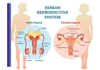
  • 2. The Reproductive System Slide 16.1 Copyright © 2003 Pearson Education, Inc. publishing as Benjamin Cummings • Gonads – primary sex organs • Testes in males • Ovaries in females • Gonads produce gametes (sex cells) and secrete hormones • Sperm – male gametes • Ova (eggs) – female gametes
  • 3. Male Reproductive System • Testes • Duct system • Epididymis • Ductus deferens • Urethra Slide 16.2a Copyright © 2003 Pearson Education, Inc. publishing as Benjamin Cummings
  • 4. Male Reproductive System • Accessory organs • Seminal vesicle • Prostate gland • Bulbourethral gland • External genitalia • Penis • Scrotum Slide 16.2b Copyright © 2003 Pearson Education, Inc. publishing as Benjamin Cummings
  • 5. Male Reproductive System Slide 16.2c Copyright © 2003 Pearson Education, Inc. publishing as Benjamin Cummings Figure 16.2
  • 6. Testes • Coverings of the testes • Tunica albuginea – capsule that surrounds each testis Slide 16.3a Figure 16.1 Copyright © 2003 Pearson Education, Inc. publishing as Benjamin Cummings
  • 7. Testes • Coverings of the testes (continued) • Septa – extensions of the capsule that extend into the testis and divide it into lobules Slide 16.3b Figure 16.1 Copyright © 2003 Pearson Education, Inc. publishing as Benjamin Cummings
  • 8. Testes • Each lobule contains one to four seminiferous tubules • Tightly coiled structures • Function as sperm-forming factories • Empty sperm into the rete testis • Sperm travels through the rete testis to the epididymis • Interstitial cells produce androgens such as testosterone Slide 16.4 Copyright © 2003 Pearson Education, Inc. publishing as Benjamin Cummings
  • 9. Epididymis • Comma-shaped, tightly coiled tube • Found on the superior part of the testis and along the posterior lateral side • Functions to mature and store sperm cells (at least 20 days) • Expels sperm with the contraction of muscles in the epididymis walls to the vas deferens Slide 16.5 Copyright © 2003 Pearson Education, Inc. publishing as Benjamin Cummings
  • 10. Ductus Deferens (Vas Deferens) • Carries sperm from the epididymis to the ejaculatory duct • Passes through the inguinal canal and over the bladder • Moves sperm by peristalsis • Spermatic cord – ductus deferens, blood vessels, and nerves in a connective tissue sheath Slide 16.6a Copyright © 2003 Pearson Education, Inc. publishing as Benjamin Cummings
  • 11. Ductus Deferens (Vas Deferens) • Ends in the ejaculatory duct which unites with the urethra • Vasectomy – cutting of the ductus deferens at the level of the testes to prevent transportation of sperm Slide 16.6b Copyright © 2003 Pearson Education, Inc. publishing as Benjamin Cummings
  • 12. Urethra • Extends from the base of the urinary bladder to the tip of the penis • Carries both urine and sperm • Sperm enters from the ejaculatory duct Slide 16.7a Copyright © 2003 Pearson Education, Inc. publishing as Benjamin Cummings
  • 13. Urethra Slide 16.7b Copyright © 2003 Pearson Education, Inc. publishing as Benjamin Cummings • Regions of the urethra •Prostatic urethra –surrounded by prostate •Membranous urethra – from prostatic urethra to penis •Spongy (penile) urethra – runs the length of the penis
  • 14. Seminal Vesicles • Located at the base of the bladder • Produces a thick, yellowish secretion (60% of semen) • Fructose (sugar) • Vitamin C • Prostaglandins • Other substances that nourish and activate sperm Slide 16.8 Copyright © 2003 Pearson Education, Inc. publishing as Benjamin Cummings
  • 15. Prostate Gland • Encircles the upper part of the urethra • Secretes a milky fluid • Helps to activate sperm • Enters the urethra through several small ducts Slide 16.9 Copyright © 2003 Pearson Education, Inc. publishing as Benjamin Cummings
  • 16. Bulbourethral Glands • Pea-sized gland inferior to the prostate • Produces a thick, clear mucus • Cleanses the urethra of acidic urine • Serves as a lubricant during sexual intercourse • Secreted into the penile urethra Slide 16.10 Copyright © 2003 Pearson Education, Inc. publishing as Benjamin Cummings
  • 17. Semen • Mixture of sperm and accessory gland secretions • Advantages of accessory gland secretions • Fructose provides energy for sperm cells • Alkalinity of semen helps neutralize the acidic environment of vagina • Semen inhibits bacterial multiplication • Elements of semen enhance sperm motility Slide 16.11 Copyright © 2003 Pearson Education, Inc. publishing as Benjamin Cummings
  • 18. External Genitalia • Scrotum • Divided sac of skin outside the abdomen • Maintains testes at 3°C lower than normal body temperature to protect sperm viability Slide 16.12 Copyright © 2003 Pearson Education, Inc. publishing as Benjamin Cummings
  • 19. External Genitalia • Penis • Delivers sperm into the female reproductive tract • Regions of the penis • Shaft • Glans penis (enlarged tip) • Prepuce (foreskin) • Folded cuff of skin around proximal end • Often removed by circumcision Slide 16.13a Copyright © 2003 Pearson Education, Inc. publishing as Benjamin Cummings
  • 20. External Genitalia • Internally there are three areas of spongy erectile tissue around the urethra Slide 16.13b Copyright © 2003 Pearson Education, Inc. publishing as Benjamin Cummings
  • 21. Spermatogenesis • Production of sperm cells • Begins at puberty and continues Slide 16.14 Copyright © 2003 Pearson Education, Inc. publishing as Benjamin Cummings throughout life • Occurs in the seminiferous tubules
  • 22. Processes of Spermatogenesis Slide 16.15a Copyright © 2003 Pearson Education, Inc. publishing as Benjamin Cummings • Spermatogonia (stem cells) undergo rapid mitosis to produce more stem cells before puberty • Follicle stimulating hormone (FSH) modifies spermatogonia division •One cell produced is a stem cell •The other cell produced becomes a primary spermatocyte
  • 23. Processes of Spermatogenesis Slide 16.15b Copyright © 2003 Pearson Education, Inc. publishing as Benjamin Cummings • Primary spermatocytes undergo meiosis • Haploid spermatids are produced
  • 24. Processes of Spermatogenesis • Spermiogenesis • Late spermatids are produced with distinct regions • Head – contains DNA covered by the acrosome • Midpiece • Tail • Sperm cells result after maturing of spermatids • Spermatogenesis takes 64 to 72 days Slide 16.16 Copyright © 2003 Pearson Education, Inc. publishing as Benjamin Cummings
  • 25. Processes of Spermatogenesis Figure 16.3 Slide 16.17 Copyright © 2003 Pearson Education, Inc. publishing as Benjamin Cummings
  • 26. Anatomy of a Mature Sperm Cell • The only human flagellated cell • DNA is found in the head Figure 16.5 Slide 16.18 Copyright © 2003 Pearson Education, Inc. publishing as Benjamin Cummings
  • 27. Testosterone Production • The most important hormone of the testes • Produced in interstitial cells Slide 16.19a Copyright © 2003 Pearson Education, Inc. publishing as Benjamin Cummings
  • 28. Testosterone Production • Functions of testosterone • Stimulates reproductive organ development • Underlies sex drive • Causes secondary sex characteristics • Deepening of voice • Increased hair growth • Enlargement of skeletal muscles • Thickening of bones Slide 16.19b Copyright © 2003 Pearson Education, Inc. publishing as Benjamin Cummings
  • 29. Regulation of Male Androgens (Sex Hormones) Slide 16.20 Copyright © 2003 Pearson Education, Inc. publishing as Benjamin Cummings Figure 16.6
  • 30. Female Reproductive System Slide 16.21a Copyright © 2003 Pearson Education, Inc. publishing as Benjamin Cummings • Ovaries • Duct System • Uterine tubes (fallopian tubes) • Uterus • Vagina • External genitalia
  • 31. Female Reproductive System Slide 16.21b Copyright © 2003 Pearson Education, Inc. publishing as Benjamin Cummings Figure 16.8a
  • 32. Ovaries • Composed of ovarian follicles (sac-like structures) • Structure of an ovarian follicle • Oocyte • Follicular cells Figure 16.7 Slide 16.22 Copyright © 2003 Pearson Education, Inc. publishing as Benjamin Cummings
  • 33. Ovarian Follicle Stages • Primary follicle – contains an immature oocyte • Graafian (vesicular) follicle – growing follicle with a maturing oocyte • Ovulation – when the egg is mature the follicle ruptures • Occurs about every 28 days • The ruptured follicle is transformed into a corpus luteum Slide 16.23 Copyright © 2003 Pearson Education, Inc. publishing as Benjamin Cummings
  • 34. Support for Ovaries • Suspensory ligaments – secure ovary to lateral walls of the pelvis • Ovarian ligaments – attach to uterus • Broad ligament – a fold of the peritoneum, encloses suspensory ligament Slide 16.24a Copyright © 2003 Pearson Education, Inc. publishing as Benjamin Cummings
  • 35. Support for Ovaries Slide 16.24b Copyright © 2003 Pearson Education, Inc. publishing as Benjamin Cummings Figure 16.8b
  • 36. Uterine (Fallopian) Tubes • Receive the ovulated oocyte • Provide a site for fertilization • Attaches to the uterus • Does not physically attach to the ovary • Supported by the broad ligament Slide 16.25 Copyright © 2003 Pearson Education, Inc. publishing as Benjamin Cummings
  • 37. Uterine Tube Function • Fimbriae – finger-like projections at the distal end that receive the oocyte • Cilia inside the uterine tube slowly move the oocyte towards the uterus (takes 3–4 days) • Fertilization occurs inside the uterine tube Slide 16.26 Copyright © 2003 Pearson Education, Inc. publishing as Benjamin Cummings
  • 38. Uterus • Located between the urinary bladder and rectum • Hollow organ • Functions of the uterus • Receives a fertilized egg • Retains the fertilized egg • Nourishes the fertilized egg Slide 16.27 Copyright © 2003 Pearson Education, Inc. publishing as Benjamin Cummings
  • 39. Support for the Uterus Slide 16.28a Copyright © 2003 Pearson Education, Inc. publishing as Benjamin Cummings • Broad ligament – attached to the pelvis • • Uterosacral ligaments – anchored posteriorly
  • 40. Support for the Uterus Slide 16.28b Copyright © 2003 Pearson Education, Inc. publishing as Benjamin Cummings Figure 16.8b
  • 41. Regions of the Uterus • Body – main portion • Fundus – area where uterine tube enters • Cervix – narrow outlet that protrudes into the vagina Slide 16.29 Copyright © 2003 Pearson Education, Inc. publishing as Benjamin Cummings
  • 42. Walls of the Uterus • Endometrium • Inner layer • Allows for implantation of a fertilized egg • Sloughs off if no pregnancy occurs (menses) • Myometrium – middle layer of smooth muscle • Serous layer – outer visceral peritoneum Slide 16.30 Copyright © 2003 Pearson Education, Inc. publishing as Benjamin Cummings
  • 43. Vagina • Extends from cervix to exterior of body • Behind bladder and in front of rectum • Serves as the birth canal • Receives the penis during sexual intercourse • Hymen – partially closes the vagina until it is ruptured Slide 16.31 Copyright © 2003 Pearson Education, Inc. publishing as Benjamin Cummings
  • 44. External Genitalia (Vulva) Slide 16.32a Copyright © 2003 Pearson Education, Inc. publishing as Benjamin Cummings • Mons pubis • Fatty area overlying the pubic symphysis • Covered with pubic hair after puberty Figure 16.9
  • 45. External Genitalia (Vulva) • Labia – skin folds • Labia majora • Labia minora Slide 16.32b Copyright © 2003 Pearson Education, Inc. publishing as Benjamin Cummings Figure 16.9
  • 46. External Genitalia • Vestibule • Enclosed by labia majora • Contains opening of the urethra and the greater vestibular glands (produce mucus) • Clitoris • Contains erectile tissue • Corresponds to the male penis Slide 16.33 Copyright © 2003 Pearson Education, Inc. publishing as Benjamin Cummings
  • 47. Oogenesis • The total supply of eggs are present at birth • Ability to release eggs begins at puberty • Reproductive ability ends at menopause • Oocytes are matured in developing ovarian follicles Slide 16.34 Copyright © 2003 Pearson Education, Inc. publishing as Benjamin Cummings
  • 48. Oogenesis • Oogonia – female stem cells found in a developing fetus • Oogonia undergo mitosis to produce primary oocytes • Primary oocytes are surrounded by cells that form primary follicles in the ovary • Oogonia no longer exist by the time of birth Slide 16.35 Copyright © 2003 Pearson Education, Inc. publishing as Benjamin Cummings
  • 49. Oogenesis • Primary oocytes are inactive until puberty • Follicle stimulating hormone (FSH) causes some primary follicles to mature • Meiosis starts inside maturing follicle • Produces a secondary oocyte and the first polar body • Meiosis is completed after ovulation only if sperm penetrates • Two additional polar bodies are produced Slide 16.36 Copyright © 2003 Pearson Education, Inc. publishing as Benjamin Cummings
  • 50. Oogenesis Figure 16.10 Slide 16.37 Copyright © 2003 Pearson Education, Inc. publishing as Benjamin Cummings
  • 51. Menstrual (Uterine) Cycle • Cyclic changes of the endometrium • Regulated by cyclic production of estrogens and progesterone • Stages of the menstrual cycle • Menses – functional layer of the endometrium is sloughed • Proliferative stage – regeneration of functional layer • Secretory stage – endometrium increases in size and readies for implantation Slide 16.38 Copyright © 2003 Pearson Education, Inc. publishing as Benjamin Cummings
  • 52. Hormonal Control of the Ovarian and Uterine Cycles Slide 16.39a Copyright © 2003 Pearson Education, Inc. publishing as Benjamin Cummings Figure 16.12a, b
  • 53. Hormonal Control of the Ovarian and Uterine Cycles Slide 16.39b Copyright © 2003 Pearson Education, Inc. publishing as Benjamin Cummings Figure 16.12c, d
  • 54. Hormone Production by the Ovaries • Estrogens • Produced by follicle cells • Cause secondary sex characteristics • Enlargement of accessory organs • Development of breasts • Appearance of pubic hair • Increase in fat beneath the skin • Widening and lightening of the pelvis • Onset of menses Slide 16.40 Copyright © 2003 Pearson Education, Inc. publishing as Benjamin Cummings
  • 55. Hormone Production by the Ovaries • Progesterone • Produced by the corpus luteum • Production continues until LH diminishes in the blood • Helps maintain pregnancy Slide 16.41 Copyright © 2003 Pearson Education, Inc. publishing as Benjamin Cummings
  • 56. Mammary Glands • Present in both sexes, but only function in females • Modified sweat glands • Function is to produce milk • Stimulated by sex hormones (mostly estrogens) to increase in size Slide 16.42 Copyright © 2003 Pearson Education, Inc. publishing as Benjamin Cummings
  • 57. Anatomy of Mammary Glands • Areola – central pigmented area • Nipple – protruding central area of areola • Lobes – internal structures that radiate around nipple • Alveolar glands – clusters of milk producing glands within lobules • Lactiferous ducts – connect alveolar glands to nipple Slide 16.43 Copyright © 2003 Pearson Education, Inc. publishing as Benjamin Cummings
  • 58. Stages of Pregnancy and Development • Fertilization • • Fetal development • Childbirth Slide 16.44 Copyright © 2003 Pearson Education, Inc. publishing as Benjamin Cummings
  • 59. Fertilization • The oocyte is viable for 12 to 24 hours after ovulation • Sperm are viable for 12 to 48 hours after ejaculation • Sperm cells must make their way to the uterine tube for fertilization to be possible Slide 16.45 Copyright © 2003 Pearson Education, Inc. publishing as Benjamin Cummings
  • 60. Mechanisms of Fertilization • Membrane receptors on an oocyte pulls in the head of the first sperm cell to make contact • The membrane of the oocyte does not permit a second sperm head to enter • The oocyte then undergoes its second meiotic division • Fertilization occurs when the genetic material of a sperm combines with that of an oocyte to form a zygote Slide 16.46 Copyright © 2003 Pearson Education, Inc. publishing as Benjamin Cummings
  • 61. The Zygote • First cell of a new individual • The result of the fusion of DNA from sperm and egg • The zygote begins rapid mitotic cell divisions • The zygote stage is in the uterine tube, moving toward the uterus Slide 16.47 Copyright © 2003 Pearson Education, Inc. publishing as Benjamin Cummings
  • 62. The Embryo • Developmental stage from the start of cleavage until the ninth week • The embryo first undergoes division without growth • The embryo enters the uterus at the 16-cell state • The embryo floats free in the uterus temporarily • Uterine secretions are used for nourishment Slide 16.48 Copyright © 2003 Pearson Education, Inc. publishing as Benjamin Cummings
  • 63. The Blastocyst • Ball-like circle of cells • Begins at about the 100 cell stage • Secretes human chorionic gonadotropin (hCG) to produce the corpus luteum to continue producing hormones • Functional areas of the blastocyst • Trophoblast – large fluid-filled sphere • Inner cell mass Slide 16.49 Copyright © 2003 Pearson Education, Inc. publishing as Benjamin Cummings
  • 64. The Blastocyst • Primary germ layers are eventually formed • Ectoderm – outside layer • Mesoderm – middle layer • Endoderm – inside layer • The late blastocyst implants in the wall of the uterus (by day 14) Slide 16.50 Copyright © 2003 Pearson Education, Inc. publishing as Benjamin Cummings
  • 65. Derivatives of Germ Layers • Ectoderm • Nervous system • Epidermis of the skin • Endoderm • Mucosae • Glands • Mesoderm • Everything else Slide 16.51 Copyright © 2003 Pearson Education, Inc. publishing as Benjamin Cummings
  • 66. Development from Ovulation to Implantation Figure 16.15 Slide 16.52 Copyright © 2003 Pearson Education, Inc. publishing as Benjamin Cummings
  • 67. Development After Implantation • Chorionic villi (projections of the blastocyst) develop • Cooperate with cells of the uterus to form the placenta • The embryo is surrounded by the amnion (a fluid filled sac) • An umbilical cord forms to attach the embryo to the placenta Slide 16.53 Copyright © 2003 Pearson Education, Inc. publishing as Benjamin Cummings
  • 68. Development After Implantation Figure 16.16 Slide 16.54 Copyright © 2003 Pearson Education, Inc. publishing as Benjamin Cummings
  • 69. Functions of the Placenta • Forms a barrier between mother and embryo (blood is not exchanged) • Delivers nutrients and oxygen • Removes waste from embryonic blood • Becomes an endocrine organ (produces hormones) and takes over for the corpus luteum • Estrogen • Progesterone • Other hormones that maintain pregnancy Slide 16.55 Copyright © 2003 Pearson Education, Inc. publishing as Benjamin Cummings
  • 70. The Fetus (Beginning of the Ninth Week) • All organ systems are formed by the end of the eighth week • Activities of the fetus are growth and organ specialization • A stage of tremendous growth and change in appearance Slide 16.56 Copyright © 2003 Pearson Education, Inc. publishing as Benjamin Cummings
  • 71. The Effects of Pregnancy on the Mother • Pregnancy – period from conception until birth • Anatomical changes • Enlargements of the uterus • Accentuated lumbar curvature • Relaxation of the pelvic ligaments and pubic symphysis due to production of relaxin Slide 16.57 Copyright © 2003 Pearson Education, Inc. publishing as Benjamin Cummings
  • 72. Effects of Pregnancy on the Mother • Physiological changes • Gastrointestinal system • Morning sickness is common due to Slide 16.58a Copyright © 2003 Pearson Education, Inc. publishing as Benjamin Cummings elevated progesterone • Heartburn is common because of organ crowding by the fetus • Constipation is caused by declining motility of the digestive tract
  • 73. Effects of Pregnancy on the Mother Slide 16.58b Copyright © 2003 Pearson Education, Inc. publishing as Benjamin Cummings • Physiological changes • Urinary System • Kidneys have additional burden and produce more urine • The uterus compresses the bladder
  • 74. Effects of Pregnancy on the Mother Slide 16.59a Copyright © 2003 Pearson Education, Inc. publishing as Benjamin Cummings • Physiological changes • Respiratory System • Nasal mucosa becomes congested and swollen • Vital capacity and respiratory rate increase
  • 75. Effects of Pregnancy on the Mother Slide 16.59b Copyright © 2003 Pearson Education, Inc. publishing as Benjamin Cummings • Physiological changes • Cardiovascular system • Body water rises • Blood volume increases by 25 to 40 percent • Blood pressure and pulse increase • Varicose veins are common
  • 76. Childbirth (Partition) • Labor – the series of events that expel the infant from the uterus • Initiation of labor • Estrogen levels rise • Uterine contractions begin • The placenta releases prostaglandins • Oxytocin is released by the pituitary • Combination of these hormones produces contractions Slide 16.60 Copyright © 2003 Pearson Education, Inc. publishing as Benjamin Cummings
  • 77. Initiation of Labor Figure 16.18 Slide 16.61 Copyright © 2003 Pearson Education, Inc. publishing as Benjamin Cummings
  • 78. Stages of Labor Slide 16.62a Copyright © 2003 Pearson Education, Inc. publishing as Benjamin Cummings • Dilation • Cervix becomes dilated • Uterine contractions begin and increase • The amnion ruptures
  • 79. Stages of Labor • Expulsion • Infant passes through the cervix and vagina • Normal delivery is head first • Placental stage • Delivery of the placenta Slide 16.62b Copyright © 2003 Pearson Education, Inc. publishing as Benjamin Cummings
  • 80. Stages of Labor Slide 16.63 Copyright © 2003 Pearson Education, Inc. publishing as Benjamin Cummings Figure 16.19
  • 81. Developmental Aspects of the Reproductive System Slide 16.64a Copyright © 2003 Pearson Education, Inc. publishing as Benjamin Cummings • Gender is determined at fertilization • Males have XY sex chromosomes • Females have XX sex chromosomes • Gonads do not begin to form until the eighth week
  • 82. Developmental Aspects of the Reproductive System • Testes form in the abdominal cavity and descend to the scrotum one month before birth • The determining factor for gonad differentiation is testosterone Slide 16.64b Copyright © 2003 Pearson Education, Inc. publishing as Benjamin Cummings
  • 83. Developmental Aspects of the Reproductive System • Reproductive system organs do not function until puberty • Puberty usually begins between ages 10 and 15 • The first menses usually occurs about two years after the start of puberty • Most women reach peak reproductive ability in their late 20s Slide 16.65 Copyright © 2003 Pearson Education, Inc. publishing as Benjamin Cummings
  • 84. Developmental Aspects of the Reproductive System • Menopause occurs when ovulation and menses cease entirely • Ovaries stop functioning as endocrine organs • There is a no equivalent of menopause in males, but there is a steady decline in testosterone Slide 16.66 Copyright © 2003 Pearson Education, Inc. publishing as Benjamin Cummings