Hydroxyl capped silver-gold alloy nanoparticles: characterization and their combination effect with different antibiotics against Staphylococcus aureus

Objective(s): Metal nanoparticles (NPs) offer a wide variety of potential applications in pharmaceutical sciences due to the unique advances in nanotechnology research. In this work, bimetal Ag-Au alloy NPs were prepared and their combinations with other antibiotics were tested against Staphylococcus aureus. Materials and Methods: Firstly, Ag-Au alloy NPs with Au/Ag molar ratio of 1:1 was fabricated and was purified by agarose gel electrophoresis system. The morphology and size of the purified NPs were confirmed by transmission electron microscopy. Chemical composition and surface chemistry of these NPs were studied with atomic absorption spectophotometry and Fourier transforms infrared spectroscopy, respectively. The size of purified Ag-Au alloy NPs was less than 200 nm. Also the presence of organic compounds with a hydroxyl residue was detected on the surface of these purified NPs. In next step the effect of purified Ag-Au alloy NPs on the antibacterial activity of different antibiotics was evaluated at sub-inhibitory content (5 μg/disk) using disk diffusion method against S. aureus. Ag NPs and Au NPs were also tested at same content (5 μg) using mentioned method. Results: The most enhancing effect of Ag-Au alloy NPs was observed for penicillin G and piperacillin. No enhancing effects on the antibacterial activity of different antibiotics were observed at 5 μg/disk for the mono-metal nanoparticles (Ag NPs and Au NPs) against S. aureus. Conclusion: These results signify that the Ag-Au alloy NPs potentiates the antimicrobial action of certain antibiotics suggesting a possible utilization of this nano material in combination therapy against resistant S. aureus.

Recomendados
Recomendados
Mais conteúdo relacionado
Mais procurados
Mais procurados (20)
Semelhante a Hydroxyl capped silver-gold alloy nanoparticles: characterization and their combination effect with different antibiotics against Staphylococcus aureus
Semelhante a Hydroxyl capped silver-gold alloy nanoparticles: characterization and their combination effect with different antibiotics against Staphylococcus aureus (20)
Mais de Nanomedicine Journal (NMJ)
Mais de Nanomedicine Journal (NMJ) (20)
Último
Último (20)
Hydroxyl capped silver-gold alloy nanoparticles: characterization and their combination effect with different antibiotics against Staphylococcus aureus
- 1. Nanomed J, Vol. 1, No. 3, Spring 2014 155 Received: Oct. 19, 2013; Accepted: Dec. 2, 2013 Vol. 1, No. 3, Spring 2014, page 155-161 Online ISSN 2322-5904 http://nmj.mums.ac.ir Original Research Hydroxyl capped silver-gold alloy nanoparticles: characterization and their combination effect with different antibiotics against Staphylococcus aureus Katayon Bahrami1 , Pardis Nazari1 , Mahshid Nabavi2 , Marjan Golkar2 , Ali Almasirad2 , Ahmad Reza Shahverdi1* 1 Department of Pharmaceutical Biotechnology and Biotechnology Research Center, Faculty of Pharmacy, Tehran University of Medical Sciences, Tehran, Iran 2 Department of Medicinal Chemistry, Pharmaceutical Sciences Branch, Islamic Azad University, Tehran, Iran Abstract Objective(s): Metal nanoparticles (NPs) offer a wide variety of potential applications in pharmaceutical sciences due to the unique advances in nanotechnology research. In this work, bimetal Ag-Au alloy NPs were prepared and their combinations with other antibiotics were tested against Staphylococcus aureus. Materials and Methods: Firstly, Ag-Au alloy NPs with Au/Ag molar ratio of 1:1 was fabricated and was purified by agarose gel electrophoresis system. The morphology and size of the purified NPs were confirmed by transmission electron microscopy. Chemical composition and surface chemistry of these NPs were studied with atomic absorption spectophotometry and Fourier transforms infrared spectroscopy, respectively. The size of purified Ag-Au alloy NPs was less than 200 nm. Also the presence of organic compounds with a hydroxyl residue was detected on the surface of these purified NPs. In next step the effect of purified Ag-Au alloy NPs on the antibacterial activity of different antibiotics was evaluated at sub-inhibitory content (5 µg/disk) using disk diffusion method against S. aureus. Ag NPs and Au NPs were also tested at same content (5 µg) using mentioned method. Results: The most enhancing effect of Ag-Au alloy NPs was observed for penicillin G and piperacillin. No enhancing effects on the antibacterial activity of different antibiotics were observed at 5 µg/disk for the mono-metal nanoparticles (Ag NPs and Au NPs) against S. aureus. Conclusion: These results signify that the Ag-Au alloy NPs potentiates the antimicrobial action of certain antibiotics suggesting a possible utilization of this nano material in combination therapy against resistant S. aureus. Keywords: Antibiotic resistance, Gold-silver alloy nanoparticles, Staphylococcus aureus *Corresponding author: Ahamd Reza Shahverdi, Department of Pharmaceutical Biotechnology, Faculty of Pharmacy and Biotechnology Research Center, Tehran University of Medical Sciences, Tehran, Iran. Tel.: +98 2166482708. E.mail: shahverd@sina.tums.ac.ir
- 2. Hydroxyl capped silver-gold alloy nanoparticles 156 Nanomed J, Vol. 1, No. 3, Spring 2014 Introduction Bacterial resistance limits the using of antimicrobials agents and is still considered as a main drawback in health and medicine (1-3). Recently, different silver nanoparticles (Ag NPs) formulations have been developed as potent anti- infective agent (4-8). Also, there are extensive reports on antibacterial effects of other metal nanoparticles or metal complex organic compounds (9-12). In addition, the combination of AgNPs with antibiotics was investigated in our previous work, where we demonstrated that antibacterial activity of penicillin G, amoxicillin, erythromycin, clindamycin, and vancomycin against Staphylococcus aureus significantly increases in the presence of silver nanoparticles (13). The effect of alloy nanomaterials such as silver gold alloy nanoparticles (Ag-Au alloy NPs) in combination with antibiotics has not been yet investigated. In this study, for the first time, the antibacterial activity of different antibiotics was evaluated against resistance S. aureus either in presence and absence of sub-inhibitory contents of Ag- Au alloy NPs, using disk diffusion assay. Ag-Au alloy NPs, were prepared with ethanol extract of black tea leaves and dextrose under microwave irradiation. These nanoparticles showed considerable enhancing effects on the antibacterial activity of different antibiotics against a resistance clinical strain of S. aureus. Materials and Methods Plant material and extraction Lahijan black tea leaves (Camellia sinensis) were supplied from the Lahijan Tea Research Center, Lahijan, Iran. The plants were pulverized (50 g), and the ethanol extract was prepared by macerating the powder for 24 h with three changes of the solvent at room temperature. The combined solvent extracts were separately evaporated to yield brownish, viscous residues. A stock solution (10 mg/ml) was prepared in ethanol and reserved in 4 o C for the next experiment. Synthesis of Ag-Au alloy NPs Ag-Au alloy NPs, were prepared by a recently optimized described methods using silver nitrate, chloroauric acid, dextrose, ethanol extract of black tea leaves as starting materials (14). All pure chemicals were purchased from Merck, Darmstadt, Germany. To obtain Ag-Au alloy NPs with an Au/Ag molar ratio (nominal value) of 1:1 the following procedure was followed. An aqueous chlorauric acid solution (10 ml, 1 mM) was added separately to the reaction vessel containing the ethanol extract of black tea (1 ml). Concomitantly, in a separate flask 200 µl of a dextrose (Merck, Germany) solution (0.1 M) and 8 µl of sodium hydroxide NaOH (0.1 M) were added to the AgNO3 solution (10 ml, 1 mM), respectively. Subsequently, two reaction mixtures were immediately added together to obtain a solution with an Au/Ag ratio of 1/1 and treated in a microwave oven (850 W) for 180 s to reduce the metal ions simultaneously. In separate experiments, gold and silver monometallic NPs were also prepared with ethanol extract of black tea leaves and dextrose, respectively (15- 17). Separation of metal alloy NPs using gel electrophoresis Separation of Au-Ag alloy NPs from the final colloid that may contain monometallic NPs (here gold or silver NPs) was performed using the gel electrophoresis system. After Au-Ag alloy NPs were fabricated using the method described in the first experimental section, as-prepared all generated metal NPs were preliminarily separated and washed from other natural ingredients or impurities by centrifuge. An aliquot of the prepared Au-Ag alloy colloids (3 ml) was centrifuged (Hettich Mikro) for 15 min at 25000 ×g and subsequently washed with distilled water. The process of centrifugation and re- dispersion in distilled water was repeated three times to ensure better separation of
- 3. Bahrami K, et al Nanomed J, Vol. 1, No. 3, Spring 2014 157 free entities from the prepared metal NPs. The re-dispersion procedure was carried out using ultrasonic waves for 10 min in an ultrasonic water bath. Finally, the pellet (about 10 mg) was mixed with 10 µl of loading buffer containing sucrose (30% w/v) in 0.5 x Tris-Borate-EDTA buffer and was subjected to agarose gel electrophoresis. The gold and silver colloids separately prepared by the ethanol extract of black tea or dextrose (15-16) were also washed with the same protocol and used as controls in this experiment. The 0.2 g of agarose powder (Sigma) was suspended in 100 ml of 0.5 x TBE-buffer pH 9 and boiled repeatedly in a microwave oven until clear. Molten agarose gel was poured in the gel casting tray. The gel was run in a horizontal electrophoresis system (Pharmacia LKB.GNA 100, electrode spacing 15 cm) for 30 min at 100 V using 0.25x TBE-buffer pH 9. The visible red band corresponding to Au-Ag alloy NPs was carefully separated using a surgical knife. The semi-solid agar containing the Au-Ag alloy was suspended in hot distilled water and centrifuged at 25000 ×g. The separated alloy fraction was washed again and subsequently re-suspended in distilled water by using ultrasonic wave treatment to form a stable colloid. The final colloid was subjected to further physicochemical characterization. Characterization of the purified Au-Ag alloy NPs The reduction of the Au+3 and Ag+ in the Au/Ag solution by the mentioned natural reducing agents was confirmed by sampling the aqueous component (2 ml) and measuring the UV-visible spectrum of the solutions. The sample was diluted three times with distilled water, and the UV- visible spectra of these samples were measured on a Labomed Model UVD- 2950 UV-VIS Double Beam PC Scanning spectrophotometer, operated at a resolution of 2 nm. The technique outlined above proved to be very useful for analyzing of NPs (18). For transmission electron microscopy,aqueoussuspension containing the purified Au-Ag alloy NPs was dispersed ultrasonically, and a drop of suspension was located on carbon-coated copper TEM grids and dried under an infrared lamp. Micrographs were achieved using a TEM (ZIESS 902A, Germany) operated at an accelerating voltage at 80 kV. The surface chemistry of the separated Au-Ag alloy NPs was further studied with Fourier transform infrared spectroscopy (Nicolet Magna 550). The Au/Ag ratio of the purified alloy NPs was confirmed with a Varian 220 atomic absorption spectrometer (AAS) equipped with a deuterium-arc lamp background corrector that was used for absorbance measurements at wavelengths of 242.2 nm and 328.1 nm for Au and Ag, respectively. Corresponding hollow-cathode lamps for Au and Ag determination (Hitachi Mitorika, Ibaraki, Japan) were used and operated at 15 mA for Au and 8 mA for Ag, respectively. Purified NPs (10 mg) were dissolved in freshly prepared concentrated HCl/HNO3 solution (3/1). The mixture was further diluted with distilled water and filtered before aspiration into the nebulizer of the atomic absorption spectrometer. Different standard solutions of Au+3 and Ag+ (0.5-5 ppm) were prepared as standard solutions. A proper amount of HAuCl4 3 H2O (Merck) and AgNO3 (Merck) was added into an aqueous solution of nitric acid (1%) to prepare these different standard solutions. Other electrophoresis bands were also separated and analyzed with the above-mentioned protocol to determine Au and Ag content in monometallic fractions. Disk diffusion assay The disk diffusion susceptibility test was carried out on Müeller-Hinton agar (Difco, Germany) plates in order to examine the antibacterial activity of candidate antibiotics against test strain. A clinical isolate of S. aureus from our culture collection used as test strain throughout this investigation. Standard antibiotics
- 4. Hydroxyl capped silver-gold alloy nanoparticles 158 Nanomed J, Vol. 1, No. 3, Spring 2014 disks listed in Table 1 were purchased from Mast Co., UK. To determine the combined effects, each standard paper disk was further aseptically loaded with Ag-Au alloy NPs at a sub-inhibitory amount of 5 µg/disk. A single colony of test strain was grown overnight in Müeller-Hinton liquid medium (Difco) on a rotary shaker (200 rpm) at 35°C. The inoculum was prepared by diluting the overnight cultures with 0.9% NaCl to a 0.5 McFarland standard and applied to the plates with a swab to ensure uniform distribution of test strain. Standard antibiotics disks, antibiotic disks containing Ag-Au alloy NPs (5 µg) and control disks (Ag-Au alloy NPs alone) were overlaid on the surface of inoculated media. After incubation at 35°C for 18 h, the inhibition zones were measured. Mean surface area of the inhibition zone (mm2 ) was calculated from mean diameter of each tested antibiotic. The above disk diffusion method was repeated for Ag NPs and Au NPs at same content (5 µg/disk). The percent of increase in the inhibition zone areas for different antibiotics against S. aureus were calculated as (b2 - a2 )/a2 ×100, where a and b are the inhibition zones for A and B, respectively. Results Synthesis of Ag-Au alloy NPs and their purification The combination of Au ions and Ag ions at a molar ratio of 1/1 was treated using ethanol extract of black tea and dextrose at alkaline condition in a microwave (850 W) oven for 180 s. The appearance of red in the reaction vessel suggested the formation of Ag-Au alloy NPs (20). The upper inset in the left illustration in Fig. 1 shows the vessel containing the Au3+ /Ag+ mixture after the reaction with the ethanol extract of black tea and dextrose in the microwave oven. The Au3+ /Ag+ -containing solution (tube not shown), which was transparent yellow at first, turned into red on completion of the reaction by the black tea ethanol extract and dextrose (upper inset in the left illustration of Figure 1). These freshly prepared NPs were subjected to agarose gel electrophoresis described in above section. As mentioned earlier, gold and silver NPs were separately synthesized and used as control. Three distinguished color bands were observed after agarose gel electrophoresis of the Au-Ag alloy NPs sample (lower inset in the left illustration Figure1). These different distinguished bands containing metal NPs showed different electrophoresis mobility in semi- solid agarose gel. By comparing to control bands contributing to Ag NPs or gold NPs (not shown in inst in Figure 1), the lower band and the upper band correspond to gold NPs and silver NPs, respectively. Both the silver and gold elements in samples derived from three distinguished bands (AuNPs, bimetal Ag-Au alloy NPs, AgNPs) were determined using atomic absorption spectroscopy. We did not detect any silver residue in lower band (corresponding to AuNPs) or gold impurities in upper band (corresponding to AgNPs) with atomic absorption spectroscopy. Middle band containing the Au-Ag alloy NPs was gently separated from the agarose gel and subjected to atomic absorption spectroscopy. The results of this experiment show the presence of both metals (gold and silver) in the purified bimetal NPs at a molar ratio of about 1 to 1. The bimetal NPs were further analyzed with UV-visible spectrophotometry and transmission electron microscopy. The plasmon resonance peaks of metal alloy fraction after purification with gel electrophoresis can be observed in the left -hand illustration in Figure 1. As can be seen in this illustration, a strong, surface plasmon resonance maximum of the Au-Ag alloy NPs was centered at 496 nm. The right- hand picture in Figure 1 shows the representative Transmission electron microscopy images recorded from the drop-coated film of the purified Au/Ag alloy NPs that were synthesized by treating the Au3+ /Ag+ -containing solution with an ethanol extract of black tea and
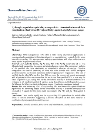
- 5. Bahrami K, et al Nanomed J, Vol. 1, No. 3, Spring 2014 159 dextrose at an alkaline condition in a microwave oven (850 W, 130 s). TEM images show spherical NPs with a size of less than 200 nm. The surface chemistry of the purified Au/Ag alloy NPs was studied using infra-red (IR) spectroscopy and the IR peaks centered at 3440 cm-1 confirmed the presence of a hydroxyl group on the surface of the Au/Ag alloy NPs (Figure 2). Figure 1. Transmission electron images of prepared Ag-Au alloy NPs (with different magnifications) synthesized by a biological method and purified using gel electrophoresis technique (Right pictures). Left illustration shows Uv-visible spectrum of purified. The insets in Fig.1 is photograph of the reaction mixture after reduction by ethanol extract of tea leaves and dextrose mixture (upper inset) and image of Ag-Au alloy NPs crude mixture run on the 0.2 % (w/v) agarose gel (lower inset). The positions of separated nanoparticles have been marked in this image. Figure 2. IR spectrum of purified Ag-Au alloy NPs prepared by ethanol extract of black tea leaves and dextrose shows hydroxyl groups on the surface. Antibacterial activity The antibacterial activity of hydroxyl capped Ag-Au alloy NPs alone was primarily investigated by disk diffusion method at the concentrations of 5, 10, and 20 µg/disk against S. aureus. A sub-MIC content (5 μg) was selected for further antibacterial assay in combination with different antibiotics against mentioned test strain. The diameters of inhibition zones (mm) in antibiotic disks either in presence or lack of Ag-Au alloy NPs are outlined in Table 1. The Ag-Au alloy NPs increased the antibacterial activity of amoxicillin, penicillin G, piperacillin, gentamycin erythromycin, bacitracin and tetracycline against S. aureus. The increase in surface areas of inhibition zones varied from 17% up to the considerable amount of 193% in presence of Ag-Au alloy NPs (Table 1). The most enhancing effect of Ag-Au alloy NPs was observed for penicillin G and piperacillin. In the second round of the test -in order to investigate the effect of mono-metal Nps (AgNPs and Au NPs) on antibacterial activity of different antibiotics listed in Table 1- the experiment was repeated; this time the antibacterial activity of different antibiotics in presence and absence of AgNPs or Au NPs were evaluated at content of 5 µg/disk against S. aureus (Table 1). As clear from the Table 1, no enhancing effects on the antibacterial activity of different antibiotics were observed for these mono-metal NPs at content of 5 µg/disk against S. aureus. Previously we demonstrated that AgNPs at content of 10 µg/disk and AuNPs at content of 40 µg/disk could increase the antibacterial activity of many antibiotics against S. aureus (13,16). The mono-metal NPs (AgNPs and AuNPs) at level of 5 µg/disks were chosen to guarantee that the enhancing effect produced was due to the bimetal alloy NPs and not to the effect of the AgNPs or AuNps themselves. Discussion In this study, the antibacterial activity of Ag-Au alloy NPs was tested against S. aureus. 0 0.1 0.2 0.3 0.4 300 400 500 600 700 Wavelength (nm) Absorbance(a.u.)
- 6. Hydroxyl capped silver-gold alloy nanoparticles 160 Nanomed J, Vol. 1, No. 3, Spring 2014 Table 1. The combination effect of Au-Ag alloy NPs (B). a All experiments were done in triplicate. b Mean surface area of the inhibition zone (mm2 ) was calculated for each tested antibiotic from the mean diameter. The percent of increases of inhibition zone area for different antibiotics against S. aureus were calculated as (b2 -a2 )/a2 ×100. The Ag-Au alloy NPs sub-inhibitory level of 5 µg/disks was selected to assure that the effect produced was due to the combination effect of Ag-Au alloy NPs with antibiotics and not to the effect of the Ag-Au alloy NPs themselves. Candidate antibiotics were carefully chosen since they represent major classes of antibiotics (penicillins, cephallosporins, macrolides, aminoglicosides, tetracyclines and fluoroquinolones). However, different antibiotics showed different activities in presence of Ag-Au alloy NPs. In detail, antibacterial activity of amoxicillin, penicillin G, piperacillin, gentamycin, erythromycin, bacitracin and tetracycline against S. aureus increased, while that the Remaining antibiotics were almost indifferent to the presence of Ag-Au alloy NPs. Many efforts have been made to overcome the emerging problem of antibiotic resistance among a variety of disease causing bacteria and advances in the field of nanobiotechnology may offer a great opportunity of research in this field. Therefore, studies on combination of antibiotic agents and synthetic and natural organic or inorganic nanomaterials are of great importance. The potential advantages of using organic or inorganic NPs as drug carriers are well reviewed in the literature (18-20). Here, for the first time, we report that antibacterial activity of amoxicillin, penicillin G, piperacillin, gentamycin erythromycin, bacitracin and tetracycline against a clinical test strains -S. aureus improves in presence of Ag-Au alloy NPs. Conclusion Due to its potential synergistic effect with mentioned antibiotics, Ag-Au alloy NPs may be considered as a valuable adjuvant in combination therapy of amoxicillin, penicillin Zone of inhibition (mm) Increase in area (%)a Antibiotics (µg/disk or unit /disk) Antibiotic Only(A)b Antibiotic plus metal nanoparticles Au-Ag alloy NPs (B) Ag NPs (B) Au NPs (B) Au-Ag alloy NPs Ag NPs Au NPs Amoxicillin 10 8 12 8 8 163.2 0 0 Penicillin G unit - 12 - - 193.8 0 0 Piperacillin 30 - 12 - - 193.8 0 0 Gentamicin 10 - 8 - - 30.6 0 0 Erythromycin 5 11 12 11 11 46.9 0 0 Bacitracin 0.04 unit 9 12 9 9 128.6 0 0 Tetracyclin 30 - 9 - - 65.3 0 0 Vancomycin 30 22 22 22 22 0 0 0 Ofloxacin 5 10 10 10 10 0 0 0 Methicillin 5 - - - - 0 0 0 Cefixime 5 - - - - 0 0 0 Ciprofloxacin 5 - - - - 0 0 0 Nalidixic acid 30 - - - - 0 0 0 Co-trimoxazole 25 - - - - 0 0 0 Cephalexin 30 - - - - 0 0 0 Kanamycin 30 - - - - 0 0 0 Carbenicillin 100 - - 0 0 0
- 7. Bahrami K, et al Nanomed J, Vol. 1, No. 3, Spring 2014 161 G, piperacillin, gentamycin erythromycin, bacitracin and tetracycline. However, the antibacterial activity of other antibiotics such as ciprofloxacin and kanamycin against S. aureus was remained constant in the presence of Ag-Au alloy NPs; therefore, the combination of Ag-Au alloy NPs with these antibiotics cannot be recommended for possible combination therapy. Acknowledgments This study was supported by School of Advanced Medical Technologies, Tehran University of Medical Sciences, Tehran, Iran. References 1. Wilke MH. Multiresistant bacteria and current therapy - the economical side of the story. Eur J Med Res. 2010; 15: 571-576. 2. Bennett PM. Plasmid encoded antibiotic resistance: acquisition and transfer of antibiotic resistance genes in bacteria. Br J Pharmacol. 2008; 153: S347-S357. 3. Fraimow HS, Tsigrelis C. Antimicrobial resistance in the intensive care unit: mechanisms, epidemiology, and management of specific resistant pathogens. Crit Care Clin. 2011; 27: 163-205. 4. Chaloupka K, Malam Y, Seifalian AM. Nanosilver as a new generation of nanoproduct in biomedical applications. Trends Biotechnol. 2010; 11: 580-588. 5. Ahamed M, Alsalhi MS, Siddiqui MK. Silver nanoparticle applications and human health.Clin Chim Acta. 2010; 411: 1841- 1848. 6. Nam KY. In vitro antimicrobial effect of the tissue conditioner containing silver nanoparticles. J Adv Prosthodont. 2011; 3: 20-24. 7. Babapour A, Yang B, Bahang S, Cao W. Low-temperature sol-gel-derived nanosilver- embedded silane coating as biofilm inhibitor. Nanotechnology. 2011; 22: 155602. 8. Yu B, Leung KM, Guo Q, Lau WM, Yang J. Synthesis of Ag-TiO2 composite nano thin film for antimicrobial application. Nanotechnology. 2011; 22: 115603. 9. Jin T, Sun D, Su JY, Zhang H, Sue HJ. Antimicrobial efficacy of zinc oxide quantum dots against Listeria monocytogenes, Salmonella Enteritidis, and Escherichia coli O157:H7. J Food Sci. 2009; 74: M46-52. 10. Borah BJ, Yadav A, Dutta DK. Au (degrees)- nanoparticles: control size and morphology stabilized by tripodal phosphine based ligands and their antimicrobial activity. J Biomed Nanotechnol. 2011; 7: 152-153. 11. Sahithi K, Swetha M, Prabaharan M, Moorthi A, Saranya N, Ramasamy K, Srinivasan N, Partridge NC, Selvamurugan N. Synthesis and characterization of nanoscale-hydroxyapatite- copper for antimicrobial activity towards bone tissue engineering applications. J Biomed Nanotechnol. 2010; 6: 333-339. 12. Li F, Mulyana Y, Feterl M, Warner JM, Collins JG, Keene FR. The antimicrobial activity of inert oligonuclear polypyridylruthenium(ii) complexes against pathogenic bacteria, including MRSA. Dalton Trans. 40: 5032-5038. 13. Shahverdi AR, Fakhimi A, Shahverdi HR, Minaian S. Synthesis and effect of silver nanoparticles on the antibacterial activity of different antibiotics against Staphylococcus aureus and Escherichia coli. Nanomedicine. 2007; 3: 168-171. 14. Mokhtari Nori N, Abdi K, Khoshayand MR, Ahmadi S, Lamei N, Shahverdi A.R. Microwave-assisted biosynthesis of gold– silver alloy nanoparticles and determination of their Au/Ag ratio by atomic absorption spectroscopy. J Exp Nanosci. 2013; 8: 442- 450 15. Banoee M, Mokhtari N, Akhavan Sepahi A, Jafari Fesharaki P, Monsef-Esfahani HR, Ehsanfar Z, Khoshayand MR, Shahverdi AR. The green synthesis of gold nanoparticles using the ethanol extract of black tea and its tannin free fraction. Iran. J. Meter. Sci. Eng. 2010; 7: 48-53. 16. Banoee M. Green synthesis of gold nanoparticles and its combination effect with different antibiotics. A Master dissertation. Science and Technology branch, Azad University. 2008. 17. Raveendran P, Fu J, Wallen S L, A simple and ‘‘green’’ method for the synthesis of Au, Ag, and Au–Ag alloy Nanoparticles. Green Chem. 2006; 8: 34-38. 18. Henglein A. Physicochemical properties of small metal particles in solution: “microelectrode” reactions, chemisorption, composite metal particles, and the atom-to- metal transition. J Phys Chem B. 1993; 97: 5457-5471. 19. Cho K, Wang X, Nie S, Chen Z, Shin DM. Theraputic nanoparticles for drug delivery in cancer. Clin Cancer Res. 2008; 14: 1310- 1316. 20. Gelperina S, Kisich K, Iseman MD, Heifets L. The potential advantages of nanoparticle drug delivery systems in chemotherapy of tuberculosis. Am J Respir Crit Care Med. 2005; 172: 1487-1490. 21. Jurgons R, Seliger C, Hilpert A, Trahms L, Odenbach S, Alexiou C. Drug loaded magnetic nanoparticles for cancer therapy. J Phys Condens Matter. 2006; 18: 2893-2902.