Degenerative Spine Disease.pptx
•Transferir como PPTX, PDF•
7 gostaram•2,574 visualizações

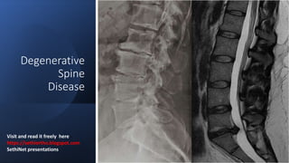
Degenerative spine disease is caused by the body's response to injury over a lifetime which results in the degradation of the functional spinal unit. This leads to changes in the intervertebral discs, endplates, facet joints, and ligaments. Risk factors include age, inheritance, trauma, and metabolic diseases. Degeneration typically occurs in the lumbar and cervical spine. Common signs are disc bulging and herniation, osteophyte formation, endplate damage, ligamentum flavum hypertrophy, and facet joint osteoarthritis - all of which can cause spinal stenosis and neurological symptoms. Treatment involves managing pain and symptoms while preserving spinal function.

Recomendados
Mais conteúdo relacionado
Mais procurados
Mais procurados (20)
Semelhante a Degenerative Spine Disease.pptx
Semelhante a Degenerative Spine Disease.pptx (20)
Mais de SethiNet presentations
Mais de SethiNet presentations (12)
Último
Último (20)
Degenerative Spine Disease.pptx
- 1. Degenerative Spine Disease Visit and read it freely here - https://sethiortho.blogspot.com SethiNet presentations
- 4. Functional spinal unit Spine is a multi articular structure comprising … • Two adjacent vertebrae • Intervertebral disc • Spinal ligaments
- 5. Functions of cervical vertebra Rotation Flexion / Extension Lateral flexion 1. Weight transmission 2. Multidirectional Movement- mainly C5/C6 and C6/C7
- 6. Thoracic vertebrae • Less mobile due to rib cage • Less frequently get degenerated
- 7. Functions lumbar vertebra 1. Weight transmission – L4/L5 and L5/S1 2. Multidirectional Movement –T12/L1
- 8. What is Degeneration ? Definition • Response to injury – Mechanical or Metabolic • Is a product of lifelong degradation of FSU with synchronized remodeling Simultaneous adaptation of the disc structures to changes in physical loading and response to the occasional injury Normal spine Destruction Repair Nutrient Weight bearing Movement Instability Remodeling Mechanical Metabolic
- 9. Risk factors Inheritance Age Abnormal mechanical axial stress Trauma Metabolic causes • Mucopolysaccharidoses – Cartilage and bone development • Hunter syndrome • Sanfilipo syndrome • Diabetes mellitus – Decrease synthesis proteoglycan and hexosamine
- 10. Where does Degeneration occur? 1. Intervertebral disk 2. End plates 3. Vertebral body 4. Posterior elements 1. Ligamentum flavum 2. Facet joint
- 12. Intervertebral disc Nucleus pulposis - Gelatenous substance - Proteoglycan - 80% water content Annulus fibrosus -Fibrocartilage -Closely attach to the end plate -Contain laminated Layers
- 13. Blood supply and Innervation
- 14. Degenerative Changes - Nucleus pulposis Degenerative process starts from Nucleous pulposus. The nucleous pulposus becomes dry and replaced by fibrous tissue Reduced intradiscal pressure, thus passing the mechanical load on to the annulus fibrosus. Annulus fibrosus has to hold greater mechanical load
- 16. Degenerative Changes- Annulus fibrosus Increased stress on the annulus fibrosus leads to development of cracks and cavities later progress to clefts and fissures. Annulus fibrosus fissures can be 1. Circumferential 2. Radial 3. Peripheral rim
- 17. Degenerative Changes- Annulus fibrosis • This loss of structural integrity of annulus fibrosus results in disc herniation. • Structural weakness may lead to the inability of the disc to maintain anatomical alignment and position progressing to instability and/or spondylolisthesis. .
- 18. Degenerative changes in End plate • End plate damage is the hallmark of degenerative changes • End plates play a crucial role in the maintenance of the mechanical environment • Participate the proper nutrition of avascular discs.
- 20. Degenerative Changes in End plate • End plate fractures lead to sudden depressurisation of the nucleous pulposus and the migration of the nucleous pulposus material into the vertebral body. • This elicits an inflammatory response and oedema • Very large end plate damage with a large volume of migrated nucleous pulposus material usually indicates Schmorl’s nodules
- 22. Degenerative changes Vertebral body - Modic Changes • Extract pathology – Related to Mechanical stress. • The abnormal uneven distribution load will affect vertebral end plates and the microenvironment of adjacent vertebral bone marrow, resulting in histological changes • There are three main forms of degenerative change involving the bone marrow of the adjacent vertebral bodies.
- 23. Type 1- Modic changes • Correspond to inflammatory stage of bone marrow leads to oedema and vascularized fibrous tissues • Signal changes may mimic or suggestsuggest infection. • Slow progressive degenerative disc disease produces a well-defined border response. • Strongly associated with nonspecific backpain and instability
- 24. Type 2- Modic changes • Type 2 changes reflect the presence of yellow marrow in the vertebral bodies • Fatty changes – Local fatty replacement of bone marrow
- 25. Type 3- Modic changes • Reactive osteosclerosis of adjacent to the endplates • Type 3 changes represent dense woven bone and the absence of marrow. • These changes are potentially stable and almost always asymptomatic
- 27. Degenerative changes - Facet joints True synovial joints Degenerative Changes are 1. Cartilage lining loses water content and wears away 2. Narrowing of the joint cavity 3. Osteophyte formation 4. Synovial cyst formation
- 28. Degenerative changes – Facet joints •Hypertrophic facet joint osteoarthritis can result in narrowing of the central canal, lateral recesses and foramina. •Osteophytes protruding ventrally from the anteromedial aspect of the facet joints may narrow the lateral recesses and intervertebral foramina causing central or lateral spinal canal stenosis
- 29. Degenerative changes – Facet joints •Bulging of the synovium through the facet joint capsule, especially in the presence of instability, may result in synovial cysts. •The majority (about 90%) of synovial cysts are found at the L4–L5 level and present clinically with lumbar radiculopathy.
- 30. Ligamentum flavum hypertrophy • It extends from the 2nd vertebra to the 1st sacral vertebra, connecting the two adjacent laminae • The ligamentum flavum tends to become hypertrophic with the degeneration of the elastic fibres and the proliferation of type II collagen. • Abnormal motions and instability within the involved segments are potential aetiologies of ligamentum flavum hypertrophy as the body tries to stabilise the diseased segment by making it
- 31. • Surgical removal is the only therapeutic manoeuvre for patients with symptoms caused by ligamentum flavum hypertrophy (Fig. 26b, c).
- 33. Spondylosis Spondylosis is common nonspecific term used to describe hypertrophic changes of the end plates (osteophytes) and facet joints. They result from increased flexibility between the vertebral bodies and the production of inhomogeneous mechanical stress on the annulus fibrosus and edges of the vertebral body, with subsequent sclerotic or hyperplastic changes occurring on the edges of the vertebral bodies. There are three types of true degenerative osteophytes:
- 34. Spondylosis …. Traction osteophytes Increase shear stress across the disk 2–3-mm bony spikes Osteophytes with a gap between the endplate and the base of the osteophytes and with the tip not protruding beyond the horizontal plane of the vertebral end plate
- 35. Spondylosis Claw osteophytes Are associated with horizontal instability. Arising from the vertebral margin with no gap and having an obvious claw appearance
- 36. Spondylosis…… A wraparound bumper osteophytes A wraparound bumper develops along the capsular insertion of the facet joints and is believed to be associated with instability
- 37. Disk Herniation
- 38. Disc Herniation Displacement of disc material beyond the limits of the IVD space
- 39. Disc bulging • Early sign of degeneration • A rapid increase in intradiscal pressure in the setting of bulging leads to the development of annular fissures and herniation. • Features • Height of the disc preserved • annulus fibrosus is intact • Often seen in asymptomatic individuals
- 40. Focal herniation - Protrusion Focal displacement of disc material with no or minimal disruption of the fibres of the overlying annulus fibrosus and intact PLL Localised (>25% of the circumference of the disc) displacement of disc material
- 42. Extrusion Extrusion is the displacement of disc material with a full thickness disruption of the annulus fibrosus fibres Usually PLL remains intact
- 43. Extrusion with sequestration • When extruded disc material that has no continuity with the disc of origin. • Fragment of disk may stay at the level of the disc or may migrate superiorly or inferiorly. • Pain and neurological symptoms may fluctuate with the migration of the free fragment within the spinal canal. • The acute displacement of a free fragment from the disc into the spinal canal may cause acute cauda equina syndrome
- 46. Acute disc Herniation - <4weeks • It occurs at the early stages of degeneration • Trauma /lifting heavy weight • when the intradiscal pressure is high displacement of NP through compromised AF fibers • Fibers of Annulus fibrosus get rupture and elict acute local inflammation. NP AF
- 47. Subacute Disc herniation < 4-12 weeks Classic Mechanical backache Pain usually arises only when the disc material migrates peripherally with increasing intradiscal pressure Pain improves when the intradiscal pressure drops. The remaining intact fibres of the annulus fibrosus recoil to bring the extruded material back into the disc space. MRI- Prone position – disk comes back to normal positions
- 48. Chronic disc herniation < 12weeks Stable displacement of the disc material - because of high intradiscal pressure pushing the nucleous pulposus material out of the disc AF – get calcified loss of recoiling effect Excess axial stress – 1. Tearing of annular fibers – Acute stage pain
- 49. Disc Herniation Cervical spine Axial neck pain Root • Occipital headache • Pain in the Trapezius region • Cervical Radiculopathy pain Cord • Upper- Lower limb weakness Lumbar Spine Axial Backache Root Sciatica /Radiculopathy pain Cord Cauda equina syndrome Complications of disc displacement
- 50. Degenerative spondylolisthesis Common - lumbar spine Less common – C.spine Never occurs in the thoracic spine.
- 51. Grading - • Based on the ratio of the overhanging part of the superior vertebral body to the anteroposterior length of the adjacent inferior vertebral body