
Recomendados
Mais conteúdo relacionado
Mais procurados
Mais procurados (20)
Semelhante a breech presentation.pptx
Semelhante a breech presentation.pptx (20)
Mais de HamzaAbid26
Último
Último (20)
breech presentation.pptx
- 2. Classification of Breech Presentation Normally, the increasing bulk of the buttocks seeks the more spacious fundus. Breech presentation persists at term in 3 to 4 % of singleton deliveries.
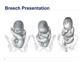
- 3. FRANK COMPLETE INCOMPLETE Classification of Breech Presentation
- 4. Diagnosis Risk Factors: Abnormal amnionic fluid volume High parity with uterine relaxation Prior breech delivery Hydrocephaly Anencephaly Pelvic tumors Uterine anomalies Multifetal gestation Early gestational age Placenta previa Fundal placental implantation
- 5. Diagnosis Examination Leopold maneuvers – to ascertain fetal presentation L1: hard, round, ballotable head L2: back & small parts L3: movable breech in the pelvic inlet L4: firm breech beneath the symphisis Accuracy of palpation varies – Sonographic evalution is indicated
- 6. Diagnosis Vaginal examination Frank Breech Ischial tuberosity, sacrum, anus, external genetalia Fetal sutures & fontanels Muscular resistance of the anus ± meconium Less yielding jaws Straight line: Ischial tuberosity & anus Triangular: Malar eminences & Mouth Complete breech Feet along side the buttocks Footling One or both feet inferior to the buttocks Breech Cephalic
- 7. Diagnosis or Fetal positions LSA = Left sacrumanterior RSA = Right sacrumanterior LSP = Left sacrum posterior RSP = Right sacrum posteri ST – Sacrum transverse LSP Designated to reflect the relations of the fetal sacrum to the maternal pelvis.
- 8. Route of Delivery Determining Factors: Pelvic dimensions Operator experience Patient preference Hospital capabilities Fetal characteristics Coexistent pregnancy complications
- 9. Route of Delivery Term Studies are conflicting regarding superior perinatal outcomes for planned cesarean delivery of a singleton term breech. Superior outcomes with CS No Difference* Term Breech Trial Presentation and Mode of Delivery (n=8,000) WHO (n=100,000) Lille Breech Group * French studies
- 10. Route of Delivery Preterm No randomized studies. Planned cesarean delivery appears to confer a survival advantage. Limited data regarding any preferable delivery route for preterm breech between 32 and 37 weeks – fetal weight is likely most important: SOGC: recommend that vaginal breech delivery is reasonable when the estimated fetal weight is > 2500 g
- 11. Delivery Complications Maternal Extend hysterectomy incision Vaginal & cervical wall laceration Deep perineal tears Increase infection risks Anesthesia » Uterine relaxation » Uterine atony » Postpartum hemorrhage
- 12. Delivery Complications Perinatal Fractures of the humerus, clavicle & femur Brachial plexus stretching » Upper extremity paralysis Skull depression or fracture Spinal cord injury Vertebral fracture Umbilical cord prolapse
- 13. Imaging Sonography Provides the best confirmation; type of breech & degree of neck flexion or extension Ensure that CS is not performed under emergency conditions for anomalous fetuses with no chances of survival Pelvimetry For assessment of bony pelvis prior to vaginal delivery. Computed tomography is favored because of its accuracy, low radiation, and wide availability.
- 14. Inlet transverse diameter ≥ 12 cm Inlet transverse diameter minus fetal BPD is ≥ 2.5 cm Midpelvic interspinous diameter ≥ 10 cm Midpelvis interspinous diameter minus the BPD is ≥ 0 Inlet anteroposterior diameter ≥ 10.5 cm Pelvic Inlet Pelvimetry
- 15. Obstetrical conjugate minus the fetal BPD is ≥ 1.5 cm Three A-P diameter of the Pelvic inlet Pelvimetry
- 16. Decision Making The decision regarding the mode of delivery should depend on the experience of the health care provider. Planned vaginal delivery of a term singleton breech fetus may be reasonable under hospital-specific protocol guidelines. -American College of Obstetrics and Gynecology
- 17. Decision Making Factors Favoring CS of Breech Fetus Prior cesarean delivery Patient request for CS Hyperextended head Lack of operator experience Large fetus >3800 to 4000 g Severe fetal-growth restriction Fetal anomaly incompatible with NSD Incomplete or footling breech presentation Prior perinatal death or neonatal birth trauma Pelvic contraction or unfavorable pelvic shape Apparently healthy and viable preterm with active labor or indicated delivery
- 18. Methods of Vaginal Delivery 1. Spontaneous breech 2. Partial breech extraction 3. Total breech extraction General methods: Necessary staff: Obstetrician Midwife/nurse/intern assist Pediatrician trained in newborn resuscitation Anesthesia (when needed)
- 19. Management of Labor Mother Start IV access for anesthesia/resuscitation. IE to assess cervical dilatation, effacement & station of the presenting part. Satisfactory progress in labor is the best indicator of pelvic adequacy. Fetus Fetal heart rate recorded at least every 15 minutes. If a nonreassuring, decision must be made regarding the necessity of cesarean delivery.
- 20. Cardinal Movements with Breech Delivery Engagement Bitrochanteric diameter Descent Of the Breech.Anterior hip usually descends more rapidly Internal rotation Internal rotation of 45o, when the resistance of the pelvic floor is met, bringing the anterior hip toward the pubic arch.
- 21. Cardinal Movements with Breech Delivery Flexion Lateral flexion of the fetal body, the posterior hip is forced over the perineum. External rotation Slight external rotation, after the birth of the breech, i.e. the back turns anteriorly as the shoulders are brought into relation with one of the oblique diameters of the pelvis. Expulsion
- 22. Partial Breech Extraction Ideally, the breech is allowed to deliver spontaneously to the umbilicus. Once the breech has passed beyond the vaginal introitus, the abdomen, thorax, arms, and head must be delivered promptly either spontaneously/assisted. Episiotomy
- 23. Partial Breech Extraction The posterior hip will deliver, usually from the 6 o’clock position. Anterior hip then delivers, followed by external rotation to a sacrum anterior position. Legs are sequentially delivered by splinting the medial aspect of each femur with the operator’s fingers positioned parallel to each femur, and by exerting pressure laterally to sweep them away from the midline.
- 24. Partial Breech Extraction Then the fetal bony pelvis is grasped with both hands, using a cloth towel moistened with warm water. Operators fingers on the anterior superior iliac crests and thumbs on the sacrum. The mother is encouraged to push in conjunction with downward traction.
- 25. Partial Breech Extraction The cardinal rule Employ steady, gentle, downward traction until the lower halves of the scapulas are delivered, making no attempt at delivery of the shoulders and arms until one axilla becomes visible.
- 26. Partial Breech Extraction The appearance of one axilla indicates that its time to deliver the shoulders. There’s no difference which shoulder is delivered first.
- 27. Partial Breech Extraction With the scapulas visible, the trunk is rotated in such a way that the anterior shoulder and arm appear at the vulva and can easily be released and delivered first.
- 30. Partial Breech Extraction The body of the fetus is then rotated 180 degrees in the reverse direction to deliver the other shoulder and arm.
- 31. Partial Breech Extraction If trunk rotation is unsuccessful. Posterior shoulder is delivered first. Feet are grasped in one hand and drawn upward over the inner thigh of the mother, toward which the ventral surface of the fetus is directed.
- 32. Partial Breech Extraction By depressing the body of the fetus, the anterior shoulder emerges beneath the pubic arch, the arm and hand usually follow spontaneously.
- 33. Nuchal Arm By rotating the fetus through a half circle in such a direction that the friction exerted by the birth canal will serve to draw the elbow toward the face. Should rotation of the fetus fail to free the nuchal arm(s), it may be necessary to push the fetus upward in an attempt to release it.
- 34. Nuchal Arm If still unsuccessful, try hooking a finger(s) over it and force the arm over the shoulder, and down the ventral surface.
- 35. Modified Prague Maneuver Rarely, the back of the fetus fails to rotate to the anterior. Rotation of the back to the anterior may be achieved using stronger traction on the fetal legs or bony pelvis.
- 36. Modified Prague Maneuver If unsuccessful try MP. Modified Prague maneuver, consists of two fingers of one hand grasping the shoulders of the back-down fetus from below while the other hand draws the feet up and over the maternal abdomen.
- 37. Delivery of the Aftercoming Head Mauriceau maneuver The index and middle finger of one hand are applied over the maxilla, to flex the head, while the fetal body rests on the palm of the hand and forearm.
- 38. Mauriceau maneuver Operator uses both hands simultaneously and in tandem to exert continuous downward traction simultaneously on the fetal neck and on the maxilla.
- 39. Mauriceau maneuver Two fingers are hooked over the fetal neck, and grasping the shoulders. Downward traction is concurrently applied until the suboccipital region appears under the symphysis.
- 40. Mauriceau maneuver Gentle suprapubic pressure simultaneously applied by an assistant helps keep the head flexed.
- 41. Mauriceau maneuver The body then is elevated toward the maternal abdomen, and the mouth, nose, brow, and eventually the occiput emerge successively over the perineum.
- 42. Forceps to Aftercoming Head Piper forceps, may be applied electively or when MM cannot be accomplished. The blades of the forceps should not be applied to the aftercoming head until it has been brought into the pelvis by gentle traction, combined with suprapubic pressure, and is engaged.
- 43. Forceps to Aftercoming Head Suspension of the body of the fetus in a towel effectively holds the fetus up and helps keep the arms and cord out of the way as the forceps blades are applied.
- 44. Forceps to Aftercoming Head Piper forceps have a downward arch in the shank to accommodate the fetal body and lack a pelvic curve.
- 45. Forceps to Aftercoming Head The blade to be placed on the maternal left is held in the operator’s left hand. The right hand slides between the fetal head and left maternal vaginal sidewall to guide the blade inward and around the parietal bone.
- 46. Forceps to Aftercoming Head The opposite blade mirrors this application.
- 47. Forceps to Aftercoming Head Once in place, the blades are articulated, and the fetal body rests across the shanks. The head is delivered by pulling gently outward and raising the handle simultaneously.
- 48. Forceps to Aftercoming Head This rolls the face over the perineum, while the occiput remains beneath the symphysis until after the brow delivers. Ideally, the head and body move in unison to minimize trauma.
- 49. Entrapment of the Aftercoming Head Especially with a small preterm, the incompletely dilated cervix will constrict around the neck and impede delivery of the head. Assume that there is significant and even total cord compression. Time management is essential! Try putting gentle traction on the fetal body, this may manually slip the cervix over the occiput.
- 50. Entrapment of the Aftercoming Head If this is not successful, then Dührssen incisions may be necessary. Incision at 2 o’clock,, followed by a second incision at 10 o’clock. Infrequently, additional incision is required at 6 o’clock.
- 51. Entrapment of the Aftercoming Head Incisions are so placed to minimize the bleeding from the laterally located cervical branches of the uterine artery. Last resort: replacement of the fetus higher into the vagina and uterus, followed by cesarean delivery.
- 52. Total Breech Extraction A hand is introduced through the vagina, and both fetal feet are grasped. The ankles are held with the second finger lying between them. With gentle traction, the feet are brought through the introitus.
- 53. Total Breech Extraction Complete & Incomplete If difficulty is experienced in grasping both feet, first one foot should be drawn into the vagina but not through the introitus. Then the other foot is advanced in a similar fashion. Both feet are grasped and pulled through the vulva simultaneously.
- 54. Total Breech Extraction Complete & Incomplete As the legs begin to emerge through the vulva, downward gentle traction is continued. When the breech appears at the vaginal outlet, gentle traction is applied until the hips are delivered.
- 55. Total Breech Extraction Complete & Incomplete The thumbs are then placed over the sacrum and the fingers over the hips, and partial breech extraction is done.
- 56. Total Breech Extraction Frank Breech Moderate traction is exerted by a finger in each groin and aided by a generous episiotomy.
- 57. Total Breech Extraction Frank Breech Once the hips are delivered, each hip and knee is flexed to deliver them from the vagina. Once the breech is pulled through the introitus, the steps described for partial breech extraction are then completed.
- 58. Frank breech decomposition (Pinard maneuver) This procedure involves manipulation within the birth canal to convert the frank breech into a footling breech. It aids in bringing the fetal feet within reach of the operator.
- 59. Frank breech decomposition (Pinard maneuver) Accomplished more readily if the membranes have ruptured only recently. Minimal amnionic fluid » uterus may become tightly contracted around the fetus.
- 60. Frank breech decomposition (Pinard maneuver) Pharmacological relaxation General anesthesia Magnesium sulfate IV Betamimetic agent such as Terbutaline, 250 μg SQ
- 61. Frank breech decomposition (Pinard maneuver) Two fingers are carried up along one extremity to the knee to push it away from the midline. Spontaneous flexion usually follows, and the foot of the fetus is felt to impinge on the back of the hand. The fetal foot then may be grasped and brought down.
- 62. ANALGESIA AND ANESTHESIA Disadvantage: May increase the need for labor augmentation and may prolong second-stage labor. Advantage: Better pain relief and increased pelvic relaxation should extensive manipulation be required. Continuous epidural analgesia, is advocated by some as ideal for women in labor with a breech presentation.
- 63. ANALGESIA AND ANESTHESIA Nitrous oxide plus oxygen inhalation provides further relief from pain. If general anesthesia is required, it must be induced quickly. Anesthesia for breech decomposition and extraction must provide sufficient relaxation to allow intrauterine manipulations.
- 64. External Cephalic Version With external version, the manipulations are performed exclusively through the abdominal wall. With internal version, they are accomplished inside the uterine cavity. Cephalic version – head is made the presenting part Podalic version – breech is made the presenting part
- 65. External Cephalic Version Complications Placental abruption Uterine rupture Fetomaternal hemorrhage, Alloimmunization Preterm labor, Fetal compromise, Amnionic fluid embolism.
- 66. External Cephalic Version Preparations: Done in a facility equipped for emergency cesarean delivery. Sonographic examination Confirm breech presentation Document amnionic fluid volume Exclude obvious fetal anomalies Identify placental location.
- 67. External Cephalic Version External monitoring to assess fetal heart rate. Anti-D immune globulin is given to Rh-D negative mother.
- 68. External Cephalic Version A forward roll is attempted first. Each hand grasps one fetal pole, and the fetal buttocks are elevated from the maternal pelvis and displaced laterally.
- 69. External Cephalic Version The buttocks are then gently guided toward the fundus, while the head is directed toward the pelvis. Backward flip is attempted, if forward roll is unsuccessful
- 70. External Cephalic Version Discontinue if with: Excessive discomfort Persistently abnormal fetal heart rate Multiple failed attempts.
- 71. Internal Podalic Version Used only for the delivery of a second twin. Membranes preferably intact. A hand is inserted into the uterine cavity to turn the fetusmanually. Operator seizes one or both feet and draws them through the cervix, while the other hand transabdominally push the upper portion of the fetal body in the opposite direction. Partial breech extraction then follows.