
Recomendados
Recomendados
Mais conteúdo relacionado
Mais procurados
Mais procurados (20)
Semelhante a 2 diagnosis of colic in equines prof.dr hamed attia
Semelhante a 2 diagnosis of colic in equines prof.dr hamed attia (20)
Mais de HamedAttia3
Mais de HamedAttia3 (20)
Último
Último (20)
2 diagnosis of colic in equines prof.dr hamed attia
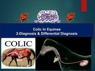
- 1. Colic In Equines 2-Diagnosis & Differential Diagnosis
- 2. ا . د عطية حامد الحيوان طب استاذ – البيطرى الطب كلية الزقازيق جامعة - مصر الخبربالرياض اوميجا بمؤسسة الفنى المستشار البيطرية لألدوية الخيول فى المغص الثانى الجزء : التفريقى والتشخيص التشخيص
- 3. الخيول فى المغص الثانى الجزء : التفريقى والتشخيص التشخيص طرق السونار باستخدام التشخيص اكس أشعة بأستخدام التشخيص البطن منظار باستخدام التشخيص المعملى التشخيص البطن فتح طريق عن التشخيص االستكشافى التفريقى التشخيص الحالة على الحكم . الحالة تاريخ األكلينيكى الفحص النبض قياس الجفاف درجة قياس المخاطية األغشية فحص السماعة باستخدام التشخيص ال اللى انبوبة باستخدام التشخيص معدى الشرجى الفحص طريق عن التشخيص
- 5. Normal Temp. Pulse & Resp. Temp 37.5 Pulse 28-40 Resp 8-18
- 7. 1-Case history • A-How sever has the pain been? • B-When did the horse last defecate? And what was the character of the feces? • C-Has the horse shown specific behavior such as playing with water?
- 8. 1-Case history • D-Could the horse have graine access highly fermentable food? • What is the horse’s past medical history?
- 9. Clinical Signs • Flank-watching or -biting, • pawing, • trying to roll, • an elevated heart rate, • a lack of gut sounds or bowel movements. • While these signs and others are pretty clear, common indicators your horse is colicking,
- 11. 2- Auscultation of the heart
- 12. 2- Examination of heart: • The heart rate and the character of pulse are important in determining the degree of severity of colic. • Rates greeter than 80 bpm should be considered the result of sever lesion or disease • عن القلب ضربات زيادة 60 مؤشر يعتبر الدقيقة فى ضربة خطر
- 13. 3-Estimation to Degree of Dehydration: الجفاف درجة تحديد A- Skin Pinch Test. B-Capillary Refile Time(CRT).
- 14. A-Skin-pinch test • Pinch the skin near the point of the shoulder. • Skin snaps quickly back into place = sufficient hydration. • Skin stays tented for 2-4 seconds = moderate dehydration. • Skin remains lifted from the flesh for 4- 6 seconds = severe dehydration.
- 15. B-Capillary Refile Time(CRT) • Pressing a finger or thumb on the upper gum, above an incisor, for a second or two. • Color returns to the gum in one to two seconds: horse is amply hydrated. • Gums remain blanched for longer than two seconds: horse is likely dehydrated.
- 16. Degree dehydration 1- Mild • Mild: • 5-7% body weight in water loss. Symptoms: • Depression. • Dry mucous membranes. • Slow capillary refill time (>2 seconds).
- 17. 2-Moderate Dehydration • Moderate: • 8-10% water loss. • Signs: • Depression, • Weak pulse, • Elevated heart rate • Prolonged capillary refill time (2-4 seconds).
- 18. 3-Severe Dehydration • Greater than 10% water loss. • Symptoms: • Cold limbs • lethargy. • persistent skin “tenting.” • Horses may be near death with multiple organ failure.
- 19. 4 Colure of the MM
- 20. • In simple dehydration the oral mm is slight bluish. • in mild dehydration it becomes brick red or cyanosed.
- 21. • In Sever Dehydration: • it becomes pale blue-grey color. • Severe dehydration normally occurs when the damaged gut is no longer capable of absorbing fluid into the body.
- 22. 5- Auscultation of the abdomen
- 23. Motilty may be one the following • 0 - Silent, no motility heard during 30 seconds. • 1 - Less than normal motility. • 2 - Normal motility. • 3 - Hypermotile, more gut sounds than usual. • الرمل حركة صوت نسمع القولون فى رمال وجود حالة فى ورقة على
- 24. السماعة وضع مكان ومن اليمين جهة من اليسار جهة اليمين اليسار
- 26. Listen to the upper right quadrant • Listen to the upper right quadrant where gas is often heard since this roughly corresponds to the base of the cecum. • Move down to the lower right quadrant • عدم سماع صوت حركة األمعاء فى الجزء العلوى من ناحي ة اليمين احتمال امتالء االعور بالغازات او الطعام • فى حالة الغازات نسمع الصوت المعدنى • عدم سماع صوت حركة األمعاء فى الجزء السفلى دليل وجود لكمة فى القولون او رمال
- 27. Listen to the upper& lower left quadrant • Listen to the upper left quarter • Drop the stethoscope down about 8 inches and listen to the lower left quadrant. ضعف حركة االمعاء فى الجزء العلوى من اليسار دليل على حدوث شلل فى االمعاء عدم سماع صوت فى الجزء السفلى من اليسار يدل على احتمال حدوث انزياح للقولون اليسار السفلى من مكانه
- 28. 6-Nasogastric intubation والعالج للتشخيص المعدى اللى انبوبة
- 29. 1-Passing a Nasogastric (Stomach) Tube استخدام دواعى المعدى اللى انبوبة الخيول فى
- 30. المعدى اللى انبوبة ادخال خطوات 1 - االولى الخطوة
- 31. المعدى اللى انبوبة ادخال خطوات 1 - الثانية الخطوة
- 32. المعدى اللى انبوبة ادخال خطوات 1 - الثالثة الخطوة انها للتأكد االنبوب نفخ المعدة داخل
- 33. Introduction of naso -gastric intubation
- 34. Introduction of naso -gastric intubation Diagnosis Therapy
- 35. Gastric reflux • : ظاهرة ارتداد السوائل فى المعدة : • A blockage in the bowel (usually the small intestine) that causes the build up of fluid in front of it.
- 36. Complications • Hemorrhage • Aspiration Pneumonia • Gastric rupture • Esophageal necrosis
- 37. Complications • Hemorrhage • Aspiration Pneumonia • Gastric rupture • Esophageal necrosis
- 38. 7-Rectal palpation • :اآلتية االعضاء فحص يمكن
- 39. 7-Rectal palpation • :اآلتية االعضاء فحص يمكن • Caudal portions of the large intestine, • Caudal edge of spleen, • Left kidney, • Aorta, • Mesenteric root, • Reproductive tract of mares, • Inguinal rings in stallions.
- 40. Precautions • Administration of sedatives: • (Buscopan :hyosin hydrobromide) • VET:(N-Butylscopolammonium bromide) • Xylazine: • Doses of up to 0.5mg/kg of xylazine IV can be useful for short durations (15 to 30 minutes)
- 41. Precautions • Glove lubricated with lubricant. • Short nails • Application of a nose twitch • Lifting one of the fore limb
- 42. Normal anatomical structure viewed from the caudal aspect of the equine abdomen.
- 43. Examination of colon & cecum A-In large colon impaction: is characterized by an enlarged, firm, filled viscous located on the pelvic floor. B-Cecal impaction : • palpation of a firm, impacted cecum or a grossly distended • fluid-filled cecum per rectum.
- 45. Indications 1- Intestinal motility and distension can be evaluated for both the large and small intestine. 2-The thickness of the intestinal wall can be evaluated and measured.
- 46. Indications 3-some abdominal abscesses. 4- Intussusception. 5-Peritoneal effusion(Volume and type). 6-Gastric dilatation
- 47. Indications • 7-Adhesions, masses, • 8-Left dorsal displacement of the large colon, • 9-Hemoperitoneum • 10-Splenic abnormalities.
- 48. Trans-abdominal ultrasonography showing thickened loops of small intestine due to inflammatory bowel disease.
- 49. 9- Radiography
- 50. 8-Radiology • Sand accumulation • Enteroliths
- 51. 10- Endoscopy
- 52. Endoscopic examination • Through esophagus and the rectum can be performed to evaluate for: • the presence of obstructions, • tears or other perforations. • Inflammation. • ulcers . • in association with a colic episode,
- 53. Gastric ulcer • Sand accumulation • Enteroliths Gastric ulceration – endoscopic appearance of grade 1 ulcers.
- 55. A-Faecal examination • Ascaris • Strongyloides are the most common causes of colic in equines.
- 56. B- CBC ▪ Blood picture revealed increase in case of: ▪ Hb and PCV percentage in all the three types of colic. ▪ هذا دليل على حدوث جفاف فى معظم أنواع المغص ويتم تحديد كمية المحاليل بناءا على نسبة الج فاف
- 58. Bile acid concentrations ▪ Can be increased in some horses with intestinal disorders, such as colic, enteritis,
- 59. Bile acid concentrations ▪ Horses with displacement of the left colon to the right occasionally have increases in: ▪ GGT activity . ▪ Direct (conjugated) bilirubin. ▪ Bile acids. ▪ ( resulting from obstruction of bile flow)
- 60. Plasma volume contraction ▪ An Increase in: ▪ hematocrit PCV. ▪ electrolyte derangements (hyponatremia- hypochloremia, and hypomagnesemia). ▪ Occurs secondarily to fluid sequestration and loss via the intestines.
- 61. Total protien ▪ Lower than total protein (especially albumin) concentration . ▪ indicates protein loss from the diseased bowel.
- 62. Analysis of peritoneal fluid Reflects these changes. • Leukocytosis • Increase of protein • ( in colonic impaction & strangulation.)
- 63. SURGICAL DIAGNOSTICS Exploratory celiotomy and laparoscopy are both surgical procedures. This done when all other methods of diagnosis are failed to reach the actual cause of colic.
- 67. Colon impaction
- 70. Prognosis
- 71. Causes of Endotoxemia in Horses • Toxin called lipopolysaccharide (LPS), which is present in the cell walls of gram-negative bacteria. • Some types of gram-negative bacteria are naturally in the gut flora and don’t cause any harm unless the horse is sick for some other reason .
- 72. Causes of Endotoxemia in Horses • and these bacteria excessively proliferate and then breach the intestinal wall, thus entering the bloodstream. • When these bacteria die, their cell walls rupture, releasing the LPS into the bloodstream and causing endotoxemia. • E. coli, Salmonella, and Enterobacter are common Gram-negative bacteria that cause endotoxemia.
- 73. Symptoms of Endotoxemia in Horses • Fever • Dehydration • Dark mucous membranes . • Sweating • Increased heart and respiratory rate • Laminitis • Pain
- 74. Endotoxin shock • Acute strangulation. • Intussusception. • Salmonellosis. • Neonatal bacteremia and septicemia
- 75. The value of measurement blood pressure in prognosis of colic
- 78. a 17- to 21-gauge catheter is placed into a metatarsal, metacarpal, facial, or transverse facial artery and is connected to a monitor
- 79. Prognosis • The most notable prognostic indicators include : • heart rate. • packed cell volume. • plasma lactate. • creatinine. • blood glucose concentrations
- 80. Pad prognosis • Increase in heart rate.(more than 80/m) • Increase packed cell volume.(more than 40) • Increase plasma lactate. • Increase creatinine. • Increase blood glucose concentrations. • Hypotension ( less than 60 mmHg).
- 81. Need Surgery سريع جراحى تدخل الى تحتاج الحاالت هذه • 1- Clean rectum( No feces in the rectum). • 2- pulse rate over 80/M. • 3- Cyanosed mm. • 4- Absence of intestinal sounds • 5- Gastric reflux • 6- Sandy colic
- 82. Any Question?