
Recomendados
Mais conteúdo relacionado
Mais procurados
Mais procurados (20)
Semelhante a Testis carcinoma- staging & tumour markers
Semelhante a Testis carcinoma- staging & tumour markers (20)
Mais de GovtRoyapettahHospit
Mais de GovtRoyapettahHospit (20)
Último
Último (20)
Testis carcinoma- staging & tumour markers
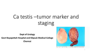
- 1. Ca testis –tumor marker and staging Dept of Urology Govt Royapettah Hospital and Kilpauk Medical College Chennai 1
- 2. Moderators: Professors: • Prof. Dr. G. Sivasankar, M.S., M.Ch., • Prof. Dr. A. Senthilvel, M.S., M.Ch., Asst Professors: • Dr. J. Sivabalan, M.S., M.Ch., • Dr. R. Bhargavi, M.S., M.Ch., • Dr. S. Raju, M.S., M.Ch., • Dr. K. Muthurathinam, M.S., M.Ch., • Dr. D. Tamilselvan, M.S., M.Ch., • Dr. K. Senthilkumar, M.S., M.Ch. Dept of Urology, GRH and KMC, Chennai. 2
- 3. Introduction • Non-seminomatous germ cell tumors (NSGCT)-mostly • 85% of NSGCT will secrete at least one tumor marker. • Germ cells in the testis have the potential to transform into a variety of cell types. • Germ cells turn into cancer cells, • Secrete proteins • Help in the diagnosis, prognosis, treatment and monitoring of testis cancer. 3 Dept of Urology, GRH and KMC, Chennai.
- 4. Markers •AFP (alpha fetoprotein) •HCG (human chorionic gonadotropin) •LDH (lactate dehydrogenase) 4 Dept of Urology, GRH and KMC, Chennai.
- 5. ALPHA-FETOPROTEIN (AFP) • NORMAL RANGE <40 micrograms/L • T ½ is 5-7 days • AFP levels are elevated in 50% to 70% of low stage (CS I, IIA, and IIB) NSGCT and • 60% to 80% of advanced (CS IIC and III) NSGCT • secreted by the • fetal yolk sac • liver • gastrointestinal tract • AFP can be secreted by NSGCT that contain • embryonal carcinoma, yolk sac tumor or teratoma. 5 Dept of Urology, GRH and KMC, Chennai.
- 6. • Malignancies including with • Hepatocellular (liver) carcinoma • Cancer of the stomach • Pancreas, biliary tract • Lung • Non-malignant diseases • Diseases of the liver • Ataxic telangiectasia • Hereditary tyrosinemia. 6 Dept of Urology, GRH and KMC, Chennai.
- 7. HUMAN CHORIONIC GONADOTROPIN (HCG) • NORMAL RANGE <5 IU/L • HALF-LIFE 24-36 hours • HCG is a glycoprotein produced by the placenta to maintain the corpus luteum during pregnancy. • Including both seminomas and NSGCT, cancerous cells can transform into syncytiotrophoblasts (a normal component of the placenta) and secrete HCG. 7 Dept of Urology, GRH and KMC, Chennai.
- 8. • 20% to 40% of low-stage NSGCT • 40% to 60% of advanced NSGCT • Levels greater than 5,000 IU are usually indicative of NSGCT • Non seminoma • higher levels of HCG are associated with a worse prognosis. • Seminoma • HCG-producing (approximately 15% of seminomas) has the same prognosis as seminoma that does not produce HCG. • Other malignancies • cancers of the liver • lung • pancreas and stomach 8 Dept of Urology, GRH and KMC, Chennai.
- 9. False positive • Cross-reactivity of the hCG assay with luteinizing hormone • Primary hypogonadism. • Elevated serum hCG results caused by hypogonadism normalize within 48 to 72 hours after administration of testosterone • Marijuana use may also cause false-positive hCG results. 9 Dept of Urology, GRH and KMC, Chennai.
- 10. LACTATE DEHYDROGENASE (LDH) • NORMAL RANGE 1.5-3.2 MIU/L • T ½ is 24 hours • Cellular enzyme found in every tissue in the body. • Highest concentrations in • muscle (including skeletal, cardiac and smooth muscle) • liver • brain. 10 Dept of Urology, GRH and KMC, Chennai.
- 11. LDH • LDH is expressed on chromosome 12p, which is often amplified in testis cancer cells. • LDH is less specific for testis cancer than HCG or AFP. • Elevated in approximately 20% of low-stage GCT and 20% to 60% of advanced GCT • Correlated to high tumor burden in seminoma and recurrence in NSGCT. • Lymphoma may also cause elevated LDH levels. 11 Dept of Urology, GRH and KMC, Chennai.
- 12. OTHERS • PLACENTAL ALKALINE PHOSPHATASE • Elevated levels present in as many as 40% patients with advanced disease • Most useful as a marker for “bulk’disease • Elevated in seminoma • GGT (Gamma glutamyl transpeptidase): • Marker of seminoma testis • Marker for “bulk” disease 12 Dept of Urology, GRH and KMC, Chennai.
- 13. Ca testis … 13 Dept of Urology, GRH and KMC, Chennai.
- 14. 14 Dept of Urology, GRH and KMC, Chennai.
- 15. TNM staging • The extent of primary tumor is classified after radical orchiectomy • pTX --Primary tumor cannot be assessed. (If no radical orchiectomy has been performed, TX is used.) • pT0-- No evidence of primary tumor • pTis-- Intratubular germ cell neoplasia (carcinoma in situ) • pT1--Tumor limited to the testis and epididymis without vascular/lymphatic invasion. • Tumor may invade into the tunica albuginea but not the tunica vaginalis 15 Dept of Urology, GRH and KMC, Chennai.
- 16. • pT2--Tumor limited to the testis and epididymis with vascular/lymphatic invasion, or • tumor extending through the tunica albuginea with involvement of the tunica vaginalis • pT3--Tumor invades the spermatic cord with or without vascular/lymphatic invasion • pT4--Tumor invades the scrotum with or without vascular/lymphatic invasion 16 Dept of Urology, GRH and KMC, Chennai.
- 17. REGIONAL LYMPH NODES • Clinical (as Determined by Noninvasive) • NX Regional lymph nodes cannot be assessed • N0 --No regional lymph node metastasis • N1 Metastasis with lymph node mass ≤2 cm in greatest dimension; • or multiple lymph nodes, none more than 2 cm in greatest dimension 17 Dept of Urology, GRH and KMC, Chennai.
- 18. • N2 Metastasis with lymph node mass, >2 cm • but not more than 5 cm in greatest dimension; or • multiple lymph nodes, • any one mass >2 cm but not more than 5 cm in greatest dimension • N3 Metastasis with lymph node mass >5 cm in greatest dimension 18 Dept of Urology, GRH and KMC, Chennai.
- 19. LYMPHATIC DRAINAGE • The retroperitoneum is the initial site of metastatic spread in 70% to 80% of patients with GCT. • For right testis tumors • Inter-aortocaval lymph nodes inferior to the renal vessels,followed by the paracaval and para- aortic nodes. • left testis tumors • The para-aortic lymph nodes, followed by the inter-aortocaval nodes . • The pattern of lymph drainage in the retroperitoneum is from right to left. • Contralateral spread is common with right-sided tumors • Rarely seen with left-sided tumors and usually is associated with bulky disease. • Retroperitoneal lymphatics drain into the cisterna chyli behind the right renal artery and right crus of the diaphragm. • From there, lymphatic spread occurs via the thoracic duct to the posterior mediastinum and left supraclavicular fossa. 19 Dept of Urology, GRH and KMC, Chennai.
- 20. Pathologic (pN) • (as Determined by Pathologic Findings of RPLND without Prior Chemotherapy or Radiotherapy) • pNX Regional lymph nodes cannot be assessed • pN0 No regional lymph node metastasis • pN1 Metastasis with lymph node mass ≤2 cm in greatest dimension and ≤5 nodes positive, none more than 2 cm in greatest dimension 20 Dept of Urology, GRH and KMC, Chennai.
- 21. • pN2 Metastasis with lymph node mass >2 cm • not more than 5 cm in greatest dimension; • or >5 nodes positive, none more than 5 cm; • or evidence of extranodal extension of tumor • pN3 Metastasis with lymph node mass >5 cm in greatest dimension 21 Dept of Urology, GRH and KMC, Chennai.
- 22. DISTANT METASTASIS(M) • MX -- Distant metastasis cannot be assessed • M0 --No distant metastasis • M1 -- Distant metastasis • M1a Non regional nodal or pulmonary metastasis • M1b Distant metastasis at site other than non regional lymph nodes or lung 22 Dept of Urology, GRH and KMC, Chennai.
- 23. SERUM TUMORMARKERS (S) • SX Marker studies unavailable or not performed 23 Dept of Urology, GRH and KMC, Chennai.
- 24. 24 Dept of Urology, GRH and KMC, Chennai.
- 25. 25 Dept of Urology, GRH and KMC, Chennai.
- 26. 26 Dept of Urology, GRH and KMC, Chennai.
- 27. CLINICAL STAGING • CS I -- clinically confined to the testis • CS II -- presence of regional (retroperitoneal) lymph node metastasis • CS III --represents non regional lymph node and/or visceral metastasis. 27 Dept of Urology, GRH and KMC, Chennai.
- 28. THANK YOU 28 Dept of Urology, GRH and KMC, Chennai.
- 29. 29 Dept of Urology, GRH and KMC, Chennai.