
Recomendados
Recomendados
Mais conteúdo relacionado
Semelhante a Introduction to Microscopy and Cell Morphology
Semelhante a Introduction to Microscopy and Cell Morphology (20)
Último
Último (20)
Introduction to Microscopy and Cell Morphology
- 1. PART I Microscopy and Cell Morphology 1
- 2. Introduction to Microscopy •Visualization of cells requires the use of either light microscope or electron microscope. •Light microscopes are used to look at intact cells at low magnification. •Electron Microscope are used to look at internal structures or details of cell surfaces. •All microscopes use lenses to magnify objects. •In addition to magnification there is resolution ie the ability to distinguish two adjacent objects as distinct and separate. •Magnification can be increased almost without limit but resolution cannot. •Resolution is dictated by the physical properties of light. •It is thus resolution and not magnification that dictates what we are able to see with a microscope. 2
- 3. Light Microscopy Various types of light microscopes exist, including bright- field, dark-field, phase contrast, and fluorescence microscopes. With bright-field microscope specimens are visualized because of the difference in contrast that exist between them and the surrounding medium. Contrast differences arises because cells absorb or scatter light to various degrees. All compound light microscopes optimize image resolution by using lenses with high light-gathering characteristics. The limit of resolution for a light microscope is about 0.2 m. 3
- 4. 4
- 5. Staining: Increasing contrast for Bright- Field Microscopy One of the limitations of bright field microscopy is insufficient contrast. dyes are used to stain cells and in increase their contrast so that they can be seen better. Dyes are organic compounds and each class of dye has an affinity for specific cellular materials. many dyes used in microbiology are positively charged (basic dyes) and combine with negatively charged cellular constituents such as nucleic acid and acidic polysaccharides. Example of basic dyes: Methylene blue, Crystal violet, and Safranin. Cell surfaces tend to be negatively charged, thus these dyes combine with high affinity to structures on the surfaces of cells and hence are excellent general purpose stains. 5
- 6. Simple stain •Simple and/or differential cell staining are used to increase contrast in bright-field microscopy. •In simple staining, dried preparations of cell suspensions are usually used. •A slide containing a dried suspension of heat-fixed cells is flooded for a minute or two with a dilute solution of dye. •The preparation is then rinsed several times with water, and blotted dry. 6
- 7. 7
- 8. 8
- 9. Differential stains: The Gram Stain •This stain is called differential because it does not stain all kinds of cells in the same colour. •The most commonly used differential stain is the Gram stain. •Based on their reaction with the gram stain, bacteria can be divided into two major groups: gram-negative and gram positive. •After gram staining, gram-positive bacteria appear purple, while gram-negative appear pink to red. •The difference in reaction with gram stain is due to differences in cell wall structure of gram negative and gram positive bacteria. •This difference in cell wall structure leads to ethanol decolorizing gram negative, but not gram-positive bacteria. 9
- 10. 10
- 11. 11
- 12. 12
- 13. Relationship of cell wall structure to the gram stain The structural differences between the cell walls of gram- positive and gram-negative Bacteria are thought to be responsible for differences in the Gram stain reaction. In the gram stain, an insoluble crystal violet-iodine complex is formed inside the cell. This complex is broken down by alcohol from gram-negative bacteria but not from gram-positive bacteria. Gram-positive bacteria have very thick cell walls consisting of several layers of peptidoglycan. These become dehydrated by the alcohol, causing the pores in the walls to close and prevent the insoluble crystal violet- iodine complex from escaping. By contrast, in gram-negative bacteria, alcohol readily penetrates the lipid-rich outer membrane and extracts the crystal violet-iodine complex from the cell. 13
- 14. 14
- 15. 15
- 16. 16
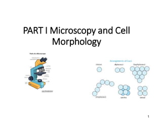
- 17. Cell Morphology Some typical bacterial morphologies include coccus, rod, spirillum, spirochete, appendaged, and filamentous. 17
- 18. 18 • How to calculate the actual size of a microscopic image. • Actual size= image size (use ruler to measure) / Magnification. • Use same units.
- 19. The Outer Membrane of Gram-Negative Bacteria In addition to peptidoglycan, gram-negative Bacteria contain an outer membrane consisting of lipopolysaccharide (LPS), protein, and lipoprotein. This layer is in fact a second lipid bilayer, but it is not solely constructed of phospholipid and protein like the cytoplasmic membrane. The outer membrane also contain polysaccharide which is linked with lipid in the outer membrane to form a lipopolysaccharide complex. And because of this, the outer membrane is often called the Lipopolysaccharide Layer (LPS) 19
- 21. Archeal membranes Archea: phylogenetically related prokaryotes distinct from bacteria. Lipids in archaea differs from those of other organisms. Unlike Bacteria and Eukarya, in which ester linkages bond fatty acids to glycerol, lipids in Archaea contain ether-linkages between glycerol and their hydrophobic side chains. In addition, archaea lipids lack fatty acids. Instead, their side chains are composed of repeating units of five carbon hydrocarbon called isopreme. However, the overall architecture of the cytoplasmic membranes of archaea, forming inner and outer hydrophilic surfaces with a hydrophobic interior, is the same as in bacteria and eukarya. 21
- 22. • Glycerol diethers and glycerol tetraethers are the major lipids present in archaea. • In the tetraether molecule, the phytanyl (composed of four linked isopremes) side chains from each glycerol molecule are covalently bonded together. • this structure thus yield a lipid monolayer instead of a lipid bilayer cytoplasmic membrane. • Lipid monolayers are quite resistant to peeling apart. • This membrane structure is thus widespread among hyperthermophile archea that grow at very high temperatures. 22
- 23. 23
- 24. Cells with no walls Peptidoglycan, a signature molecule of species of bacteria, can be destroyed by certain agents e.g. by Lysozyme- a protein that breaks the β-1,4-glycosidic bonds between N- acetylglucosamine and N-acetylmuramic acid in the peptidoglycan, thereby weakening the wall. Water then enters the cell, and the cell swells and eventually burst, a process called lysis. Lysozymes are found in animal secretions including tears, saliva, and other body fluids, and presumably functions as a major line of defence against infection by Bacteria. 24
- 25. • If a solute that does not penetrate the cell, such as sucrose, is added to a cell suspension, the solute concentration outside the cell balances that inside (a condition called isotonic). • Under this conditions, lysozyme still digests peptidoglycan, but water does not enter the cell and lysis does not occur. • Instead a protoplast (a bacterium that has lost its cell wall) is formed. • If such sucrose-stabilized protoplasts are then placed in water, lysis occurs immediately. 25
- 26. 26
- 27. •Although most prokaryotes cannot survive in nature without their cell walls, some are able to do so. •These include mycoplasmas, a group that causes certain infectious diseases, and the Thermoplasma group (Archaea that naturally lack cell walls). •These prokaryotes are free-living protoplasts that survive without cell walls because they have unusually tough membranes or live in osmotically protected habitats, such as the animal body. •Some mycoplasmas have sterols in their cell membranes, which lends strength and rigidity to this structure. 27
- 28. Pseudopeptidoglycan Some species of archaea contain cell walls constructed of a polysaccharide very similar to that of peptidiglycan called pseudopeptidoglycan. The backbone of pseudopeptidoglycan is composed of alternating repeats of N-acetylglucosamine and N-acetyltalosamin uronic acid (the later replaces the N-acetylmuramic acid of peptidoglycan). The backbone of pseudopeptidoglycan is linked by b-1,3 bonds instead of the b-1,4 bonds found in true peptidoglycan. Some cell walls of archaea lacks both peptidoglycan and pseudopeptidoglycan and consist of polysaccharide, glycoprotein, or protein. e.g. Methanosarcina species contain thick polysaccharide walls composed of glucose, glucuronic acid, galactosamine and acetate. Extremely halophilic (salt-loving) archaea such as Halococcus contain cell wall similar to that of Methanosarcina but in addition contain sulfate residues 28
- 29. 29
- 30. S-layers in Archaea The most common cell wall type among Archaea is the paracrystalline surface layer (S-layer). The S-layer consists of protein or glycoprotein and usually has a hexagonal symmetry. S-layers have been found among species of groups of Archaea, the extreme halophiles, the methanogens, and the hyperthermophiles. Some species of bacteria also have S-layers on their outer surfaces. Species of archaea have a variety of cell wall chemistries, varying from molecules that closely resemble peptidoglycan to walls totally lacking a polysaccharide component. 30
- 31. • With a rare exceptions, all archaea contain a cell wall of some sort, and as in bacteria, the archeal cell wall functions to prevent osmotic lysis and to define cell shape. • In addition, because they lack peptidoglycan in their cell walls, all archaea are naturally resistant to the action of lysozymes and penicillin, agents that destroy peptidoglycan or prevent its proper synthesis. 31
- 32. S-Layer in bacteria Prokaryotes may contain cell surface layers composed of a two-dimensional array of protein called an S-layer. S-layers have been detected in representatives of almost every phylogenetic grouping of bacteria and are widespread among archaea. In some species of archaea the S-layer is also the cell wall. S-layers have a crystaline appearance and show various symmetries such as hexagonal, tetragonal, or trimeric, depending upon the number and structure of the protein or glycoprotein subunits of which they are composed. 32
- 33. The major function of S-layers is unknown, however as the interface between the cell and its environment, it is likely that: S-layers function as a selective sieve, allowing the passage of low-molecular-weight substances while excluding large molecules and structures (such as viruses). The S-layer would also then function to retain proteins near the cell much like the outer membrane does in gram-negative bacteria. Evidence also exists that in pathogenic bacteria that contain S-layers, this structure may provide protection against certain host defense mechanisms. 33
- 34. 34 In G- bacteria the S layer adheres to the outer membranes whereas in G+ bacteria it is associated with the peptidoglycan surface.
- 35. The Outer Membrane of Gram-Negative Bacteria In addition to peptidoglycan, gram-negative Bacteria contain an outer membrane consisting of lipopolysaccharide (LPS), protein, and lipoprotein. This layer is in fact a second lipid bilayer, but it is not solely constructed of phospholipid and protein like the cytoplasmic membrane. The outer membrane also contain polisaccharide which is linked with lipid in the outer membrane to form a liposaccharide complex. And because of this, the outer membrane is often called the Lipopolysaccharide Layer (LPS) 35
- 36. Chemistry of LPS The chemistry of LPS has been best studied in Salmonella species. The polysaccharide portion of the LPS consists of, Lipd A, ketodeoxyoctonate (KDO), core polysaccharide and the O- polysaccharide. The core polysaccharide consists of (KDO), seven-carbon sugars (heptoses), glucose, galactose, and N-acetylglucosamine. O-specific polysaccharide is connected to the core, and normally contains galactose, glucose, rhamnose, and manose (all hexoses), plus one or more unusual dideoxy sugars such as abequose, colitose, paratose, or tyvelose. These sugars are connected in four or five-membered sequences, which are usually branched. When the sequences repeat, the long O-polysaccharide is formed. 36
- 37. • The lipid portion of the lipopolysacharide known as Lipid A, is not a glycerol lipid, but instead is the fatty acids are connected by amine ester linkage to a disaccharide composed of N-acetylglucosamine phosphate. • The disaccharide is attached to the core polysacharide through KDO. • Fatty acids commonly found in in Lipid A include caproic, lauric, myristic, palmitic, and stearic acids. • In the outer membrane, LPS associates with several proteins to form the outer leaflet of the membrane. • A lipoprotein complex is found on the inner leaflet of the LPS of a number of gram-negative bacteria. • The lipoprotein functions as an anchor between the outer membrane and the and the peptidoglycan. • Also, in the outer leaflet of the outer membrane, LPS replaces phospholipids. • Phospholipids are found only in the inner leaflet. • Thus, even though the outer membrane can be considered a lipid bilayer, its structure is quite distinct from that of the cytoplasmic membrane. 37
- 38. 38
- 39. 39
- 40. Porins and periplasm Unlike the cytoplasmic membrane, the outer membrane of gram-negative bacteria is relatively permeable to small molecules even though its is a lipid bilayer. This permeability is due to proteins called porins which are present in the outer membrane. Porins allow for permeability across the outer membrane by creating channels that traverse the membrane and allow the entrance and exit of hydrophilic low molecular weight substances. Several different porins are known, including both specific and nonspecific classes. Non-specific porins form water-filled channels through which any small substance can pass. Some porins are highly specific because they contain a specific binding site for one or a group of structurally related substances. Structural studies have shown that most porins are proteins that contain three identical units. Porins are transmembrane proteins and associate to form small holes about 1 nm in diameter in the outer membrane. They are beta barrel proteins that cross a cellular membrane and act as a pore, through which molecules can diffuse. 40
- 41. 41
- 42. Even though the outer membrane is permeable to small molecules, it is not permeable to enzymes or other large molecules. One major functions of the outer membrane is to keep proteins that are present outside the cytoplasmic membrane from diffusing away from the cell. The space between the membranes is the periplasm, which contains various proteins/ enzymes involved in important cellular functions. The contents of the periplasm are gel-like in consistency because of the abundance of periplasmic proteins found there. The periplasm can contain several proteins, depending on the organism. This include: hydrolytic enzymes, which function in the initial degradation of food molecules; binding proteins, which begin the process of transporting substrates, and chemoreceptors, which are proteins involved in the chemotaxis response. 42
- 43. 43
- 44. Endotoxin properties of LPS Even though the major function of the outer membrane is structural, it also has a biologically important function, that of toxicity to animals. Gram-negative bacteria that are pathogenic to humans and other mammals include members of the genera Salmonella, Shigella and Escherichia, and some of the symptoms that they elicit in their hosts are due to their toxic outer membrane. The toxic properties are associated with part of the LPS, especially Lipid A. The term endotoxin refers to this toxic component of the LPS. Some toxins cause violent symptoms in humans including severe gastrointestinal distress (gas, diarrhea, and vomiting). Endotoxins are responsible for several bacterial infection e.g. Salmonella food infection. LPS from several nonpathogenic bacteria have also been shown to have endotoxin activity. This means that an organism does not need to be pathogenic for it to contain toxic outer membrane components. 44
- 45. Bacterial Cell Surface Structures Prokaryotic cells often contain various surface structures, including fimbriae and pili, S- layers, capsules, and slime layers. A key function of these structures is in attaching cells to a solid surface. 45
- 46. Fimbrae and Pili Short protein filaments used for attachment are fimbriae. These proteins extends from the surface of a cell. Fimbriae enable organisms to stick to surfaces, including animal tissues in the case of some pathogenic bacteria, or to form pellicles or biofilms on surfaces. Well known pathogens that have fimbriae structure include Salmonella typhimurium (salmonelosis), Neisseria gonorrhoeae (gonorrhea), and Bordetella pertussis (whooping cough). Pili are structurally similar to fimbriae but are typically longer, and only one or a few pili are present on the surface. Because they serve as receptors for certain types of viruses, pili can be seen under electron microscope when they become coated with virus particles. Although possibly involved in attachment, as for fimbriae, pili are mainly involved in the process of conjugation (a form of genetic exchange) in prokaryotes 46
- 47. 47
- 48. 48
- 49. There are many classes of fimbriae/pili, and they can be distinguished by their structure and function e.g. Type IV fimbriae/pili. Type IV fimbriae/pili is involved in an unusual form of motility in certain bacteria called twitching motility. Twitching motility is a type of movement on solid surfaces, where it is thought that rapid and reversible extension and retraction of the fimbriae allow the cell to crawl along the surface. Unlike other fimbriae, type IV fimbriae/pili are found only at the poles of the cells, and besides motility, have been implicated as key host colonization factors in a variety of pathogens including Vibrio cholerae (cholera), and Neisseria meningitidis (bacterial meningitis). Type IV fimbriae are also though to mediate genetic transfer by the process of transformation in a wide variety of bacteria. 49
- 50. Capusules and slime layers Many prokayotic organisms secrete slimy or gummy materials on their surfaces. A variety of these structures consist of polysaccharide and a few consist of protein. The terms capsule and slime layer are frequently used to describe these polysaccharide layers. The composition of these layers varies in different organisms but may be thick or thin, rigid or flexible, depending on their chemical nature. The rigid layers are organized in a tight matrix that excludes small particles, such as india ink and this form is called a capsule. If the layer is easily more deformed, it will not exclude particles and is more difficult to see, and this form is called slime layer. 50
- 51. Polysaccharide layers have several functions in bacteria. Surface polysaccharides assist in the attachment of microorganisms to solid surfaces. Pathogenic Mos that enter the animal body by specific routes usually do so by first binding specifically to surface components of host tissues. This binding is often mediated by surface polysaccharides on the bacterial cell. Many non-pathogenic bacteria also bind to solid surfaces in nature, sometimes forming a thick layer of cells called biofilm, and polysaccharides plays a key role in the development of biofilms. Slime layers perform other roles as well, for example: Encapsulated pathogenic bacteria are typically more difficult for phagocytic cells of immune system to recognize and subsequently destroy. In addition, because outer polysaccharide layers bind a significant amount of water, it is likely that these layers play some role in resistance to desiccation. 51
- 52. 52 A slime layer (right) is loosely associated with the bacterium and can be easily washed off, whereas a capsule (left) is attached tightly to the bacterium and has definite boundaries.
- 53. 53
- 54. Gliding motility in Flavobacterium johnsoniae In F. johnsoniae movement, no slime is sectreted. Instead, the movement of proteins on the cell surface has been identified as the likely mechanism of gliding. In F. johnsoniae, specific motility proteins anchored in the cytoplasmic and outer membranes are thought to propel the cell forward in a type of continuous ratcheting mechanism. The movement of the cytoplasmic membrane proteins is driven by energy released from the proton motive force, and they somewhat transmit this energy to outer membrane proteins located along the cell surface. It is hypothesized that the movement of the proteins against the solid surface literally pulls the cell forward. 54
- 55. 55
- 56. Cell Inclusions Prokaryotic cells often contain internal granules that function as energy reserves or as storage of structural building materials. Most cellular inclusions are enclosed by a thin non-unit membrane consisting of lipid separating the inclusion from the proper cytoplasm. 56
- 57. Carbon Storage Polymers Poly-b-hydroxybutyrate (PHB) is a common storage material of prokaryotic cells. PHB is a lipid that is formed from β-hydroxybutyric acid units. The monomers of this acid are connected by ester linkages, forming the long PHB polymer, and these polymers aggregate into granules. The length of the monomer in the polymer can vary considerable, from as short as C4 to as long as C18 in certain organisms. Poly-b-hydroxyalkanoates (PHAs) and glycogen are produced as storage polymers when carbon is in excess. PHAs are synthesized when carbon is in excess and are broken down for use as carbon skeletons for biosynthesis or to make ATP when conditions warrant. 57
- 58. Glycogen is a polymer of glucose Like PHAs, glycogen is a storage depot for carbon and energy. Glycogen is produced when carbon is in excess in the environment and is consumed when carbon is limiting. Glycogen resembles starch, the major storage reserve in plants, but differs from starch in the way the glucose units are linked together. 58
- 59. Other storage materials and inclusions Many MOs accumulate inorganic phosphate (P) in form of granules of polyphosphate. These granules can be degraded and used as sources of phosphate for nucleic acid and phospholipids. In addition, many prokaryotes are capable of oxidizing reduced sulfur compounds such as hydrogen sulfide. Oxidation of hydrogen sulfide is linked to energy production for use in metabolism and biosynthesis. Elemental sulfur can accumulate inside the cell in visible globules. This globules of elemental sulfur remain as long as the source of reduced sulfur is still present. When the reduced source of sulfur is in low levels, the sulfur in the granules is oxidized to sulfate, and the granules disappear as the reaction proceeds. 59
- 60. Sulfur globules reside in the periplasma instead of the cytoplasm (sulfur-oxidizing MOs are usually gram-negative bacteria). The periplasm expands outward to accommodate the globules as hydrogen sulfide is oxidized to elemental sulfur. It is thought that in gram negative, some other intra- cytoplasmic inclusions may be periplasmic as well. Thus, it appears that at least poly-β-hydroxyalkanoates fall into this category (of cell inclusion). 60
- 61. Magnetosomes are intracellular particles of the iron mineral magnetite (Fe3O4) that allow organisms to respond to a magnetic field. Magnetosomes impart a magnetic dipole to a cell, allowing it to respond to a magnetic field. Bacteria that produce magnetosomes exhibit magnetotaxis (the process of orienting and migrating along Earth’s magnetic field lines). Even though a suffix –taxis is used in the word magnetotaxis, there is no evidence that magnetotactic bacteria employ sensory system of chemotactic bacteria or phototactic bacteria. Instead the alignment of magnetosomes in the cell simply imparts magnetic properties to it, which then orient the cell in a particular direction in its environment. 61
- 62. The major functions of magnetosomes are unknown. Magnetosomes have been found in a variety of aquatic bacteria that grows best at low oxygen concentrations. Thus it has been hypothesized that magnetosomes functions to guide this aquatic cells towards sediments (since the major magnetic field line points downwards) where oxygen levels are low. Magnetosomes are surrounded by a membrane containing phospholipids, proteins, and glycoproteins. This membrane is not a unit membrane like a cytoplasmic membrane, instead it is a non-unit membrane, like that surrounding granules of PHB. Magnetosome membrane proteins probably play a role in precipitating Fe3+ (which is brought into the cell in soluble form by chelating agents) as Fe3O4 in the developing magnetosome. The morphology of magnetosomes appears to be species-specific, varying in shape from square to rectangular to spike-shaped in certain bacteria, forming into chains inside the cell. 62
- 63. Endospores The endospore (the prefix endo means within) is a highly resistant differentiated bacterial cell produced by certain gram-positive Bacteria (the normally- growing cell that forms the endospore is called a vegetative cell). Endospores are highly resistant to heat and are difficult to destroy even by harsh chemicals or radiation. Highly resistant bacterial endospores can survive heating to temperature as high as 150 °C. Endospores are also resistant to other harmful agents such as drying, ultraviolet radiation, strong acids or bases, and chemical disinfectants, and can remain dormant for extremely long periods. Endospores are produced by certain bacteria during a process called sporulation. The biological function of endospores is to enable an organism to endure and survive difficult times, this includes: extreme temperature, drying and nutrient depletion. Endospores are also ideal structures for dispersal by wind, water or through the animal gut. Endospore-forming bacteria are mainly found in the soil, and the genera Bacillus and Clostridium are the best studied of the endospore-forming bacteria. . 63
- 64. Endospore Structure The structure of endospore differs distinctly from that of a vegetative cell. The endospore is structurally more complex in that it has many layers that are absent in a vegetative cell. The outermost layer is called exporium, which is a thin protein covering. Within the exporium, there are spore coats, composed of layers of spore-specific proteins. Below the spore coat is the cortex, which consists of loosely cross- linked peptidoglycan. Inside the cortex is the core or spore protoplast, which contains the core wall, cytoplasmic membrane, cytoplasm, nucleoid, ribosomes, and other cellular essentials. Thus, the endospore differs structurally from a vegetative cell primarily in the kinds of structures found outside the core wall 64
- 65. 65
- 66. Dipicolinic acid is one of the chemical substances that is present in endospores but absent in vegetative cell. It has been found in endospores from all endospore-forming bacteria examined and is located in the core. Endospores are also very rich in calcium ions, most of which are combined with dipicolinic acid. The calcium-dipicolinic acid complex of the core represents about 10% of the dry weight of the endospore. The complex functions to reduce water availability within the endospore (binds free water molecules, causing dehydration) In addition, the complex intercalates (inserts between bases) in DNA, and in so doing, protects it from heat denaturation. 66
- 67. 67
- 68. Endospores differ significantly from the vegetative, or normally functioning, cells. 68
- 69. Properties of endospore core The core of a mature endospore differs greatly from the vegetative cell from which it was formed. Apart from the high levels of calcium-dipicolinate, which reduces the water content of the core, the core becomes further dehydrated during the sporulation process. The core of a mature endospore contains only 10-15% of the water content of the vegetative cell, and thus the consistency of the core is that of a gel. The dehydration of a core greatly enhances the heat resistance of the endospore. The dehydration has also been shown to confer resistance in the endospore to chemicals, such as hydrogen peroxide, and causes enzymes remaining in the core to become inactive. 69
- 70. In addition to the low water content of the endospore, the pH of the core is about one unit lower than that of the vegetative cell cytoplasm. Furthermore, the core contains high levels of proteins called small acid- soluble proteins (SASPs). The SASPs are made during the sporulation process and have at least two functions: SASPs bind tightly to DNA in the core and protect it from potential damage from UV radiation, desiccation, and dry heat. UV radiation protection is conferred when SASPs change the molecular structure of DNA from the normal B form to a more compact A form. A-form DNA is more resistant to the formation of pyrimidime dimers by UV radiation, a means of mutation, and to denaturing effects of dry heat. In addition, SASPs function as a carbon and energy source for the outgrowth of a new vegetative cell from the endospore, a process called germination. 70
- 71. Endospore Formation During endospore formation, a vegetative cell is converted to a non-growing, heat resistant structure. Sporulation involves a complex series of events in cellular differentiation. Bacterial sporulation does not occur when cells are dividing exponentially, but only when growth ceases due to the exhaustion of an essential nutrient. Thus, cells of Bacillus, a typical endospore-forming bacterium, cease vegetative growth and begin sporulation when a key nutrient such as the carbon or nitrogen source becomes limiting. Many genetically directed changes in the cell underlie the conversion from vegetative growth to sporulation. 71
- 72. Genetic studies of mutants of Bacillus, each blocked at one of the stages of sporulation indicates that more than 200 genes are involved in the sporulation process. Sporulation requires that the synthesis of many proteins involved in vegetative cell functions cease and that specific endospore proteins be made. This is accomplished by activation of a variety of endospore- specific genes including spo, ssp, (which encodes SASPs), and many other genes in response to to an environmental trigger to sporulate. The proteins encoded by these genes catalyze the series of events leading from a moist, metabolizing vegetative cell to a relatively dry, metabolically inert but extremely resistant, endospore. 72
- 73. 73
- 74. Endospore Germination Endospores can remain dormant indefinitely but germinate quickly when the appropriate trigger is applied. This process involves three steps: activation, germination, and outgrowth. Activation is most easily accomplished by heating freshly formed endospore for several minutes at an elevated but sub lethal temperature. Activated endospores are then conditioned to germinate when placed in the presence of specific nutrients, such as certain amino acids (e.g. alanine is a good trigger of endospore germination). Germination is usually a rapid process, and it involves loss of microscopic refractibility of the endospore, increased ability to be stained by dyes, and loss of resistance to heat and chemicals. 74
- 75. Loss of calcium dipicolinate and cortex components from the endospores occurs during the germination stage, and the SASPs are degraded. Outgrowth is the final stage of endospore germination, and it involves visible swelling due to water uptake and synthesis of new RNA, proteins and DNA. The cell emerges from the broken spore coat and eventually begins to divide. The cell then remains in vegetative growth until environmental signals 75
- 76. Microbial Locomotion Many cells can move under their own power. Motility allows cells to reach different regions of their environment. In a struggle for survival, movement to a new location may offer a cell new resources and growth opportunities and spell the difference between life and death. 76
- 77. Flagella and Motility Motility in most microorganisms is accomplished by flagella. In prokaryotes, the flagellum is a complex structure made of several proteins, most of which are anchored in the cell wall and cytoplasmic membrane. 77
- 78. Bacterial flagella Bacterial flagella are long thin appendages free at one end and attached to the cell at the other end. Flagella are very thin (about 20 nm) that a single flagellum can only be seen with a light microscope after staining with special flagella stains that increase their diameter. Flagella are arranged differently on different bacteria. In polar flagellation, the flagella are attached at one or both ends of the cell. Occasionally, a tuft (group) of flagella may arise at one end of the cell, an arrangement called lophotrichous (lopho means tuft, and trichous means hair). Tufts of flagella of this type can be seen in living cells by dark-field microscopy, where the flagella appear light and are attached to light-colored cells against a dark background. In peritrichous flagellation, the flagella are inserted at many locations around the cell surface (peri means around). The type of flagellation on a bacteria is used as one of the characterization for classification of bacteria. 78
- 79. 79
- 80. 80
- 81. Flagellar structure flagella are not straight but are helically shaped. When flattened, flagella show a constant distance between adjacent curves, called the wavelength, and this wavelength is characteristic for any given species. The filament of bacterial flagella is composed of subunits of a protein called flagellin. The shape and wavelength of the flagellum are in part determined by the structure of the flagellin protein and also to extent by the direction of rotation of the filament. In bacterial species, flagellin is highly conserved, suggesting that flagellar motility has deep evolutionary roots within this domain. A flagellum consists of several components and functions by rotation, much like a propeller in a motor boat. The base of the flagellum is different in structure from that of the filament. There is a wider region at the base of the flagellum called the hook. 81
- 82. The hook consists of a single type of protein and functions to connect the filament to the motor portion of the flagellum. The motor is anchored in the cytoplasmic membrane and cell wall. The motor consists of a small central rod that passes through a series of rings. In gram-negative bacteria, an outer ring, called the L ring, is anchored in the lipopolysaccharide layer. A second ring, called the P ring, is anchored in the peptidoglycan layer of the cell wall. A third set of rings, called the MS and C rings, are located within the cytoplasmic membrane and the cytoplasm, respectively. In gram-positive bacteria, which lack an outer membrane, only the inner pair of rings is present. Surrounding the inner ring and anchored in the cytoplasmic membrane are a series of proteins called Mot proteins. A final set of proteins, called Fli proteins function as the motor switch, reversing the direction of rotation of the flagella in response to intracellular signals. 82
- 83. 83
- 84. Flagellar Movement The flagellum is a tiny rotary motor. Rotary motors contain two components: The rotor and the stator. In flagellar motor, the rotor consists of the C, MS, and P rings. Collectively, these structures make up the basal body. The stator consists of Mot proteins that surround the Ms and C rings and functions to generate torque. The rotary motion of the flagellum is imparted from the basal body. The energy required for rotation of the flagellum comes from the proton motive force. Proton movement across the cytoplasmic membrane through the Mot complex drives rotation of the flagellum, and calculations have shown that about 1000 protons must be translocated per single rotation of the flagellum. 84
- 85. A proton turbine model has been proposed to explain the available experimental data. In this model, protons flowing through channels in the stator exert electrostatic forces on helically arranged charges on the rotor proteins. Attractions between positive and negative charges would cause the basal body to rotate as protons flow through the stator. 85
- 86. 86
- 87. Flagellar Synthesis Several genes are required for flagellar synthesis and subsequent motility. In E. coli and Salmonella typhimurium, where studies have been most extensive, over 40 genes are necessary for motility. These genes have several functions, including encoding structural proteins of the flagellar apparatus, export of flagellar components through the cytoplasmic membrane to the outside of the cell, and regulation of the many biochemical events surrounding the synthesis of new flagella. An individual flagellum does not grow from its base, as does an animal hair, but from the tip. The MS ring is synthesized first and inserted into the cytoplasmic membrane. 87
- 88. Other anchoring proteins are synthesized along with the hook before filament formation occurs. Flagellin molecules synthesized in the cytoplasm pass up through a 3-nm channel inside of the filament and add on at the terminus to form the mature flagellum. At the end of the growing flagellum, a protein cap exists. Cap proteins assist flagellin molecules that have diffused through the channel to organize at the flagellum termini to form new filament. Growth of the flagellum occurs more or less continuously until the flagellum reaches its final length. Broken flagella still rotate and can be repaired with new flagellin units passed through the filament channel to replace the lost ones. 88
- 89. 89
- 90. Cell Motion as a Behavioral Response: Chemotaxis and Phototaxis Prokaryotes often encounter gradients of physical or chemical agents in nature, and cells have evolved means to respond to these gradients by moving either toward or away from the signal molecule, depending on weather it is a useful or harmful substance. Such directed movement are called taxes. Chemotaxis, a response to chemicals, and phototaxis, a response to light, are the most well studied taxes. 90
- 91. Chemotaxis Motile bacteria can respond to chemical and physical gradients in their environment. While moving, prokaryotes compare the chemical or physical state of their environment with that sensed a few seconds before. This means that bacteria respond to the temporal gradient of signal molecules as they swim along. Most of the research on chemotaxis has been done with the peritrichously flagellated bacteria, E. coli. In the absence of a gradient, cells of E. coli move in a random fashion that includes runs, where the cell is swimming forward in a smooth fashion, and tumbles, when the cell stop and jiggles about. Following a tumble, the direction of the next run is random. Thus, by means of runs and tumbles, the cell moves about randomly in its environment but does not really go anywhere. 91
- 92. However, if a gradient of a chemical attractant is present, these random movements becomes biased. As the organism senses that it is moving toward higher concentrations of the attractant (through periodic sampling of the concentration of the chemical in its environment), runs become longer and tumbles less frequent. The net result of this behavioral response is that the organism moves up the concentration gradient of the attractant. 92
- 93. If the organism is sensing a repellent, the same general mechanism applies, although in this case it is the decrease in concentration of the repellent (rather than the increase in concentration of an attractant) that promotes runs. Forward movement in a run occurs when flagella motor is rotating counterclockwise. When flagella rotate clockwise, the bundles pushes apart, forward motion ceases, and the cell tumbles. Many polarly flagellated bacteria, such as Pseudomonas species, can reverse the direction of rotation of their flagella like peritrichously flagellated cells and reverse direction in this fashion. 93
- 94. 94
- 95. 95
- 96. Chemotaxis in Rhodobacter sphaeroides In R. sphaeroides, which has only a single flagellum inserted subpolarly, rotation of the flagellum stops periodically. During this time the cell becomes randomly reoriented by Brownian motion. As the flagellum begins to rotate once again, the cell moves off in new direction. Cells of R. sphaeroides are strongly chemotactic to a variety of carbon compounds and also show tactic responses to oxygen and light. And although it cannot reverse its flagellar motor, chemotaxis in R. sphaeroides works in a similar way to that of E. coli: An increased gradient of attractants keeps the R. sphaeroides flagellum rotating while a decreased gradient of an atractant or an increased gradient of a repellent tends to make the cells stop and reorient. 96
- 97. Phototaxis Phototaxis is the movement of phototrophic organisms toward an increasing intensity of light. The advantage of phototaxis is that it allows a phototrophic organism to orient itself most efficiently to receive light for photosynthesis. This can be shown if a light spectrum is spread across a microscope slide on which there are motile phototrophic bacteria. On such a slide, the bacteria accumulate at wavelengths at which their photosynthetic pigments absorb. There are two different types of taxes observed in phototrophic prokaryotes. Scotophobotaxis is observed only microscopically and occurs when a phototrophic bacterium by chance happens to swim outside the illuminated field of view of the microscope into darkness. Entering darkness negatively affects the energy state of the cell and signals the cell to tumble, reverse direction, and once again swim in a run, thus re-entering the light. 97
- 98. In addition to scotophobotaxis, phototrophic MOs can carry out true phototaxis, a directed movement up a light gradient toward an increasing intensity of light. This is analogous to positive chemotaxis except that the attractant is light instead of a chemical. In some species, such as the highly motile phototrophic organism Rhodospirillum centenum, entire colonies of cells show phototaxis and move in unison toward the light. There is a good reason to believe that several parts of the regulatory system that govern chemotaxis are also involved in phototaxis. These include in particular cytoplasmic proteins (Che proteins) that control the direction of flagella rotation.
- 99. This conclusion has come from study of mutants of phototrophic bacteria defective in phototaxis; such mutants typically have defective chemotaxis systems as well. A photoreceptor, analogous to a chemoreceptor but able to sense a gradient of light instead of chemicals, orchestrates the phototaxis response. The photoreceptor interacts with cytoplasmic proteins that affect flagella rotation to maintain the cell in a run if it is swimming toward an increasing intensity of light. Thus, although the stimulus in chemotaxis and phototaxis is different, the same cytoplasmic machinery is employed to process both types of signals.
- 100. Regulation of Chemotaxis • Prokaryotes can move towards attractants or away from repellents, a process called chemotaxis. Prokaryotes are too small to actually sense spatial gradients of a chemical, but they can respond to temporal gradients. •That is, they can sense the change in concentration of a chemical over time rather than the absolute concentration of the chemical stimulus. •Prokaryotes use a modified two-component system to sense temporal changes in attractants or repellents and process this information to regulate flagellar rotation. •In chemotaxis, there is a two-component system that directly regulates the activity of preexisting flagella, not the transcription of the genes encoding the flagella. 100
- 101. Step 1: Response to signal •The mechanism of chemotaxis is complex and depends upon a variety of different proteins. •Several sensory proteins reside in the cytoplasmic membrane and sense the presence of attractants and repellents. The sensory proteins allow the cell to monitor the concentration of various substances over time. •The sensory proteins are called methyl-accepting proteins (MCPs). •In E. coli, five different MCPs have been identified, and each is a transmembrane protein. •Each MCP can sense a variety of compounds. e.g. the Tar MCP of E. coli can sense the attractants aspartate and maltose as well as repellents such as heavy metals cobalt and nickel. 101
- 102. • MCPs bind attractants or repellents directly or in some cases indirectly through interactions with periplasmic binding proteins. • Binding of an attractant or repellent initiates a series of interactions with cytoplasmic proteins that eventually affects flagellar rotation. • If the rotation of the flagellum is ccw, the cell will move in a run, whereas if the flagellum rotates clockwise, the cell will tumble. • MCPs are in contact with the cytoplasmic proteins CheW and CheA. CheA is the sensor kinase in chemotaxis. • When an MCP binds a chemical, it changes conformation and (with help from CheW) affects the autophosphorylation of CheA to form CheA-P. • Attractants decrease the rate of autophosphorylation, whereas repellents increase this rate. • CheA-P then passes the phosphate to CheY (forming CheY-P); this is the response regulator. • CheA-P can also pass the phosphate to CheB, another response regulator, but this is a much slower rxn than phosphorylation of CheY 102
- 103. Step 2: Controlling flagellar rotation • CheY is a key protein in the system because it governs the direction of rotation of the flagellum. • CheY-P interacts with the flagellar motor to induce clockwise flagellar rotation and tumbling. • If phosphorylated, CheY cannot bind to the flagellar motor and the flagellum continues to rotate ccw; this causes the cell to continue to run. • Another protein, CheZ, dephosphorylates CheY, returning it to a form that allows runs instead of tumbles. • Because repellents increase the level of CheY-P, they lead to tumbling, wherease attractants lead to a lower level of CheY-P and smooth swimming (runs) 103
- 104. 104
- 105. Step 3: Adaptation • In addition to processing a signal and regulating flagellar rotation, the system must be able to detect a change in concentration of the attractant or repellent with time. • This problem is solved by yet another aspect of chemotaxis, the process of adaptation • Adaptation is the feedback loop necessary to reset the system. This involves covalent modification of the MCPs by CheB. • MCPs can be methylated. The cytoplasmic protein, CheR, continually adds methyl groups to the MCPs at a slow rate using S-adenosyl methionine as a methyl donor. • The response regulator CheB is a demethylase that removes methyl groups from the MCPs. • Phosphorylation of CheB greatly increases its rate of activity. 105
- 106. • The changes in level of methylation of the MCPs results in conformational changes similar to those caused by binding of attractant or repellant. • Methylation thus allows adaptation to sensory signals in the following manner: • If the level of an attractant remains high, the level of phosphorylation of CheA (and therefore of CheY and CheB) remains low, and the cell swim smoothly. • The level of methylation of the MCPs increases during this period because CheB-P is not present to demethylate them rapidily. However, MCPs no longer respond to the attractant when they are fully methylated. • Therefore, even if the level of attractant remains high, the level of CheA-P and CheB-P increases and the cell begins to tumble. • However, now the MCPs can be demethylated by CheB-P, and when this happens, the receptors are reset and can once again respond to further increase or decrease in the level of attractants. 106
- 107. • The course of events is just the opposite for repellents. • Fully methylated MCPs respond best to an increasing gradient of repellents and send a signal for cell tumbling to begin. • The cell then moves off in a random direction while MCPs are slowly demethylated. • With this mechanism for adaptation, chemotaxis succesfully achieves the ability to monitor small changes in the concentrations of both attractants and repellents over time. 107